. 2013 Aug;58(2):589-602.
doi: 10.1002/hep.26267. Epub 2013 Jun 24.

Dendritic cells limit fibroinflammatory injury in nonalcoholic steatohepatitis in mice

Affiliations

Dendritic cells limit fibroinflammatory injury in nonalcoholic steatohepatitis in mice

Justin R Henning et al. Hepatology. 2013 Aug.

Abstract

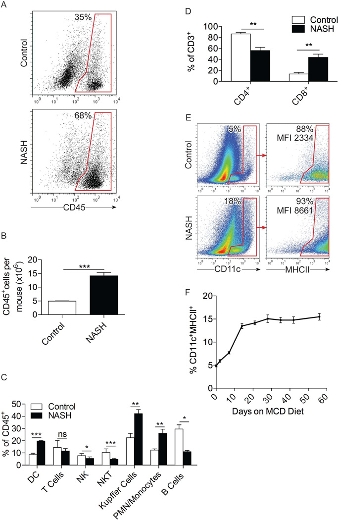
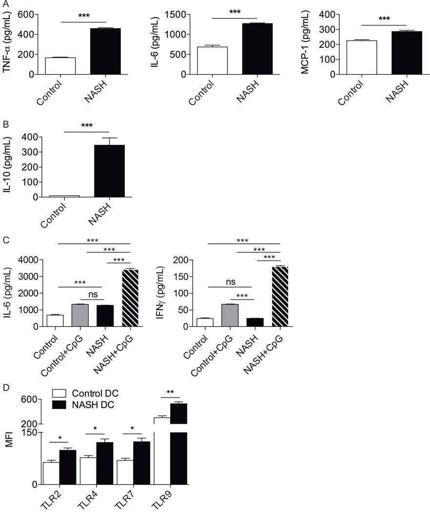
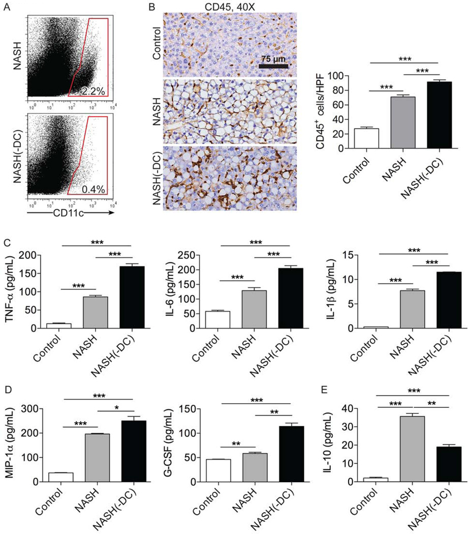
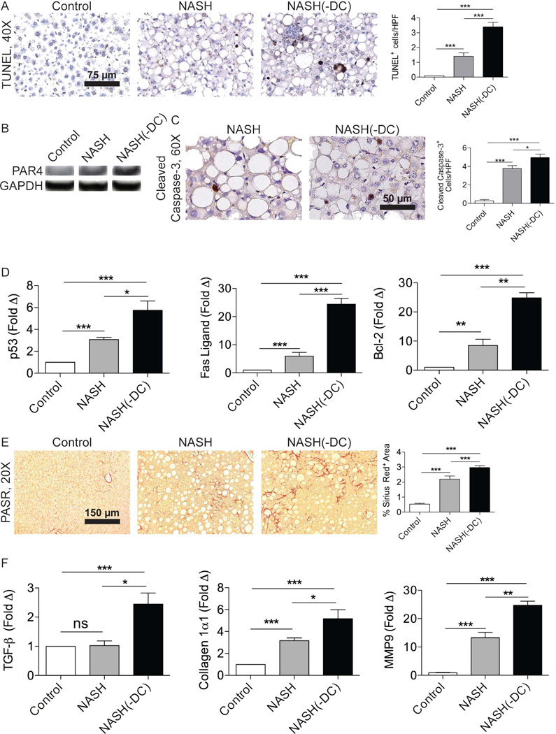
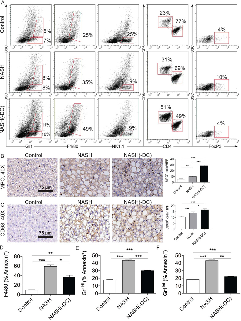
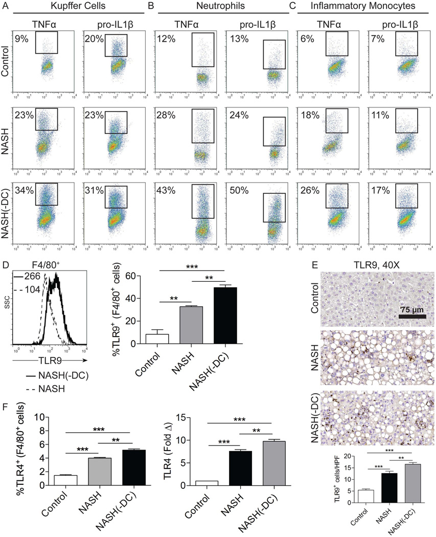
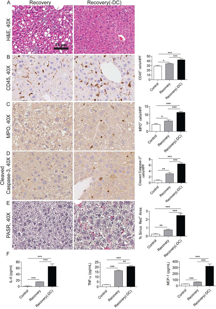
Nonalcoholic steatohepatitis (NASH) is the most common etiology of chronic liver dysfunction in the United States and can progress to cirrhosis and liver failure. Inflammatory insult resulting from fatty infiltration of the liver is central to disease pathogenesis. Dendritic cells (DCs) are antigen-presenting cells with an emerging role in hepatic inflammation. We postulated that DCs are important in the progression of NASH. We found that intrahepatic DCs expand and mature in NASH liver and assume an activated immune phenotype. However, rather than mitigating the severity of NASH, DC depletion markedly exacerbated intrahepatic fibroinflammation. Our mechanistic studies support a regulatory role for DCs in NASH by limiting sterile inflammation through their role in the clearance of apoptotic cells and necrotic debris. We found that DCs limit CD8(+) T-cell expansion and restrict Toll-like receptor expression and cytokine production in innate immune effector cells in NASH, including Kupffer cells, neutrophils, and inflammatory monocytes. Consistent with their regulatory role in NASH, during the recovery phase of disease, ablation of DC populations results in delayed resolution of intrahepatic inflammation and fibroplasia.

Conclusion: Our findings support a role for DCs in modulating NASH. Targeting DC functional properties may hold promise for therapeutic intervention in NASH.


Figures

Figure 1
Figure 1. DC expand in NASH liver
(a) The fraction and (b) total number of CD45+ leukocytes in control and NASH liver (c) as well as the fraction of specific hepatic leukocyte subsets were determined by flow cytometry at 6 weeks after beginning an MCD diet in C57BL/6 mice. (d) The fraction of CD4+ and CD8+ T cells in control and NASH liver was determined by flow cytometry. (e) Co-expression of the DC markers CD11c and MHCII in control and NASH liver at 6 weeks and (f) the time course of hepatic DC recruitment in mice fed an MCD diet were determined by flow cytometry. Experiments were repeated more than 5 times with similar results using 3–5 mice per data point (*p<0.05, **p<0.01, ***p<0.001).
Figure 2
Figure 2. NASH DC are pro-inflammatory
DC derived from control and NASH liver in C57BL/6 mice at 6 weeks after beginning an MCD diet were tested for production of (a) TNF-α, IL-6, MCP-1, and (b) IL-10 in cell culture supernatant. (c) DC production of IL-6 and IFN-γ in cell culture supernatant were analyzed after stimulation with TLR9 ligand CpG ODN1826. (d) DC from NASH and control liver were analyzed for surface expression of TLR2 and TLR4 and intracellular expression of TLR7 and TLR9. MFIs are shown for each respective TLR. Experiments were repeated at least 3 times with similar results (*p<0.05, **p<0.01, ***p<0.001).
Figure 3
Figure 3. DC depletion exacerbates inflammation in NASH liver
(a) Splenocytes from NASH and NASH(-DC) mice at 6 weeks after beginning an MCD diet were tested for expression of CD11c. (b) Paraffin-embedded hepatic sections were stained using an mAb against CD45. The number of CD45+ cells per HPF was calculated. NPC production of (c) TNF-α, IL-6, IL-1β, (d) MIP-1α, G-CSF, and (e) IL-10 in cell culture supernatant was determined in control, NASH, and NASH(-DC) liver. Experiments were repeated at least 3 times using 3–5 mice per group (*p<0.05, **p<0.01, ***p<0.001).
Figure 4
Figure 4. DC depletion exacerbates apoptosis and fibrosis in NASH liver
(a) TUNEL staining was performed in control, NASH, and NASH(-DC) liver from mice sacrificed 6 weeks after beginning an MCD diet. The number of apoptotic bodies per HPF was quantified. (b) Lysates from control, NASH, and NASH (-DC) liver were probed for expression of PAR4. (c) Cleaved Caspase-3 staining was performed on paraffin sections from control, NASH, NASH(-DC) liver and results quantified. (d) mRNA from each group was tested for p53, Fas ligand, and Bcl-2 by PCR. (e) Picric Acid Sirius Red (PASR) staining was performed in control, NASH, and NASH (-DC) liver. Fibrosis was quantified by examining 10 HPFs per liver. (f) TGF-β, Collagen Iα1, and MMP9 expression were determined by PCR. Experiments were repeated at least 3 times with similar results (*p<0.05, **p<0.01, ***p<0.001).
Figure 5
Figure 5. DC depletion increases Kupffer cell and granulocyte proliferation in NASH liver
(a) Gr1, F4/80, and NK1.1 expression in bulk liver NPC, co-expression of CD4 and CD8 on hepatic CD3+ T cells, and expression of FoxP3 on CD4+CD25+ T cells were analyzed in control, NASH, and NASH(-DC) liver from mice sacrificed 6 weeks after initiating an MCD diet. (b) MPO and (c) CD68 IHC were performed on paraffin-embedded liver sections. The number of positive cells per HPF was quantified. (d–f) The fraction of Annexin V+ apoptotic (d) Kupffer cells, (e) neutrophils and (f) inflammatory monocytes in control, NASH, and NASH (-DC) liver were determined by flow cytometry. Experiments were repeated 3 times with similar results (*p<0.05, **p<0.01, ***p<0.001).
Figure 6
Figure 6. DC depletion increases activation of Kupffer cells, neutrophils, and inflammatory monocytes in NASH liver
(a–c) Intracellular expression of TNF-α and pro-IL-1β in freshly isolated (a) F4/80+ Kupffer cells, (b) Gr1HiCD11b+ neutrophils, and (c) Gr1IntCD11b+ inflammatory monocytes from control, NASH, and NASH(-DC) liver is shown. (d) Expression of TLR9 in Kupffer cells was determined by flow cytometry. MFI is indicated and the fraction TLR9+ Kupffer cells was quantified for each group. (e) Paraffin-embedded hepatic sections were stained using an mAb directed against TLR9. The number of positive cells per HPF was quantified. (f) F4/80+ Kupffer cell expression of TLR4 on was determined by flow cytometry and whole liver tissue expression level of TLR4 was determined by PCR in control, NASH, and NASH(-DC) groups. In all experiments, mice were sacrificed 6 weeks after initiating an MCD diet. Experiments were repeated at least three times with similar results (**p<0.01, ***p<0.001).
Figure 7
Figure 7. DC depletion delays resolution of fibro-inflammation during recovery from NASH
(a) Representative H&E staining is shown in livers of mice sacrificed three days after of cessation of an MCD diet (Recovery) and in mice where DC were depleted coincident with MCD diet cessation (Recovery(-DC)). (b) CD45, (c) MPO, and (d) Cleaved Caspase-3 expression were tested by IHC in Recovery and Recovery(-DC) liver. (e) Picric Acid Sirius Red (PASR) staining was performed in Recovery, and Recovery(-DC) liver. Data were quantified by examining 10 HPFs per mouse. (f) NPC production of IL-6, TNF-α, and MCP-1 in cell culture supernatant were measured in each group (n=3–5 mice/group; *p<0.05, ** p<0.01, ***p<0.001).

Comment in

References

    1. Younossi ZM, Stepanova M, Afendy M, et al. Changes in the Prevalence of the Most Common Causes of Chronic Liver Diseases in the United States From 1988 to 2008. Clinical gastroenterology and hepatology : the official clinical practice journal of the American Gastroenterological Association. 2011;9:524.e1–530.e1. - PubMed
    1. Marrero J, Fontana R, Su G, et al. NAFLD May Be a Common Underlying Liver Disease in Patients with Hepatocellular Carcinoma in the United States. Hepatology. 2002;36:1349–1354. - PubMed
    1. Afzali A, Berry K, Ioannou GN. Excellent posttransplant survival for patients with nonalcoholic steatohepatitis in the United States. Liver Transplantation. 2012;18:29–37. - PubMed
    1. Miura K, Kodama Y, Inokuchi S, et al. Toll-Like Receptor 9 Promotes Steatohepatitis by Induction of Interleukin-1β in Mice. Gastroenterology. 2010;139:323.e7–334.e7. - PMC - PubMed
    1. Tomita K, Tamiya G, Ando S, et al. Tumour necrosis factor α signalling through activation of Kupffer cells plays an essential role in liver fibrosis of non-alcoholic steatohepatitis in mice. Gut. 2006;55:415–424. - PMC - PubMed

Publication types

MeSH terms

Substances