. 2013 Mar 8;288(10):7373-86.
doi: 10.1074/jbc.M112.434431. Epub 2013 Jan 14.

Architecture and assembly of HIV integrase multimers in the absence of DNA substrates

Affiliations

Architecture and assembly of HIV integrase multimers in the absence of DNA substrates

Ravi Shankar Bojja et al. J Biol Chem. .

Abstract

We have applied small angle x-ray scattering and protein cross-linking coupled with mass spectrometry to determine the architectures of full-length HIV integrase (IN) dimers in solution. By blocking interactions that stabilize either a core-core domain interface or N-terminal domain intermolecular contacts, we show that full-length HIV IN can form two dimer types. One is an expected dimer, characterized by interactions between two catalytic core domains. The other dimer is stabilized by interactions of the N-terminal domain of one monomer with the C-terminal domain and catalytic core domain of the second monomer as well as direct interactions between the two C-terminal domains. This organization is similar to the "reaching dimer" previously described for wild type ASV apoIN and resembles the inner, substrate binding dimer in the crystal structure of the PFV intasome. Results from our small angle x-ray scattering and modeling studies indicate that in the absence of its DNA substrate, the HIV IN tetramer assembles as two stacked reaching dimers that are stabilized by core-core interactions. These models of full-length HIV IN provide new insight into multimer assembly and suggest additional approaches for enzyme inhibition.


Figures

FIGURE 1.
FIGURE 1.
Schema for alternate apoIN assemblies. Two possible HIV apoIN dimer forms are illustrated in this schematic. The three domains common to retroviral integrase proteins are depicted in color-coded shapes identified in the monomer as N for the red NTD, C for the green CTD, and core for the blue catalytic core domain. A small white circle symbolizes the active site in the core. The arrangement of domains in the monomer and reaching dimer are adapted from the published architectures of ASV monomers and dimers (3). The arrangement of the core-core dimer is adapted from the “outer” dimers in the crystal structure of the PFV intasome (2). A crystal structure of the full-length apoIN tetramer is not available. An architecture based on our solution studies of HIV IN is proposed in the present study (cf. Fig. 10).
FIGURE 2.
FIGURE 2.
HIV IN is enzymatically active in the presence of urea. A, shown are CD spectra of wild type HIV-1 IN in the absence and indicated concentrations of urea. Fluorescence-based assays for single end processing (B) and joining (C) have been described previously (44). Reactions included the indicated concentrations of urea. D, to measure concerted integration, a modification of the conditions described by Li and Craigie (45), was used. HIV IN was first treated with 10 mm EDTA overnight at 4 °C in a buffer that included 1 m NaCl to optimize activity (19) as described in the supplemental Experimental Procedures. Positions of half-site and concerted integration products are indicated by the arrows; positions of Form I (supercoiled) and Form II (relaxed) circles of the target DNA are also shown. Lane 5, a negative control, contained all reaction components except MgCl2. Lane 6 contained only the target DNA. Markers are shown in lane 7. As with the SAXS experiments, the HIV IN analyzed in these experiments was a His6-tagged version of the wild type protein.
FIGURE 3.
FIGURE 3.
Processing activities of HIV-1 IN Phe-181- and E11K-substituted derivatives. A, shown are Phe-181-substituted proteins compared with wild type IN. Proteins were treated with 10 mm EDTA overnight at 4 °C in storage buffer to optimize activity (19) (see supplemental Experimental Procedures for details). B, shown is a comparison of wild type HIV IN and the Glu-11-substituted protein. The fluorescence-based assay for single 3′ end-processing and details of data acquisition and processing were as described (44).
FIGURE 4.
FIGURE 4.
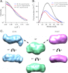
SAXS data and envelopes for wild type HIV IN and the F181T and E11K derivatives. A, shown is intensity versus Q (A−1) at similar protein concentrations (1.6 mg/ml). B, P(r) functions show distinct curves but similar Dmax. C, shown are two orthogonal views of the SAXS-determined envelopes of the wild type HIV IN tetramer and the F181T and E11K dimers. Data were obtained as described under “Experimental Procedures” (see also supplemental Fig. S2).
FIGURE 5.
FIGURE 5.
Comparison of P(r) functions of wild type HIV IN and a D64N derivative in the presence and absence of the metal cofactor (Mg2+).
FIGURE 6.
FIGURE 6.
Destabilization of the wild type HIV IN tetramer and F181T dimer assembly by EDTA treatment. SAXS envelopes were derived for proteins treated with EDTA and separated by size exchange chromatography (SEC-SAXS).
FIGURE 7.
FIGURE 7.
Inter-protein proximities in HIV IN dimers identified by chemical cross-linking coupled with mass spectrometry. A, shown is SDS-PAGE separation of EDC cross-linked monomers, dimers, and tetramers from 1:1 mixtures of isotopically labeled HIV IN proteins and isolation of the dimer bands for mass spectrometry. i, IN F181T at 1:1 mixtures of unlabeled and isotopically labeled protein at 25 μm final concentration. EDC was added at increasing molar ratios of 5:1, 20:1, 50:1, and 100:1. The dimer band was excised from the 5:1 lane as indicated. ii, IN F181T at a 1:1 mixture at 250 nm final concentration of the labeled and unlabeled proteins. Molar ratios of EDC were 20:1, 50:1, and 100:1, and the dimer band was excised from the 100:1 lane as indicated. iii, HIV IN wild type protein 1:1 mixture at 450 nm final concentration of labeled and unlabeled protein. Molar ratios of EDC were 5:1, 20:1, 50:1, and 100:1. The dimers were excised from the 5:1 lane as indicated. B, cross-linked residues were identified by mass spectrometry analysis of EDC cross-linked dimers formed with a 25 μm mixture of labeled and unlabeled IN F181T (Ai). C, cross-linked residues were identified by mass spectrometry analysis of EDC cross-linked dimers formed with the 250 nm mixture of labeled and unlabeled IN F181T (Aii). D, cross-linked residues were identified by mass spectrometry analysis of the EDC cross-linked dimers formed from a 450 nm mixture of labeled and unlabeled HIV IN wild type (Aiii). Cross-links between different domains of the labeled and unlabeled proteins are indicated with dashed black lines, and cross-links between like domains are indicated by solid lines. NTD-NTD cross-links are in red, core-core cross-links are in blue, and CTD-CTD cross-links are in green.
FIGURE 8.
FIGURE 8.
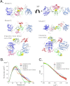
Models of the HIV IN F181T reaching dimer and selection of the best fit. A, shown is a depiction of the three HADDOCK-derived models, generated by docking with distance constraints from EDC cross-links, and published intasome models sans DNA. The best-fit Model A, which includes NTD and CTD stacking interactions at the interface, is shown in two orthogonal views. The interface of this reaching dimer includes interactions between the NTD of one monomer (red) to the NTD of the second monomer (pink) and also Trp-243:Trp-243 stacking between the subunit CTDs (yellow spheres). Model B was derived without Trp-243 stacking, and model C was derived with inclusion of NTD to core interactions. The intasome and intasome inner dimer models are derived from the published HIV intasome model (22) by omitting the viral DNAs. IN domains are colored as in Fig. 1, and the second monomer is shown in faded colors. B and C shown a comparison of the theoretical P(r) functions and scattering profiles (generated with CRYSOL) of the HADDOCK models and the intasome inner dimer with experimental data.
FIGURE 9.
FIGURE 9.
Arrangement of the HIV F181T reaching dimer and a model of the HIV E11K core-core dimer in their respective SAXS-derived envelopes. A, left, the SAXS envelope for F181T with the model A structure from Fig. 8 is shown. Right, shown is a model of E11K fit into its SAXS-derived envelope. B, shown are P(r) plots for F181T and E11K proteins compared with those derived for the E11K model shown in A. C, shown is a plot of the scattering data obtained with the E11K protein compared with plots derived for the F181T and E11K models in A.
FIGURE 10.
FIGURE 10.
Inclusion of established core-core interactions to generate a likely HIV IN tetramer assembly via direct stacking of two reaching dimers. A, shown is a proposed view of core-core interactions between the two reaching dimers, which maintains a Dmax similar to that of the F181T dimer. B, shown are two orthogonal views of the tetramer model arranged inside the SAXS determined envelope of the wild type HIV IN tetramer; for clarity, each of the monomers are colored individually. C, shown is a comparison of experimental data for the wild type HIV IN tetramer with the proposed tetramer model and an intasome tetramer model without the viral DNA substrate.

Similar articles

Cited by

References

    1. Craigie R. (2012) The molecular biology of HIV integrase. Future Virol. 7, 679–686 - PMC - PubMed
    1. Hare S., Gupta S. S., Valkov E., Engelman A., Cherepanov P. (2010) Retroviral intasome assembly and inhibition of DNA strand transfer. Nature 464, 232–236 - PMC - PubMed
    1. Bojja R. S., Andrake M. D., Weigand S., Merkel G., Yarychkivska O., Henderson A., Kummerling M., Skalka A. M. (2011) Architecture of a full-length retroviral integrase monomer and dimer, revealed by small angle x-ray scattering and chemical cross-linking. J. Biol. Chem. 286, 17047–17059 - PMC - PubMed
    1. Grabarek Z., Gergely J. (1990) Zero-length cross-linking procedure with the use of active esters. Anal. Biochem. 185, 131–135 - PubMed
    1. Kessl J. J., Eidahl J. O., Shkriabai N., Zhao Z., McKee C. J., Hess S., Burke T. R., Jr., Kvaratskhelia M. (2009) An allosteric mechanism for inhibiting HIV-1 integrase with a small molecule. Mol. Pharmacol. 76, 824–832 - PMC - PubMed

Publication types

MeSH terms