. 2013 Apr;54(4):1066-76.
doi: 10.1194/jlr.M034314. Epub 2013 Jan 14.

Micrometric segregation of fluorescent membrane lipids: relevance for endogenous lipids and biogenesis in erythrocytes

Affiliations

Micrometric segregation of fluorescent membrane lipids: relevance for endogenous lipids and biogenesis in erythrocytes

Ludovic D'Auria et al. J Lipid Res. 2013 Apr.

Erratum in

  • J Lipid Res. 2013 Jul;54(7):2029

Abstract

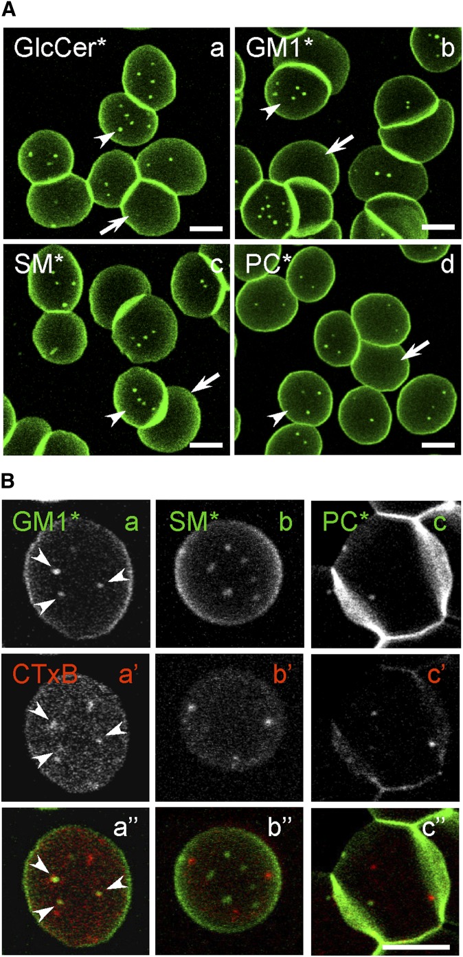
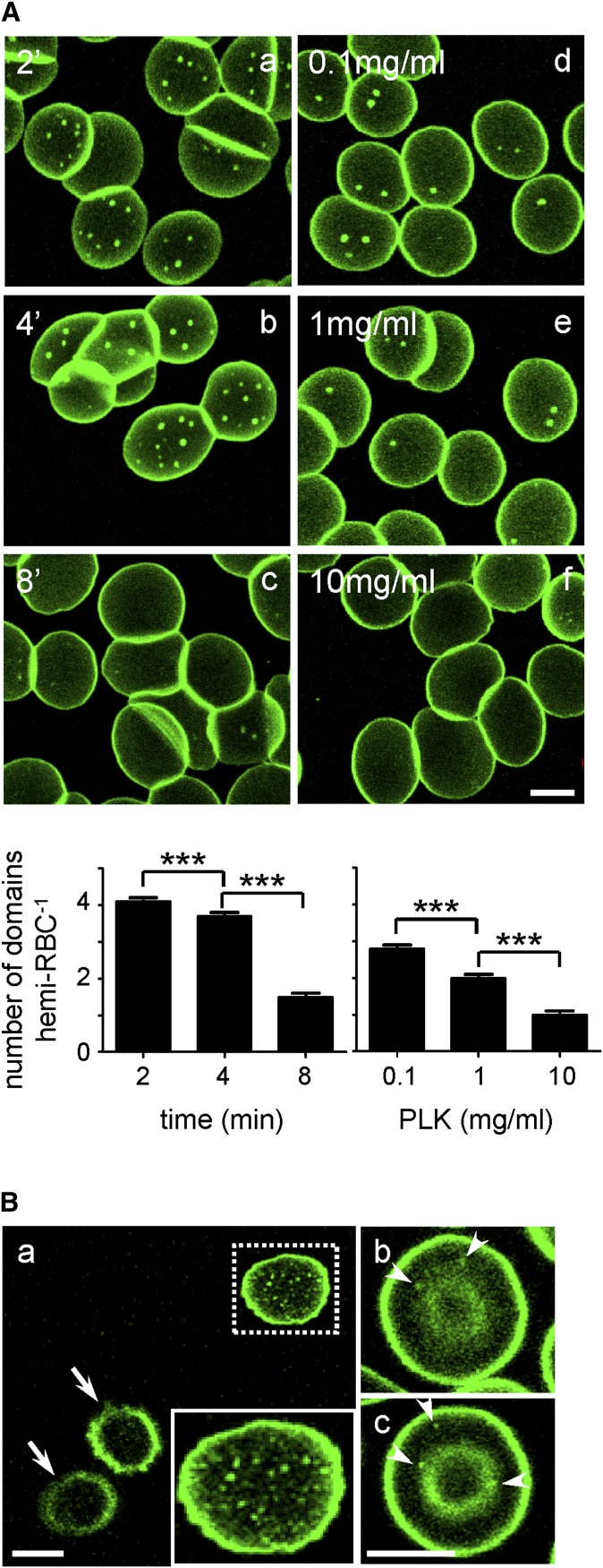
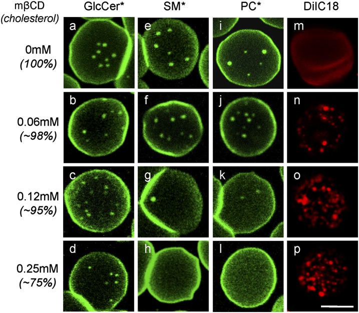
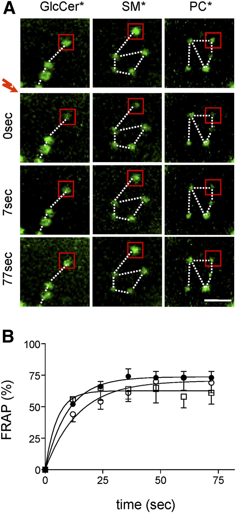
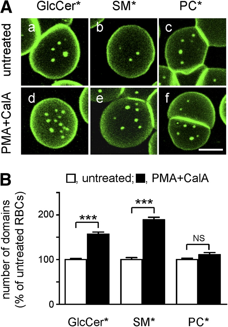
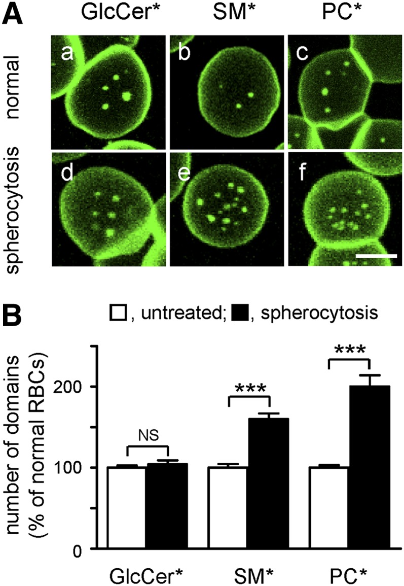
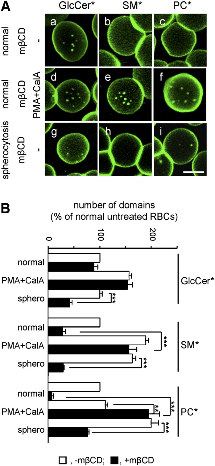
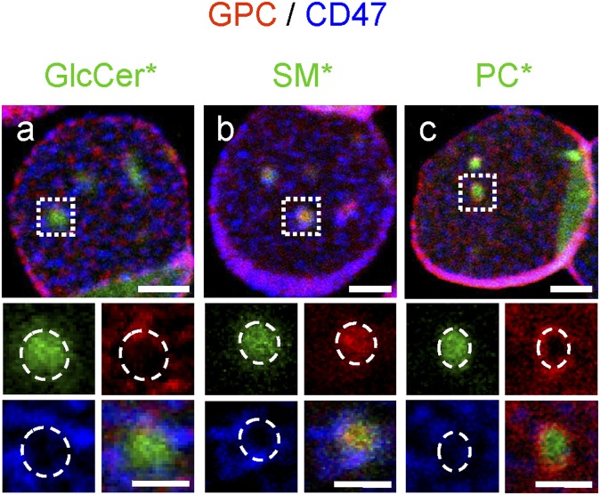
Micrometric membrane lipid segregation is controversial. We addressed this issue in attached erythrocytes and found that fluorescent boron dipyrromethene (BODIPY) analogs of glycosphingolipids (GSLs) [glucosylceramide (BODIPY-GlcCer) and monosialotetrahexosylganglioside (GM1BODIPY)], sphingomyelin (BODIPY-SM), and phosphatidylcholine (BODIPY-PC inserted into the plasma membrane spontaneously gathered into distinct submicrometric domains. GM1BODIPY domains colocalized with endogenous GM1 labeled by cholera toxin. All BODIPY-lipid domains disappeared upon erythrocyte stretching, indicating control by membrane tension. Minor cholesterol depletion suppressed BODIPY-SM and BODIPY-PC but preserved BODIPY-GlcCer domains. Each type of domain exchanged constituents but assumed fixed positions, suggesting self-clustering and anchorage to spectrin. Domains showed differential association with 4.1R versus ankyrin complexes upon antibody patching. BODIPY-lipid domains also responded differentially to uncoupling at 4.1R complexes [protein kinase C (PKC) activation] and ankyrin complexes (in spherocytosis, a membrane fragility disease). These data point to micrometric compartmentation of polar BODIPY-lipids modulated by membrane tension, cholesterol, and differential association to the two nonredundant membrane:spectrin anchorage complexes. Micrometric compartmentation might play a role in erythrocyte membrane deformability and fragility.


Figures

Fig. 1.
Fig. 1.
In adherent RBCs, fluorescent exogenous membrane lipids and cholera toxin binding to endogenous GM1 labeled submicrometric domains. A: Single vital imaging of three classes of polar BODIPY-lipid analogs (*). Freshly isolated RBCs (from a normal donor) were allowed to attach onto high-molecular mass (70–150 kDa) PLK-coverslips for 4 min and spread for another 4 min, then briefly labeled with GSLs* [BODIPY-GlcCer (a), GM1* (b)], BODIPY-SM (c), or BODIPY-PC (d), rinsed and immediately imaged. Notice that each lipid analog forms several micrometric domains on partially spread (<8 μm; arrowheads) but not on fully spread RBCs (>8 μm; arrows). For general views, see supplementary Fig. IV (upper row). B: Double vital imaging of BODIPY-lipid domains versus endogenous GM1 (CTxB labeling). RBCs were sequentially labeled in suspension with the indicated BODIPY-lipids (upper row; green at merge) then with Alexa568-CTxB (middle row; red at merge) in the continued presence of BODIPY-lipids. Cells were then attached onto PLK-coverslips before additional spreading, washed and imaged as in Fig. 1A (sequential recording in the green and red channels, with settings adjusted to best match signal intensities), then merged (lower row). Notice that CTxB patches largely colocalize with GM1* (arrowheads at left) but not with BODIPY-SM and BODIPY-PC domains. All scale bars, 5 μm. *, BODIPY.
Fig. 2.
Fig. 2.
Spontaneous formation of lipid submicrometric domains is regulated by membrane tension. A: Extended spreading onto PLK-coverslips by increased adhesion time (a–c) or increased PLK concentration (d–f) abrogate micrometric BODIPY (*) lipid domains. Upper panel, representative confocal images. Freshly isolated normal RBCs were attached for 4 min either onto coverslips precoated with 0.1 mg/ml 70–150 kDa PLK with additional spreading time as indicated (a–c), or onto coverslips coated with the indicated concentrations of 1–5 kDa PLK before additional spreading for 4 min (d–f), then labeled with BODIPY-GlcCer (as an example). Notice the highest number of BODIPY-GlcCer domains for shortest times and lowest concentrations of PLK (a, b, d, e) and their disappearance upon extended stretching (c, f). Scale bar, 5 μm. Lower panel, morphometry. Number of BODIPY-GlcCer domains are mean ± SEM of 93–1,144 RBCs from 3 to 6 independent experiments at left and of 37–346 RBCs from 2 independent experiments at right. B: Fluorescent lipids label submicrometric domains on loosely-attached RBCs. RBCs were attached either onto 0.01 mg/ml 1–5 kDa PLK-coverslips for 4 min before additional spreading for 4 min (a), or onto plastic IBIDI chambers coated or not (b) with 1–5 kDa PLK (c) and labeled with BODIPY-GlcCer. Although most RBCs appear as echinocytes [arrows in (a)], the rare flat RBCs show numerous domains with variable size at very low PLK concentration [inset in (a)]. Domains can also be found on barely-attached discocytes/stomatocytes [arrowheads in (b, c)]. Scale bars, 5 μm. *** p < 0.001.
Fig. 3.
Fig. 3.
Moderate cholesterol depletion suppresses BODIPY-SM and BODIPY-PC, but not BODIPY-GlcCer, domains and induces DiIC18 patches. Normal RBCs were treated in suspension with the indicated mβCD concentrations (for level of depletion and lack of toxicity, see supplementary Fig. IIIA), then attached onto PLK-coverslips for 4 min, spread for an additional 4 min, labeled with BODIPY-GlcCer (a–d), BODIPY-SM (e–h), BODIPY-PC (i–l), or DiIC18 (m–p), rinsed, and imaged as in Fig. 1A. Notice the disappearance of BODIPY-SM and BODIPY-PC domains under moderate (∼25%) cholesterol depletion (g, h, k, l) contrasting with better preservation of BODIPY-GlcCer domains (a–d), and conversely, induction of DiIC18 patches (m–p). Scale bar, 5 μm. For general views, see supplementary Fig. IIIB. *, BODIPY.
Fig. 4.
Fig. 4.
Micrometric BODIPY-lipid domains assume fixed positions but rapidly exchange their constituents. Normal RBCs were attached onto PLK-coverslips, spread, and labeled with BODIPY-GlcCer [left column in (A); open squares in (B)], BODIPY-SM [central column in (A); closed circles in (B)], or BODIPY-PC [right in (A); open circles in (B)], then processed for FRAP at 37°C in 5 μm2 fields centered on a micrometric domain [exemplified by red squares in (A)]. A: Representative vital imaging. Red flash indicates bleaching of areas shown by red squares. Scale bar, 2 μm. Notice domain immobility (fixed positions as in constellations, dotted lines). B: Quantification. Fluorescence recovery is expressed as percentage of signal before photobleaching, after correction for residual fluorescence immediately after bleaching. Curves are derived by monoexponential fitting. Notice the fast (t1/2 ∼10 s) and extensive fluorescence recovery of SL* (BODIPY-GlcCer, BODIPY-SM) domains in bleached areas, indicating high mobility of constituents. *, BODIPY.
Fig. 5.
Fig. 5.
Anchorage via 4.1R-based complexes restricts BODIPY-GlcCer and BODIPY-SM, but not BODIPY-PC, domains. A: Representative vital imaging. RBCs from a normal donor were attached onto PLK-coverslips and spread as in Fig. 1, then either kept untreated (a–c) or treated with PMA as PKC activator and the phosphatase inhibitor CalA to uncouple membrane:cytoskeleton at 4.1R complexes (d–f), labeled with BODIPY-GlcCer, BODIPY-SM, or BODIPY-PC and imaged (in the continued presence of PMA+CalA if appropriate). Scale bar, 5 μm. For general views, see supplementary Fig. IVb, e, h. B: Morphometry. Abundance of BODIPY-GlcCer, BODIPY-SM, and BODIPY-PC domains on untreated (open bars) and PMA+CalA-treated RBCs (filled bars) is expressed as the percentage of untreated cells and are mean ± SEM of: i) 296 (untreated) and 361 (PMA+CalA) RBCs from 3 independent experiments for BODIPY-GlcCer domains; ii) 256 and 471 from 5 experiments for BODIPY-SM domains; and iii) 238 and 117 from 2 experiments for BODIPY-PC domains. *, BODIPY; NS, not significant. *** p < 0.001.
Fig. 6.
Fig. 6.
Anchorage via ankyrin-based complexes restricts BODIPY-SM and BODIPY-PC, but not BODIPY-GlcCer, domains. A: Representative vital imaging. RBCs from a normal donor (normal; a–c) versus a spherocytotic patient (spherocytosis; d–f), both splenectomised, were attached onto PLK-coverslips, spread, and labeled with BODIPY-GlcCer, BODIPY-SM, or BODIPY-PC for vital imaging. Scale bar, 5 μm. For general views, see supplementary Fig. IV, lower row. B: Morphometry. Abundance of BODIPY-GlcCer, BODIPY-SM, and BODIPY-PC domains on normal (open bars) and spherocytotic RBCs (filled bars) is expressed by reference to normal RBCs and are mean ± SEM of: i) 200 and 558 RBCs for BODIPY-GlcCer domains; ii) 114 and 454 RBCs for BODIPY-SM domains; and iii) 114 and 398 RBCs for BODIPY-PC domains, each from 2 independent experiments. *, BODIPY; NS, not significant. *** p < 0.001.
Fig. 7.
Fig. 7.
High cholesterol promotes BODIPY-SM and BODIPY-PC domains via 4.1R complexes and BODIPY-GlcCer domains via ankyrin complexes. A: Representative vital imaging. RBCs from a normal donor (normal; a–f) versus a spherocytotic patient (P#1; spherocytosis; g–i), both splenectomised, were preincubated in suspension with 0.25 mM mβCD as in Fig. 3, attached onto PLK-coverslips, spread, labeled with BODIPY-GlcCer (a, d, g), BODIPY-SM (b, e, h), or BODIPY-PC (c, f, i), then either kept in fresh medium (a–c, g–i) or further incubated with PMA+CalA (d–f) as in Fig. 5. Scale bar, 5 μm. B: Morphometry. Abundance of BODIPY-GlcCer, BODIPY-SM, or BODIPY-PC domains in the absence (open bars) or presence of mβCD (filled bars) is expressed by reference to untreated normal RBCs and are mean ± SEM of: i) 37–361 RBCs from 1 to 5 independent experiments for BODIPY-GlcCer domains; ii) 50–471 RBCs from 2 to 6 experiments for BODIPY-SM domains; and iii) 35–300 RBCs from 2 to 6 experiments for BODIPY-PC domains. *, BODIPY. ** p < 0.01; *** p < 0.001.
Fig. 8.
Fig. 8.
Differential association of micrometric BODIPY-lipid domains with patched glycophorin C and CD47. RBCs from a normal donor were attached onto PLK-coverslips, labeled with BODIPY-lipid (green), then GPC (4.1R complex; red) and CD47 (ankyrin complex; blue) were patched by antibodies. Notice that GPC patches selectively overlapped BODIPY-SM domains (b), while circumscribing BODIPY-PC domains (c). In contrast, CD47 patches were never as tightly linked to lipid-domains, but frequently seemed in closer proximity to BODIPY-GlcCer and BODIPY-SM (a, b) than to BODIPY-PC domains (c). Scale bars, 2 μm; at insets, 1 μm. Dashed circles at insets indicate lipid* domain boundaries. *, BODIPY.

References

    1. Singer S. J., Nicolson G. L. 1972. The fluid mosaic model of the structure of cell membranes. Science. 175: 720–731. - PubMed
    1. Lingwood D., Simons K. 2010. Lipid rafts as a membrane-organizing principle. Science. 327: 46–50. - PubMed
    1. Lingwood D., Ries J., Schwille P., Simons K. 2008. Plasma membranes are poised for activation of raft phase coalescence at physiological temperature. Proc. Natl. Acad. Sci. USA. 105: 10005–10010. - PMC - PubMed
    1. Pike L. J. 2006. Rafts defined: a report on the Keystone Symposium on Lipid Rafts and Cell Function. J. Lipid Res. 47: 1597–1598. - PubMed
    1. Fujita A., Cheng J., Hirakawa M., Furukawa K., Kusunoki S., Fujimoto T. 2007. Gangliosides GM1 and GM3 in the living cell membrane form clusters susceptible to cholesterol depletion and chilling. Mol. Biol. Cell. 18: 2112–2122. - PMC - PubMed

Publication types

MeSH terms

LinkOut - more resources