

A genome-wide siRNA screen reveals positive and negative regulators of the NOD2 and NF-κB signaling pathways
- PMID: 23322906
- PMCID: PMC3887559
- DOI: 10.1126/scisignal.2003305
A genome-wide siRNA screen reveals positive and negative regulators of the NOD2 and NF-κB signaling pathways
Abstract
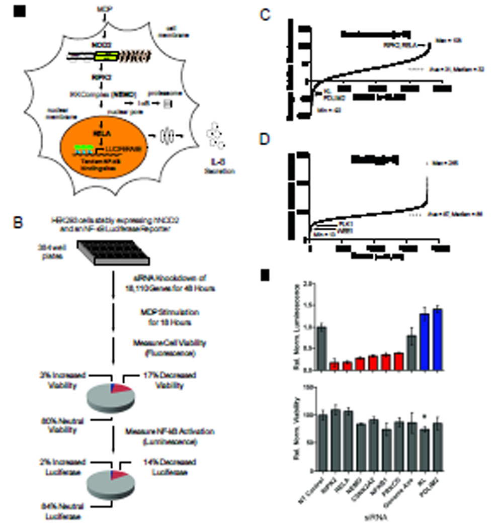
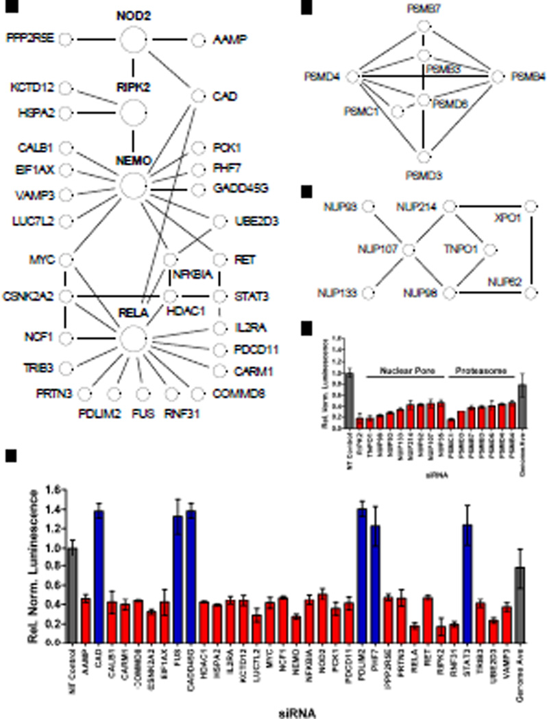
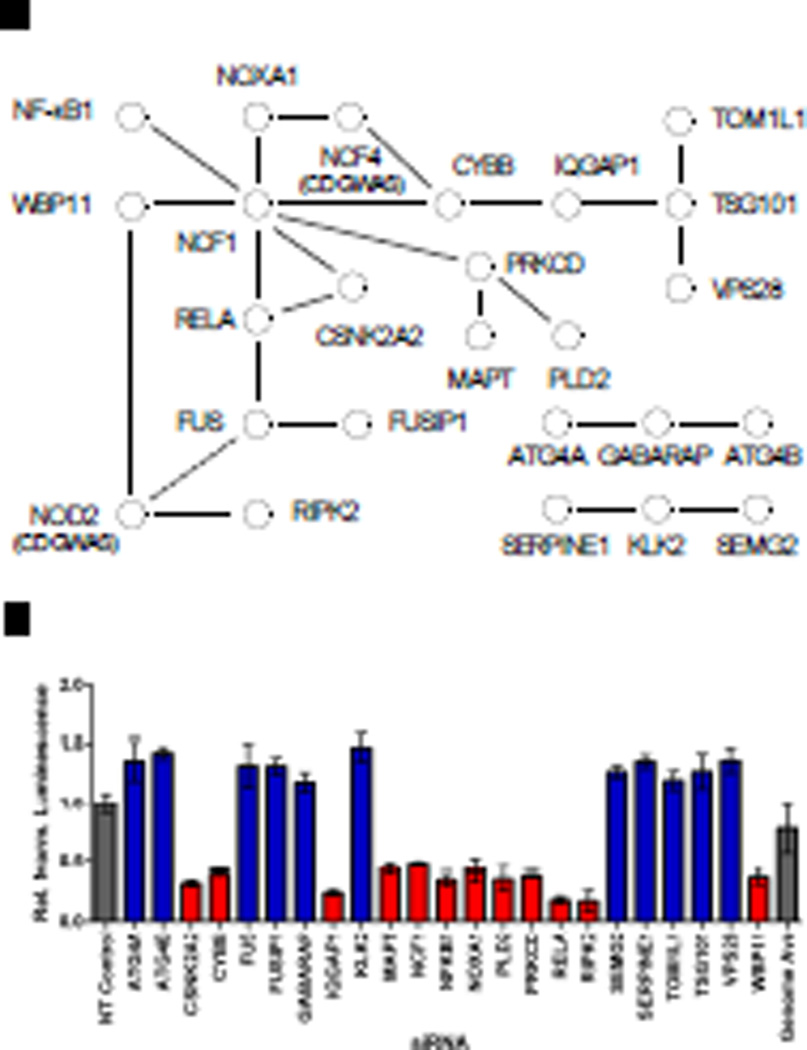
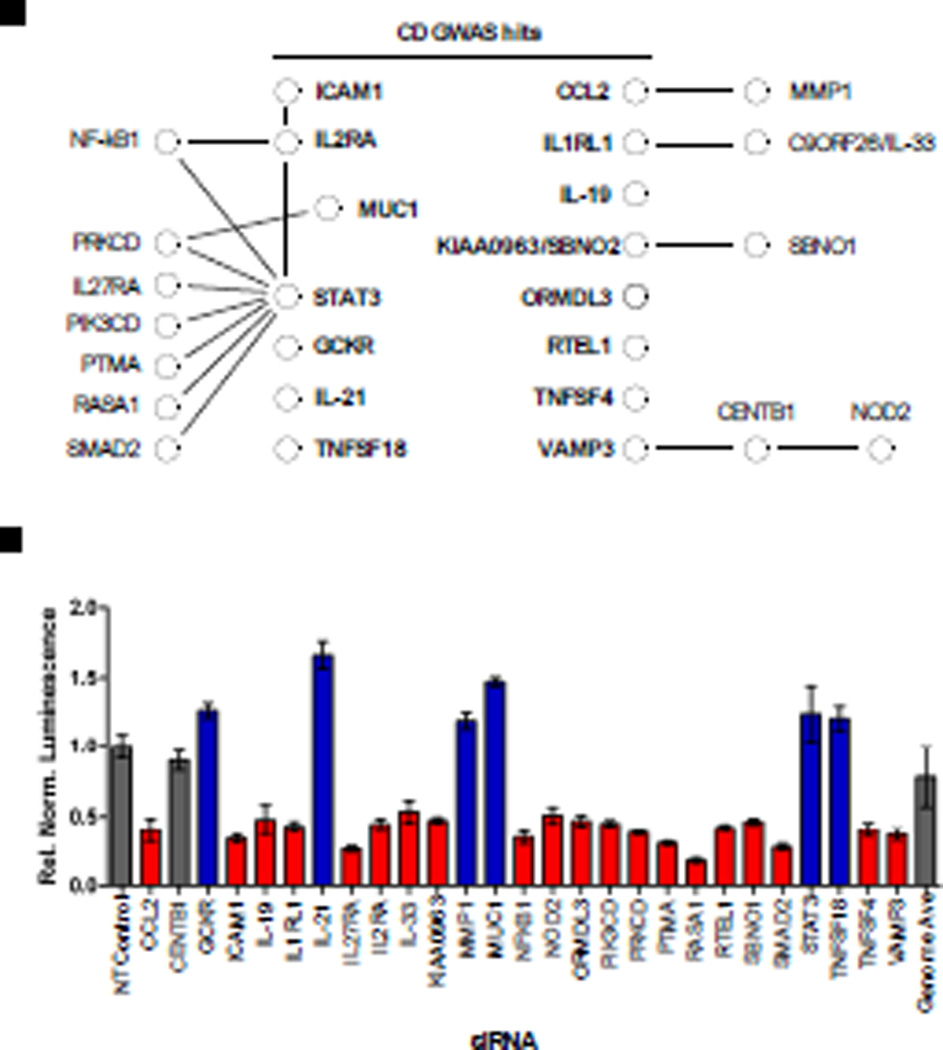
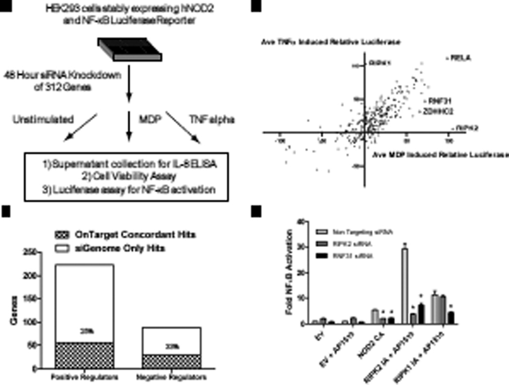
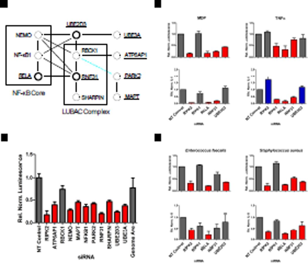
The cytoplasmic receptor NOD2 (nucleotide-binding oligomerization domain 2) senses peptidoglycan fragments and triggers host defense pathways, including activation of nuclear factor κB (NF-κB) signaling, which lead to inflammatory immune responses. Dysregulation of NOD2 signaling is associated with inflammatory diseases, such as Crohn's disease and Blau syndrome. We used a genome-wide small interfering RNA screen to identify regulators of the NOD2 signaling pathway. Several genes associated with Crohn's disease risk were identified in the screen. A comparison of candidates from this screen with other "omics" data sets revealed interconnected networks of genes implicated in NF-κB signaling, thus supporting a role for NOD2 and NF-κB pathways in the pathogenesis of Crohn's disease. Many of these regulators were validated in secondary assays, such as measurement of interleukin-8 secretion, which is partially dependent on NF-κB. Knockdown of putative regulators in human embryonic kidney 293 cells followed by stimulation with tumor necrosis factor-α revealed that most of the genes identified were general regulators of NF-κB signaling. Overall, the genes identified here provide a resource to facilitate the elucidation of the molecular mechanisms that regulate NOD2- and NF-κB-mediated inflammation.
Figures
Similar articles
-
A genome-wide small interfering RNA (siRNA) screen reveals nuclear factor-κB (NF-κB)-independent regulators of NOD2-induced interleukin-8 (IL-8) secretion.J Biol Chem. 2014 Oct 10;289(41):28213-24. doi: 10.1074/jbc.M114.574756. Epub 2014 Aug 28. J Biol Chem. 2014. PMID: 25170077 Free PMC article.
-
Blau syndrome-associated Nod2 mutation alters expression of full-length NOD2 and limits responses to muramyl dipeptide in knock-in mice.J Immunol. 2015 Jan 1;194(1):349-57. doi: 10.4049/jimmunol.1402330. Epub 2014 Nov 26. J Immunol. 2015. PMID: 25429073 Free PMC article.
-
Blau syndrome NOD2 mutations result in loss of NOD2 cross-regulatory function.Front Immunol. 2022 Sep 15;13:988862. doi: 10.3389/fimmu.2022.988862. eCollection 2022. Front Immunol. 2022. PMID: 36189261 Free PMC article.
-
Nucleotide-binding oligomerization domain containing 2: structure, function, and diseases.Semin Arthritis Rheum. 2013 Aug;43(1):125-30. doi: 10.1016/j.semarthrit.2012.12.005. Epub 2013 Jan 24. Semin Arthritis Rheum. 2013. PMID: 23352252 Review.
-
Functional consequences of NOD2 (CARD15) mutations.Inflamm Bowel Dis. 2006 Jul;12(7):641-50. doi: 10.1097/01.MIB.0000225332.83861.5f. Inflamm Bowel Dis. 2006. PMID: 16804402 Review.
Cited by
-
SYPL1 Inhibits Apoptosis in Pancreatic Ductal Adenocarcinoma via Suppression of ROS-Induced ERK Activation.Front Oncol. 2020 Sep 15;10:1482. doi: 10.3389/fonc.2020.01482. eCollection 2020. Front Oncol. 2020. PMID: 33042794 Free PMC article.
-
Linear ubiquitination in immunity.Immunol Rev. 2015 Jul;266(1):190-207. doi: 10.1111/imr.12309. Immunol Rev. 2015. PMID: 26085216 Free PMC article. Review.
-
Missense variants in NOX1 and p22phox in a case of very-early-onset inflammatory bowel disease are functionally linked to NOD2.Cold Spring Harb Mol Case Stud. 2019 Feb 1;5(1):a002428. doi: 10.1101/mcs.a002428. Print 2019 Feb. Cold Spring Harb Mol Case Stud. 2019. PMID: 30709874 Free PMC article.
-
Harnessing the Complete Repertoire of Conventional Dendritic Cell Functions for Cancer Immunotherapy.Pharmaceutics. 2020 Jul 14;12(7):663. doi: 10.3390/pharmaceutics12070663. Pharmaceutics. 2020. PMID: 32674488 Free PMC article. Review.
-
A Chinese girl of Blau syndrome with renal arteritis and a literature review.Pediatr Rheumatol Online J. 2023 Mar 13;21(1):23. doi: 10.1186/s12969-023-00804-z. Pediatr Rheumatol Online J. 2023. PMID: 36915122 Free PMC article. Review.
References
-
- Chen G, Shaw MH, Kim YG, Nunez G. NOD-like receptors: role in innate immunity and inflammatory disease. Annu Rev Pathol. 2009;4:365–398. - PubMed
-
- Inohara N, Ogura Y, Fontalba A, Gutierrez O, Pons F, Crespo J, Fukase K, Inamura S, Kusumoto S, Hashimoto M, Foster SJ, Moran AP, Fernandez-Luna JL, Nunez G. Host recognition of bacterial muramyl dipeptide mediated through NOD2. Implications for Crohn's disease. J Biol Chem. 2003;278:5509–5512. - PubMed
-
- Girardin SE, Boneca IG, Viala J, Chamaillard M, Labigne A, Thomas G, Philpott DJ, Sansonetti PJ. Nod2 is a general sensor of peptidoglycan through muramyl dipeptide (MDP) detection. J Biol Chem. 2003;278:8869–8872. - PubMed
-
- Kobayashi K, Inohara N, Hernandez LD, Galan JE, Nunez G, Janeway CA, Medzhitov R, Flavell RA. RICK/Rip2/CARDIAK mediates signalling for receptors of the innate and adaptive immune systems. Nature. 2002;416:194–199. - PubMed
Publication types
MeSH terms
Substances
Grants and funding
LinkOut - more resources
Full Text Sources
Other Literature Sources
Molecular Biology Databases
