. 2012 Dec 21;18(47):6935-42.
doi: 10.3748/wjg.v18.i47.6935.

Impaired suppressive activities of human MUTYH variant proteins against oxidative mutagenesis

Affiliations

Impaired suppressive activities of human MUTYH variant proteins against oxidative mutagenesis

Kazuya Shinmura et al. World J Gastroenterol. .

Abstract

Aim: To investigate the suppressive activity of MUTYH variant proteins against mutations caused by oxidative lesion, 8-hydroxyguanine (8OHG), in human cells.

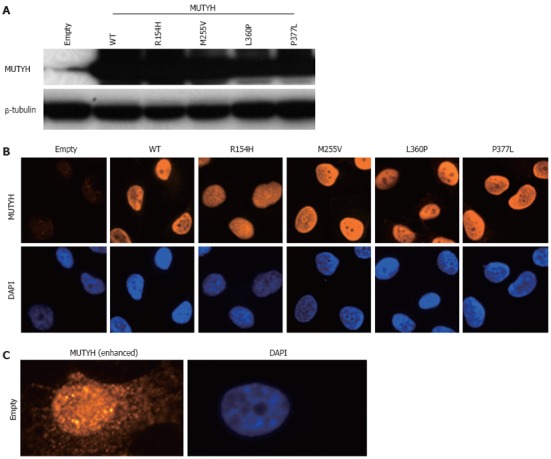
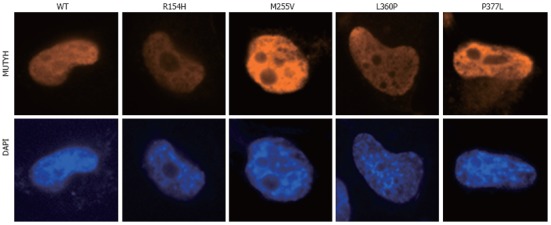
Methods: p.R154H, p.M255V, p.L360P, and p.P377L MUTYH variants, which were previously found in patients with colorectal polyposis and cancer, were selected for use in this study. Human H1299 cancer cell lines inducibly expressing wild-type (WT) MUTYH (type 2) or one of the 4 above-mentioned MUTYH variants were established using the piggyBac transposon vector system, enabling the genomic integration of the transposon sequence for MUTYH expression. MUTYH expression was examined after cumate induction using Western blotting analysis and immunofluorescence analysis. The intracellular localization of MUTYH variants tagged with FLAG was also immunofluorescently examined. Next, the mutation frequency in the supF of the shuttle plasmid pMY189 containing a single 8OHG residue at position 159 of the supF was compared between empty vector cells and cells expressing WT MUTYH or one of the 4 MUTYH variants using a supF forward mutation assay.

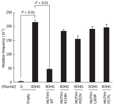
Results: The successful establishment of human cell lines inducibly expressing WT MUTYH or one of the 4 MUTYH variants was concluded based on the detection of MUTYH expression in these cell lines after treatment with cumate. All of the MUTYH variants and WT MUTYH were localized in the nucleus, and nuclear localization was also observed for FLAG-tagged MUTYH. The mutation frequency of supF was 2.2 × 10(-2) in the 8OHG-containing pMY189 plasmid and 2.5 × 10(-4) in WT pMY189 in empty vector cells, which was an 86-fold increase with the introduction of 8OHG. The mutation frequency (4.7 × 10(-3)) of supF in the 8OHG-containing pMY189 plasmid in cells overexpressing WT MUTYH was significantly lower than in the empty vector cells (P < 0.01). However, the mutation frequencies of the supF in the 8OHG-containing pMY189 plasmid in cells overexpressing the p.R154H, p.M255V, p.L360P, or p.P377L MUTYH variant were 1.84 × 10(-2), 1.55 × 10(-2), 1.91 × 10(-2), and 1.96 × 10(-2), respectively, meaning that no significant difference was observed in the mutation frequency between the empty vector cells and cells overexpressing MUTYH mutants.

Conclusion: The suppressive activities of p.R154H, p.M255V, p.L360P, and p.P377L MUTYH variants against mutations caused by 8OHG are thought to be severely impaired in human cells.

Keywords: 8-hydroxyguanine; Colorectal polyposis; MUTYH; MUTYH-associated polyposis; Mutation; Oxidative mutagenesis; piggyBac transposon; supF forward mutation assay.


Figures

Figure 1
Figure 1
Establishment of H1299 human cell lines inducibly expressing MUTYH variant proteins. A: Detection of MUTYH proteins in cumate-inducible stable cell lines expressing MUTYH in the presence of cumate using Western blotting analysis with an anti-MUTYH antibody. Lysates from empty vector-transposed cells and cells inducibly expressing wild-type (WT) MUTYH or p.R154H, p.M255V, p.L360P, or p.P377L MUTYH variants were analyzed. β-tubulin protein was also analyzed as an internal control; B: Immunofluorescence detection of MUTYH proteins expressed in the cell lines used in (A) in the presence of cumate. The MUTYH protein (red) was stained with anti-MUTYH as the primary antibody and Alexa Fluor 594-conjugated goat anti-mouse IgG as the secondary antibody. The nuclei were counterstained with 4’,6-diamidino-2-phenylindole (DAPI) (blue); C: Immunofluorescence detection of endogenous MUTYH proteins in the empty vector-transposed cells as described in (B). The intensity of the signals of MUTYH protein (red) was enhanced with image-editing software to determine the subcellular localization of endogenous MUTYH protein. The nuclei were counterstained with DAPI (blue).
Figure 2
Figure 2
Nuclear localization of MUTYH variant proteins (p.R154H, p.M255V, p.L360P, and p.P377L). H1299 cells were transiently transfected with a vector expressing various types of MUTYH proteins tagged with FLAG, and MUTYH-FLAG protein (red) was stained with anti-FLAG M2 as the primary antibody and Alexa Fluor 594-conjugated goat anti-mouse IgG as the secondary antibody. The nuclei were counterstained with 4’,6-diamidino-2-phenylindole (DAPI) (blue). WT: Wild-type.
Figure 3
Figure 3
Measurement of the mutation frequency of the supF gene in the pMY189 plasmid using a supF forward mutation assay in H1299 human cell lines inducibly expressing MUTYH variant proteins. Empty vector-transposed cells and cells inducibly expressing wild-type (WT) MUTYH or p.R154H, p.M255V, p.L360P, or p.P377L MUTYH variants in the presence of cumate were transfected with a pMY189 shuttle plasmid, and the mutation frequency of supF in these human cell lines was measured. “8-hydroxyguanine (8OHG)” indicates a pMY189 plasmid containing an 8OHG residue at position 159 of supF, while “G” indicates a pMY189 plasmid containing the WT supF gene. The data are shown as the means ± SE.

References

    1. Kasai H, Nishimura S. Hydroxylation of deoxyguanosine at the C-8 position by ascorbic acid and other reducing agents. Nucleic Acids Res. 1984;12:2137–2145. - PMC - PubMed
    1. Shibutani S, Takeshita M, Grollman AP. Insertion of specific bases during DNA synthesis past the oxidation-damaged base 8-oxodG. Nature. 1991;349:431–434. - PubMed
    1. Slupska MM, Luther WM, Chiang JH, Yang H, Miller JH. Functional expression of hMYH, a human homolog of the Escherichia coli MutY protein. J Bacteriol. 1999;181:6210–6213. - PMC - PubMed
    1. Takao M, Zhang QM, Yonei S, Yasui A. Differential subcellular localization of human MutY homolog (hMYH) and the functional activity of adenine: 8-oxoguanine DNA glycosylase. Nucleic Acids Res. 1999;27:3638–3644. - PMC - PubMed
    1. Shinmura K, Yamaguchi S, Saitoh T, Takeuchi-Sasaki M, Kim SR, Nohmi T, Yokota J. Adenine excisional repair function of MYH protein on the adenine: 8-hydroxyguanine base pair in double-stranded DNA. Nucleic Acids Res. 2000;28:4912–4918. - PMC - PubMed

Publication types