

Effect of salvianolate on intestinal epithelium tight junction protein zonula occludens protein 1 in cirrhotic rats
- PMID: 23323006
- PMCID: PMC3531692
- DOI: 10.3748/wjg.v18.i47.7040
Effect of salvianolate on intestinal epithelium tight junction protein zonula occludens protein 1 in cirrhotic rats
Abstract
Aim: To study the effect of salvianolate on tight junctions (TJs) and zonula occludens protein 1 (ZO-1) in small intestinal mucosa of cirrhotic rats.
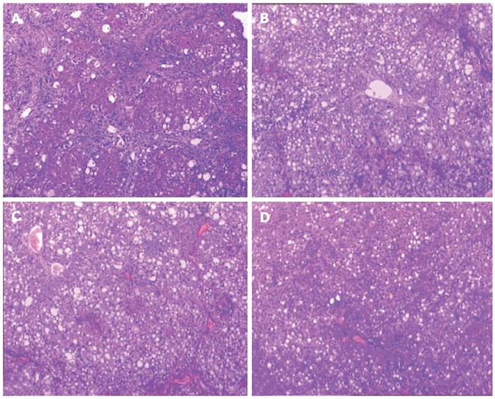
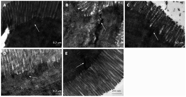
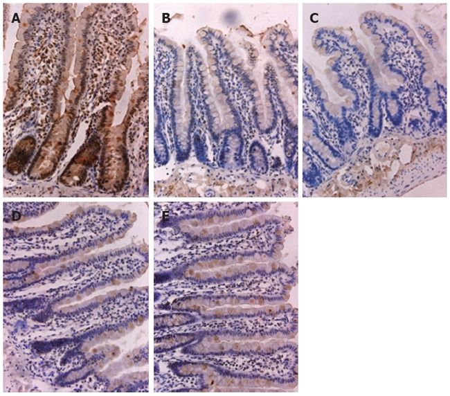
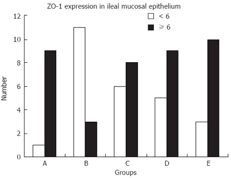
Methods: Cirrhosis was induced using carbon tetrachloride. Rats were randomly divided into the untreated group, low-dose salvianolate (12 mg/kg) treatment group, medium-dose salvianolate (24 mg/kg) treatment group, and high-dose salvianolate (48 mg/kg) treatment group, and were treated for 2 wk. Another 10 healthy rats served as the normal control group. Histological changes in liver tissue samples were observed under a light microscope. We evaluated morphologic indices of ileal mucosa including intestinal villi width and thickness of mucosa and intestinal wall using a pathological image analysis system. Ultrastructural changes in small intestinal mucosa were investigated in the five groups using transmission electron microscopy. The changes in ZO-1 expression, a tight junction protein, were analyzed by immunocytochemistry. The staining index was calculated as the product of the staining intensity score and the proportion of positive cells.
Results: In the untreated group, hepatocytes showed a disordered arrangement, fatty degeneration was extensive, swelling was obvious, and disorganized lobules were divided by collagen fibers in hepatic tissue, which were partly improved in the salvianolate treated groups. In the untreated group, abundant lymphocytes infiltrated the fibrous tissue with proliferation of bile ducts, and collagen fibers gradually decreased and damaged hepatic lobules were partly repaired following salvianolate treatment. Compared with the untreated group, no differences in intestinal villi width between the five groups were observed. The villi height as well as mucosa and intestinal wall thickness gradually thickened with salvianolate treatment and were significantly shorter in the untreated group compared with those in the salvianolate treatment groups and normal group (P < 0.01). The number of microvilli decreased and showed irregular lengths and arrangements in the untreated group. The intercellular space between epithelial cells was wider. The TJs were discontinuous, which indicated disruption in TJ morphology in the untreated group. In the treated groups, the microvilli in the intestinal epithelium were regular and the TJs were gradually integrated and distinct. The expression of ZO-1 decreased in the small intestine of the untreated cirrhotic rats. The high expression rate of ZO-1 in ileal mucosa in the untreated group was significantly lower than that in the medium-dose salvianolate group (21.43% vs 64.29%, χ(2) = 5.25, P < 0.05), high-dose salvianolate group (21.43% vs 76.92%, χ(2) = 8.315, P < 0.01) and normal group (21.43% vs 90%, χ(2) = 10.98, P < 0.01).
Conclusion: Salvianolate improves liver histopathological changes, repairs intestinal mucosa and TJ structure, and enhances ZO-1 expression in the small intestinal mucosa in cirrhotic rats.
Keywords: Cirrhosis; Gut barrier; Salvianolate; Tight junction; Zonula occludens protein 1.
Figures
Similar articles
-
Protective effect of salvianolic acid B on NASH rat liver through restoring intestinal mucosal barrier function.Int J Clin Exp Pathol. 2015 May 1;8(5):5203-9. eCollection 2015. Int J Clin Exp Pathol. 2015. PMID: 26191218 Free PMC article.
-
Effects of rhein on intestinal epithelial tight junction in IgA nephropathy.World J Gastroenterol. 2013 Jul 14;19(26):4137-45. doi: 10.3748/wjg.v19.i26.4137. World J Gastroenterol. 2013. PMID: 23864776 Free PMC article.
-
Age-related changes in small intestinal mucosa epithelium architecture and epithelial tight junction in rat models.Aging Clin Exp Res. 2014 Apr;26(2):183-91. doi: 10.1007/s40520-013-0148-0. Epub 2013 Nov 17. Aging Clin Exp Res. 2014. PMID: 24243034
-
Pharmacological Targeting of the Inflamed Intestinal Barrier.Curr Pharm Des. 2016;22(35):5400-5414. doi: 10.2174/1381612822666160726123857. Curr Pharm Des. 2016. PMID: 27464718 Review.
-
Role of zonula occludens in gastrointestinal and liver cancers.World J Clin Cases. 2022 Apr 26;10(12):3647-3661. doi: 10.12998/wjcc.v10.i12.3647. World J Clin Cases. 2022. PMID: 35647143 Free PMC article. Review.
Cited by
-
Inhibitory effect of matrine on blood-brain barrier disruption for the treatment of experimental autoimmune encephalomyelitis.Mediators Inflamm. 2013;2013:736085. doi: 10.1155/2013/736085. Epub 2013 Sep 8. Mediators Inflamm. 2013. Retraction in: Mediators Inflamm. 2025 May 29;2025:9829464. doi: 10.1155/mi/9829464. PMID: 24194630 Free PMC article. Retracted.
-
Acidic bile salts induces mucosal barrier dysfunction through let-7a reduction during gastric carcinogenesis after Helicobacter pylori eradication.Oncotarget. 2018 Apr 6;9(26):18069-18083. doi: 10.18632/oncotarget.24725. eCollection 2018 Apr 6. Oncotarget. 2018. PMID: 29719591 Free PMC article.
-
Protective effect of salvianolic acid B on NASH rat liver through restoring intestinal mucosal barrier function.Int J Clin Exp Pathol. 2015 May 1;8(5):5203-9. eCollection 2015. Int J Clin Exp Pathol. 2015. PMID: 26191218 Free PMC article.
-
Effects of rhein on intestinal epithelial tight junction in IgA nephropathy.World J Gastroenterol. 2013 Jul 14;19(26):4137-45. doi: 10.3748/wjg.v19.i26.4137. World J Gastroenterol. 2013. PMID: 23864776 Free PMC article.
-
Mosapride Stabilizes Intestinal Microbiota to Reduce Bacterial Translocation and Endotoxemia in CCl4-Induced Cirrhotic Rats.Dig Dis Sci. 2017 Oct;62(10):2801-2811. doi: 10.1007/s10620-017-4704-x. Epub 2017 Aug 16. Dig Dis Sci. 2017. PMID: 28815345
References
-
- Pascual S, Such J, Esteban A, Zapater P, Casellas JA, Aparicio JR, Girona E, Gutiérrez A, Carnices F, Palazón JM, et al. Intestinal permeability is increased in patients with advanced cirrhosis. Hepatogastroenterology. 2003;50:1482–1486. - PubMed
-
- Choi Y, Jeon WK, Hwang SJ, Kim BI, Sohn CI, Park DI, Cho YK, Kim HJ, Park JH. The role of the gut barrier function in the pathophysiology of viral liver cirrhosis. Hepatogastroenterology. 2011;58:1244–1247. - PubMed
-
- Ruiz-del-Arbol L, Urman J, Fernández J, González M, Navasa M, Monescillo A, Albillos A, Jiménez W, Arroyo V. Systemic, renal, and hepatic hemodynamic derangement in cirrhotic patients with spontaneous bacterial peritonitis. Hepatology. 2003;38:1210–1218. - PubMed
-
- Wiest R, Garcia-Tsao G. Bacterial translocation (BT) in cirrhosis. Hepatology. 2005;41:422–433. - PubMed
Publication types
MeSH terms
Substances
LinkOut - more resources
Full Text Sources
Medical
Research Materials
