

Image reporting and characterization system for ultrasound features of thyroid nodules: multicentric Korean retrospective study
- PMID: 23323040
- PMCID: PMC3542293
- DOI: 10.3348/kjr.2013.14.1.110
Image reporting and characterization system for ultrasound features of thyroid nodules: multicentric Korean retrospective study
Erratum in
- Korean J Radiol. 2013 Mar-Apr;14(2):389
Abstract
Objective: The objective of this retrospective study was to develop and validate a simple diagnostic prediction model by using ultrasound (US) features of thyroid nodules obtained from multicenter retrospective data.
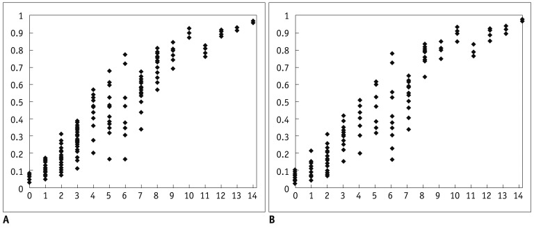
Materials and methods: Patient data were collected from 20 different institutions and the data included 2000 thyroid nodules from 1796 patients. For developing a diagnostic prediction model to estimate the malignant risk of thyroid nodules using suspicious malignant US features, we developed a training model in a subset of 1402 nodules from 1260 patients. Several suspicious malignant US features were evaluated to create the prediction model using a scoring tool. The scores for such US features were estimated by calculating odds ratios, and the risk score of malignancy for each thyroid nodule was defined as the sum of these individual scores. Later, we verified the usefulness of developed scoring system by applying into the remaining 598 nodules from 536 patients.
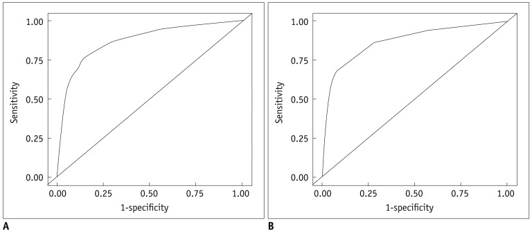
Results: Among 2000 tumors, 1268 were benign and 732 were malignant. In our multiple regression analysis models, the following US features were statistically significant for malignant nodules when using the training data set: hypoechogenicity, marked hypoechogenicity, non-parallel orientation, microlobulated or spiculated margin, ill-defined margins, and microcalcifications. The malignancy rate was 7.3% in thyroid nodules that did not have suspicious-malignant features on US. Area under the receiver operating characteristic (ROC) curve was 0.867, which shows that the US risk score help predict thyroid malignancy well. In the test data set, the malignancy rates were 6.2% in thyroid nodules without malignant features on US. Area under the ROC curve of the test set was 0.872 when using the prediction model.
Conclusion: The predictor model using suspicious malignant US features may be helpful in risk stratification of thyroid nodules.
Keywords: Thyroid; Thyroid cancer; Ultrasound.
Figures
References
-
- Park SH, Kim SJ, Kim EK, Kim MJ, Son EJ, Kwak JY. Interobserver agreement in assessing the sonographic and elastographic features of malignant thyroid nodules. AJR Am J Roentgenol. 2009;193:W416–W423. - PubMed
-
- Wienke JR, Chong WK, Fielding JR, Zou KH, Mittelstaedt CA. Sonographic features of benign thyroid nodules: interobserver reliability and overlap with malignancy. J Ultrasound Med. 2003;22:1027–1031. - PubMed
-
- Choi SH, Kim EK, Kwak JY, Kim MJ, Son EJ. Interobserver and intraobserver variations in ultrasound assessment of thyroid nodules. Thyroid. 2010;20:167–172. - PubMed
-
- Horvath E, Majlis S, Rossi R, Franco C, Niedmann JP, Castro A, et al. An ultrasonogram reporting system for thyroid nodules stratifying cancer risk for clinical management. J Clin Endocrinol Metab. 2009;94:1748–1751. - PubMed
Publication types
MeSH terms
LinkOut - more resources
Full Text Sources
Other Literature Sources
