. 2012;7(4):62-74.

Plasmodium berghei ANKA Infection in ICR Mice as a Model of Cerebral Malaria

Affiliations

Plasmodium berghei ANKA Infection in ICR Mice as a Model of Cerebral Malaria

R Basir et al. Iran J Parasitol. 2012.

Abstract

Background: Animal models with various combination of host-parasite have long been employed to study malaria pathogenesis. Here, we describe the combination of Plasmodium berghei ANKA infection in inbred ICR mice as a model of cerebral malaria (CM).

Methods: Infection in mice was initiated by intraperitoneal injection of 2 x 10(7) (0.2ml) parasitized red blood cells (PRBCs).

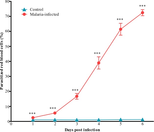
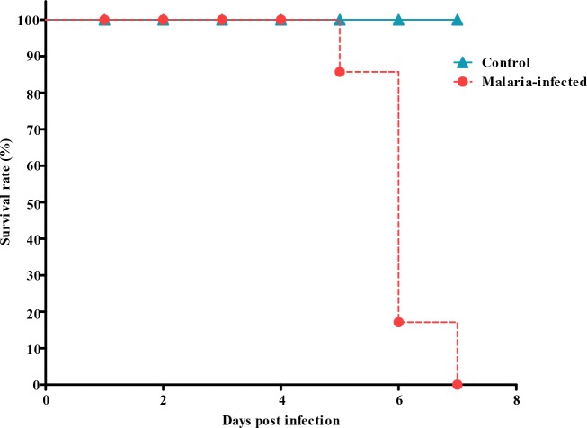
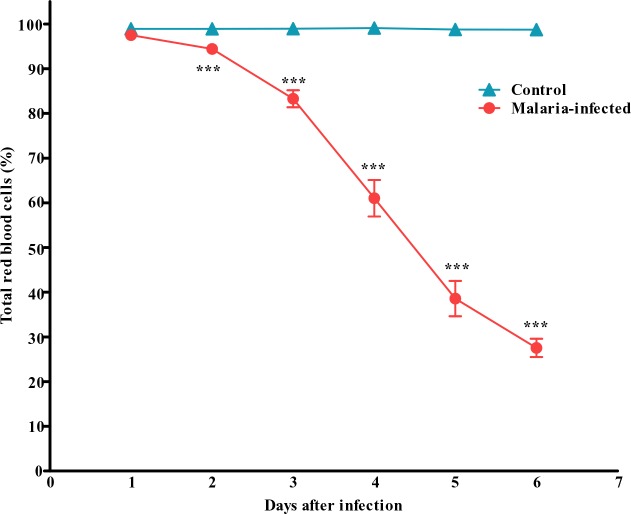
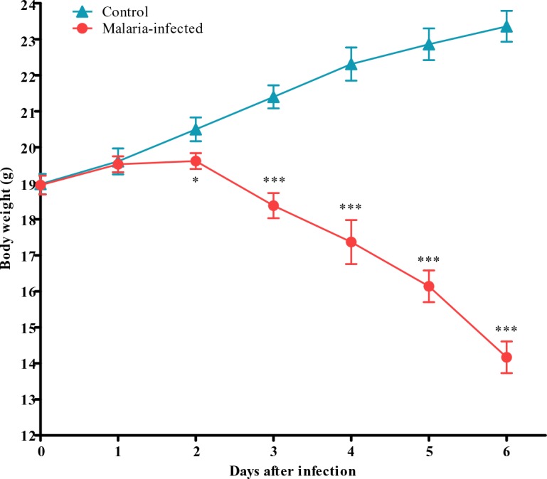
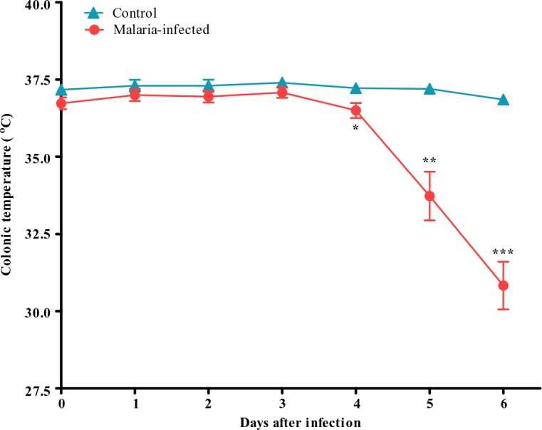
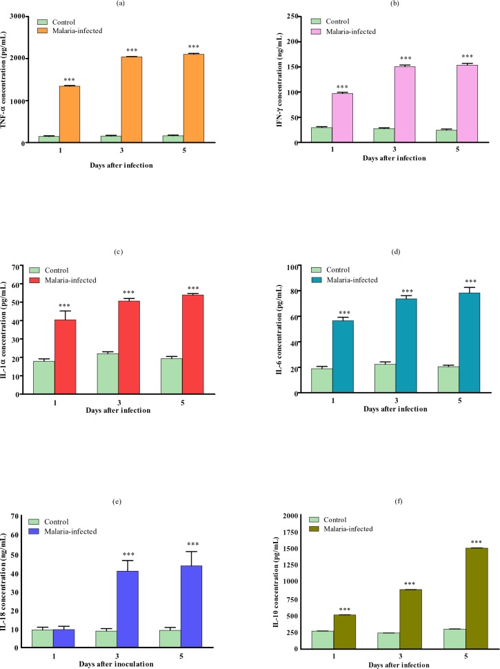
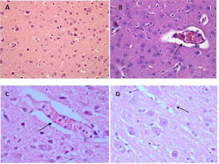
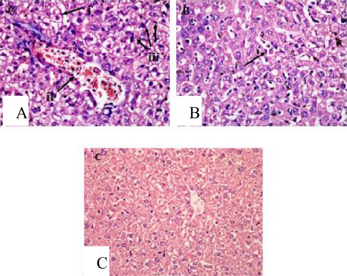
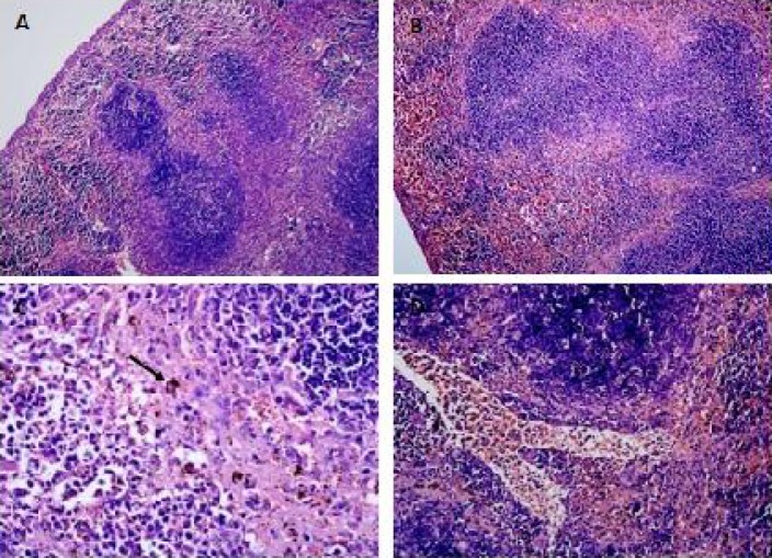
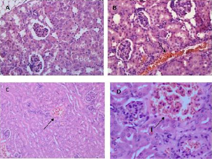
Results: This model can produce a severe degree of infection presented by the high degree of parasitaemia followed by death 6-7 days post infection. Severe anemia, splenomegaly, hepatomegaly and discolourations of major organs were observed. Histopathological findings revealed several important features mimicking human CM including, microvascular sequestration of PRBCs in major organs, particularly in the brain, hypertrophy and hyperplasia of the kupffer cells in the liver, pulmonary edema and hyaline membrane formation in the lungs and haemorrhages in the kidney's medulla and cortex. Proinflammatory cytokines TNFα, IFNγ, IL-1, IL-6 and IL-18, and anti-inflammatory cytokine IL-10 were all found to be elevated in the plasma of infected mice.

Conclusion: This model can reproduce many of the important features of CM and therefore can be used as a tool to advance our understanding of the disease pathogenesis.

Keywords: Animal model; ICR mice; Malaria; Plasmodium berghei.


Figures

Fig. 1
Fig. 1
Parasitaemia measured in the control and malaria-infected mice. Parasitized red blood cells were taken as an index of parasitaemia. Results are mean ± s. e. m with N = 8, ***P<0.0005
Fig. 2
Fig. 2
Percentage survival of control and malaria infected mice following inoculation with the parasites
Fig. 3
Fig. 3
Total red blood cells counted in the control and malaria-infected mice. Results are mean ± s.e.m of N = 8. ***P<0.0005
Fig. 4
Fig. 4
Body weight of control and malaria-infected mice. Results are mean ± s.e.m of N = 8, *P<0.05, ***P<0.0005
Fig. 5
Fig. 5
Colonic temperature of control and malaria-infected mice. Results are presented as the mean ± s.e.m of N = 8, *P<0.05, **P<0.005, ***P<0.0005
Fig. 6
Fig. 6
Plasma concentrations of a) TNFα, b) IFNγ, c) IL-1, d) IL-6, e) IL-18 and f) IL-10 of control and malaria-infected mice. Results are mean ± s.e.m of N = 8, ***P<0.0005
Fig. 7
Fig. 7
Haematoxylin-Eosin staining of brain tissue showed sequestration of PRBCs in the cerebral microvessels (B) & (C) as compared to the normal uninfected mice (A). Petechial haemorrhages were also observed in the cerebral tissue (D). Specimens were examined under light microscopy at 400x magnification
Fig. 8
Fig. 8
Hyaline membrane formation due to alveolar haemorrhage (B (i)) was seen in the lung tissue of malaria-infected mice as compared to the normal uninfected animal (A). Accumulation of malarial pigment, haemozoin, were also observed (B (ii)), and the alveolar was congested with macrophages engulfing pigments (C). Viewed under light microscopy at 400x
Fig. 9
Fig. 9
In the liver of malaria-infected mice, hyperplasia and hypertrophy of kupffer cells (A(iii)) with vacuolar degeneration and atrophy of hepatocytes (A(i)) were observed as compared to uninfected mice (C). Microvascular congestion with PRBCs also occurred (A(ii)) leading to congestion of the blood vessels in the liver. Hyperaemia were observed accompanied with capillaries dilatation (B). Viewed under light microscopy at 400x magnification
Fig. 10
Fig. 10
In the spleen of mice infected with malaria, the red and white pulp elements were enlarged accompanied by the lost of typical structure of the germinal centre (B), which is comparable to the normal uninfected mice (A). Accumulation of malarial pigments, haemozoin, was obvious in the pulp histiocytes and sinusoidal lining cells (C). Microvascular sequestrations of PRBCs were also present (D). Viewed under light microscopy at 100x (A & B) and 400x (C & D) magnification
Fig. 11
Fig. 11
The medulla tissues of the kidney from infected mice were presented with widespread sequestration of PRBCs in the microvascular and interstitium (B), compared to the normal uninfected mouse (A). Haemorrhages (C, D(i)) and congestion (D(i)) of the cortex with PRBCs were also observed. Viewed under light microscopy at 400x (A,B,D) and 200x (C) magnification

References

    1. Druilhe P, Hagan P, Rook GAW. The importance of models of infection in the study of disease resistance. Trends Microbiol. 2002;10(10):38–46. - PubMed
    1. World Malaria Report. 2011. http://www.who.int/malaria/world_malaria_report_2011/9789241564403_eng.pdf.
    1. Hunt NH, Grau GE. Cytokines: accelerators and brakes in the pathogenesis of cerebral malaria. Trends Immunol. 2003;24(9):491–499. - PubMed
    1. Combes V, de Souza JB, Renia L, Hunt NH, Grau GE. Cerebral malaria: which parasite?, Which model? Drug Disc Today. 2005;2(2):141–147.
    1. Berendt AR, Turner GDH, Newbold CI. Cerebral malaria: the sequestration hypothesis. Parasitol Today. 1994;10(10):412–414. - PubMed

LinkOut - more resources