

Prevalence of incidental maxillary sinus findings in Italian orthodontic patients: a retrospective cone-beam computed tomography study
- PMID: 23323247
- PMCID: PMC3542453
- DOI: 10.4041/kjod.2012.42.6.329
Prevalence of incidental maxillary sinus findings in Italian orthodontic patients: a retrospective cone-beam computed tomography study
Abstract
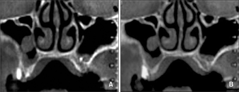
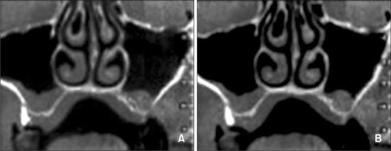
Objectives: To determine the prevalence of incidental maxillary sinus findings in a large sample of orthodontic patients by cone-beam computed tomography (CBCT) with a wide field of view and assess the relationships of such abnormalities with age and gender.
Methods: Five hundred thirteen CBCT scans obtained for orthodontic diagnosis and treatment planning in a Northern Italian population (N = 513; 292 female and 221 male subjects; 1,026 maxillary sinuses) were studied. The frequencies of pseudocysts and mucosal thickening of the maxillary sinus were recorded. Logistic regression analysis was used to determine the influence of age and gender on these abnormalities.
Results: Pseudocysts were detected in 52 patients (10.1%) and 59 sinuses (5.75%). Mucosal thickening was observed in 206 patients (40.1%) and 258 sinuses (25.1%). Gender and age were significantly associated with pseudocysts (p = 0.027) and mucosal thickening (p < 0.001), respectively.
Conclusions: Half of the orthodontic patients had incidental maxillary sinus findings. Men were more likely to show pseudocysts, and older patients (aged 41 - 60 years) were more likely to show mucosal thickening.
Keywords: Computed tomography; Three dimensional diagnosis and treatment planning.
Conflict of interest statement
The authors report no commercial, proprietary, or financial interest in the products or companies described in this article.
Figures
Similar articles
-
Prevalence of Incidental Findings and Assessment of Maxillary Sinus Pathologies and Dental Diseases Using Cone-Beam Computed Tomography (CBCT) in the Tamil Nadu Population: A Retrospective Study.Cureus. 2024 Sep 8;16(9):e68929. doi: 10.7759/cureus.68929. eCollection 2024 Sep. Cureus. 2024. PMID: 39385860 Free PMC article.
-
Incidental maxillary sinus findings in orthodontic patients: a radiographic analysis using cone-beam computed tomography (CBCT).Orthod Craniofac Res. 2011 Feb;14(1):17-24. doi: 10.1111/j.1601-6343.2010.01502.x. Epub 2010 Nov 22. Orthod Craniofac Res. 2011. PMID: 21205165
-
Prevalence of incidental maxillary sinus pathologies in dental patients on cone-beam computed tomographic images.Contemp Clin Dent. 2014 Jul;5(3):361-5. doi: 10.4103/0976-237X.137949. Contemp Clin Dent. 2014. PMID: 25191074 Free PMC article.
-
Cone-beam computed tomographic evidence of the association between periodontal bone loss and mucosal thickening of the maxillary sinus.J Periodontol. 2012 May;83(5):557-64. doi: 10.1902/jop.2011.110376. Epub 2011 Sep 12. J Periodontol. 2012. PMID: 21910593
-
Prevalence of sinus membrane thickening and association with unhealthy teeth: a retrospective review of 831 consecutive patients with 1,662 cone-beam scans.J Oral Maxillofac Surg. 2014 Dec;72(12):2454-60. doi: 10.1016/j.joms.2014.06.442. Epub 2014 Jun 27. J Oral Maxillofac Surg. 2014. PMID: 25236817 Review.
Cited by
-
Prevalence of Incidental Findings and Assessment of Maxillary Sinus Pathologies and Dental Diseases Using Cone-Beam Computed Tomography (CBCT) in the Tamil Nadu Population: A Retrospective Study.Cureus. 2024 Sep 8;16(9):e68929. doi: 10.7759/cureus.68929. eCollection 2024 Sep. Cureus. 2024. PMID: 39385860 Free PMC article.
-
Idiopathic bilateral antral exostoses: A rare case in maxillary sinus.Int J Surg Case Rep. 2014;5(9):624-7. doi: 10.1016/j.ijscr.2014.05.005. Epub 2014 Jul 29. Int J Surg Case Rep. 2014. PMID: 25128728 Free PMC article.
-
Paranasal sinus pathoses on cone beam computed tomography.J Istanb Univ Fac Dent. 2016 Jan 12;50(1):27-34. doi: 10.17096/jiufd.47796. eCollection 2016. J Istanb Univ Fac Dent. 2016. PMID: 28955552 Free PMC article.
-
The Relationship between an Accessory Maxillary Ostium and Variations in Structures Adjacent to the Maxillary Sinus without Polyps.Int Arch Otorhinolaryngol. 2022 Jan 28;26(4):e548-e555. doi: 10.1055/s-0042-1742325. eCollection 2022 Oct. Int Arch Otorhinolaryngol. 2022. PMID: 36405481 Free PMC article.
-
Incidence of Maxillary Sinus Disorders in Dental Patients Undergoing Cone-Beam Computed Tomography: A Retrospective Cross-Sectional Study.Cureus. 2024 Oct 8;16(10):e71114. doi: 10.7759/cureus.71114. eCollection 2024 Oct. Cureus. 2024. PMID: 39391257 Free PMC article.
References
-
- Horner K, Islam M, Flygare L, Tsiklakis K, Whaites E. Basic principles for use of dental cone beam computed tomography: consensus guidelines of the European Academy of Dental and Maxillofacial Radiology. Dentomaxillofac Radiol. 2009;38:187–195. - PubMed
-
- Cha JY, Mah J, Sinclair P. Incidental findings in the maxillofacial area with 3-dimensional cone-beam imaging. Am J Orthod Dentofacial Orthop. 2007;132:7–14. - PubMed
-
- Ritter L, Lutz J, Neugebauer J, Scheer M, Dreiseidler T, Zinser MJ, et al. Prevalence of pathologic findings in the maxillary sinus in cone-beam computerized tomography. Oral Surg Oral Med Oral Pathol Oral Radiol Endod. 2011;111:634–640. - PubMed
-
- Pazera P, Bornstein MM, Pazera A, Sendi P, Katsaros C. Incidental maxillary sinus findings in orthodontic patients: a radiographic analysis using cone-beam computed tomography (CBCT) Orthod Craniofac Res. 2011;14:17–24. - PubMed
LinkOut - more resources
Full Text Sources