. 2013 Feb 19;85(4):2231-8.
doi: 10.1021/ac303038j. Epub 2013 Feb 4.

Single-cell lipidomics: characterizing and imaging lipids on the surface of individual Aplysia californica neurons with cluster secondary ion mass spectrometry

Affiliations

Single-cell lipidomics: characterizing and imaging lipids on the surface of individual Aplysia californica neurons with cluster secondary ion mass spectrometry

Melissa K Passarelli et al. Anal Chem. .

Abstract

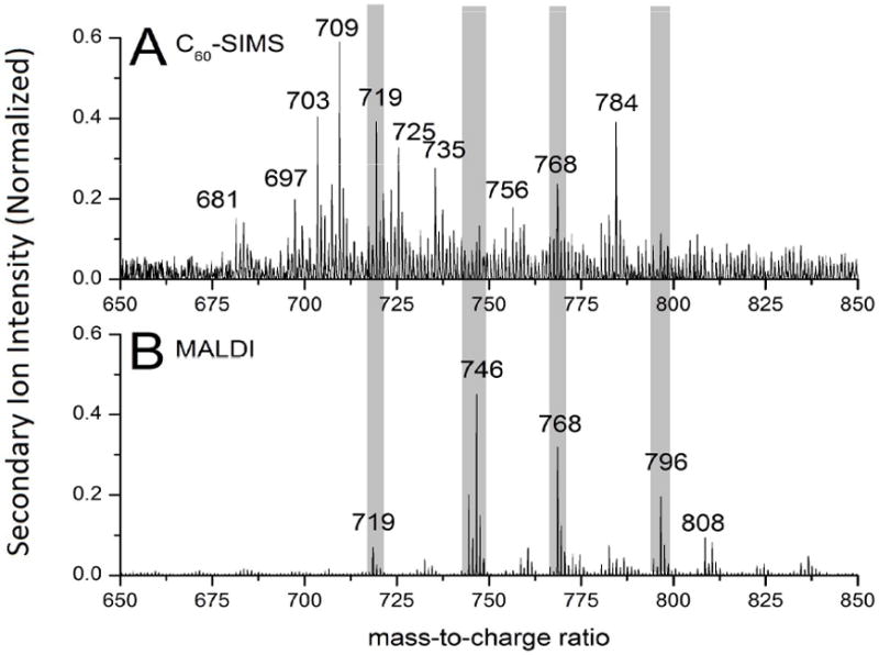
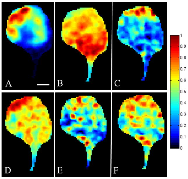
Neurons isolated from Aplysia californica , an organism with a well-defined neural network, were imaged with secondary ion mass spectrometry, C(60)-SIMS. A major lipid component of the neuronal membrane was identified as 1-hexadecyl-2-octadecenoyl-sn-glycero-3-phosphocholine [PC(16:0e/18:1)] using tandem mass spectrometry (MS/MS). The assignment was made directly off the sample surface using a C(60)-QSTAR instrument, a prototype instrument that combines an ion source with a commercial electrospray ionization/matrix-assisted laser desorption ionization (ESI/MALDI) mass spectrometer. Normal phase liquid chromatography mass spectrometry (NP-LC-MS) was used to confirm the assignment. Cholesterol and vitamin E were also identified with in situ tandem MS analyses that were compared to reference spectra obtained from purified compounds. In order to improve sensitivity on the single-cell level, the tandem MS spectrum of vitamin E reference material was used to extract and compile all the vitamin E related peaks from the cell image. The mass spectrometry images reveal heterogeneous distributions of intact lipid species, PC(16:0e/18:1), vitamin E, and cholesterol on the surface of a single neuron. The ability to detect these molecules and determine their relative distribution on the single-cell level shows that the C(60)-QSTAR is a potential platform for studying important biochemical processes, such as neuron degeneration.


Figures

Figure 1
Figure 1
Lipid profiles, mass range m/z 650-850, obtained from the surface of a single Aplysia neuron using C60-SIMS (A) and MALDI (B). The MALDI spectrum was normalized to the integrate counts of the maximum peak at m/z 746.5 and the SIMS spectrum was normalized to the integrate counts of the maximum peak at m/z 709.5.
Figure 2
Figure 2
Glycerophosphocholines, e.g. PC(16 : 0e/18 : 1), are readily adducted to biological salts, (i.e. sodium and potassium). In the gas phase, these lipid-adducted species, lose a trimethylamine group from the phosphocholine headgroup to form a high mass fragment [M + (K or Na) - TMA]. The tandem MS of the protonated lipid species, the sodiated-lipid adduct and the high mass fragment associated with the loss of TMA are shown and the common peaks are highlighted. The protonated lipid species provides less structural information than the respective sodiated and high mass lipid species.
Figure 3
Figure 3
ToF-SIMS images of obtained from a single Aplysia neuron(A) Vitamin E, alpha-tocopherol C29H50O2, M, m/z 430.3, (B) hydrocarbon (m/z 128.1), (C) cholesterol C27H45, [M + H – OH]+ (m/z 369.3) (D) phosphocholine headgroup (m/z 184), C5H15NPO4; [M + H]+ (E) PC (16 : 0 e/18 : 1) high mass fragment, (m/z 709.6, C39H75O7PNa, [M + Na – TMA]+ and (F) sum of PC (16 : 0 e/18 : 1) related peaks [m/z 709, (C39H75O7PNa, [M + Na – TMA]+, m/z 725, (C39H75O7PK, [M + K – TMA]+), m/z 746, (C42H85NO7P, [M + H]+), m/z 768 (C42H84NO7PNa, [M + Na]+) and m/z 784 (C42H84NO7PK [M + K]+)). (scale bar = 100 μm)
Figure 4
Figure 4
An optical image (A) and the ToF-SIMS image of vitamin E (B) was overlaid (C) to show the overlap of the vitamin E with the yellow-orange pigment. ToF-MS (E: top) and tandem MS of the protonated molecular ion at m/z 430.3 (E: bottom) reference spectra for vitamin E are shown. The tandem MS obtained from vitamin E reference material was used to extract and compile the vitamin E related peaks in the ToF-MS image of the Aplysia neuron in order to improve the sensitivity of the measurement (D). (scale bar = 100 μm)
Figure 5
Figure 5
The vitamin E signal (m/z 430.3) and compiled PC(16:0e/ 18:1) lipid signal overlaid on the the optical image. A region of high co-localization (threshold = 0.5) between the two chemicals can be seen in the upper portion of the soma. (scale bar = 100 μm)

Similar articles

Cited by

References

    1. Amstalden van Hove ER, Smith DF, Heeren R. J Chromatog A. 2010;1217:3946–3954. - PubMed
    1. Watrous JD, Alexandrov T, Dorrestein PC. J Mass Spectrom. 2011;46:209–222. - PMC - PubMed
    1. Trouillon R, Passarelli MK, Wang J, Kurczy M, Ewing A. Anal Chem. 2012 - PubMed
    1. Colliver TL, Brummel CL, Pacholski ML, Swanek FD, Ewing AG, Winograd N. Anal Chem. 1997;69:2225–2231. - PubMed
    1. Lin Y, Trouillon R, Safina G, Ewing AG. Anal Chem. 2011;83:4369–4392. - PMC - PubMed

Publication types

LinkOut - more resources