

Multinodular and vacuolating neuronal tumors of the cerebrum: 10 cases of a distinctive seizure-associated lesion
- PMID: 23324039
- PMCID: PMC8029170
- DOI: 10.1111/bpa.12035
Multinodular and vacuolating neuronal tumors of the cerebrum: 10 cases of a distinctive seizure-associated lesion
Abstract
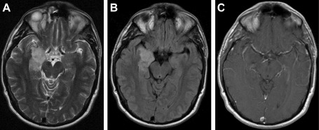
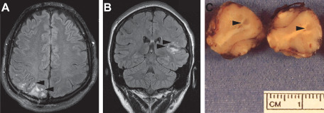
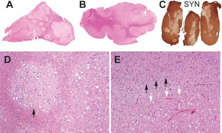
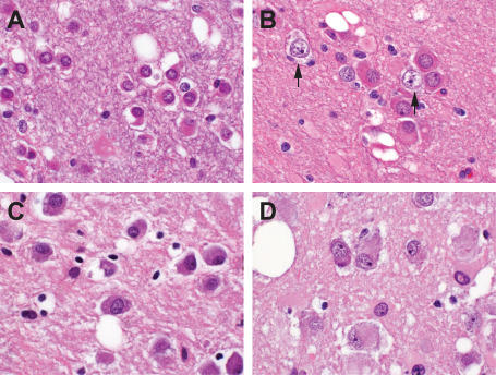
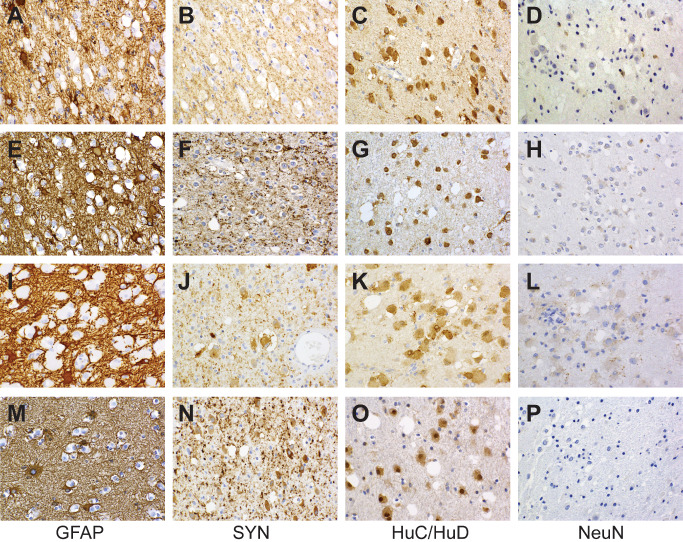
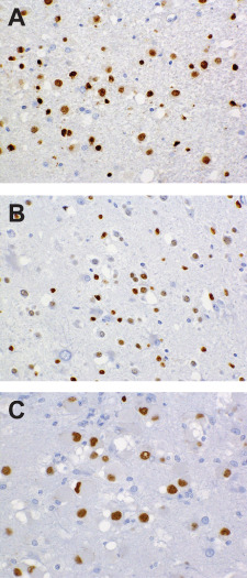
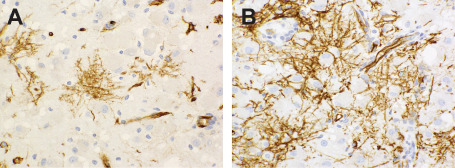
We report 10 cases of a non-neurocytic, purely neuronal tumor affecting adults. Situated in the cerebral hemispheres, with 7 of 10 confined to the temporal lobes, most presented with seizures as their principal clinical manifestations. On magnetic resosnance imaging (MRI), the tumors generally appeared solid and non-contrast enhancing with minimal diffuse infiltration, edema, or mass effect. Six examples demonstrated internal nodularity. Microscopically, the tumor cells were largely distributed into discrete and coalescent nodules exhibiting varying degrees of matrix vacuolization, principally within the deep cortical ribbon and superficial subcortical white matter. Populating elements ranged from morphologically ambiguous to recognizably neuronal, with only two cases manifesting overt ganglion cell cytology. In all cases, tumor cells exhibited widespread nuclear immunolabeling for the HuC/HuD neuronal antigens, although expression of other neuronal markers, including synaptophysin, neurofilament and chromogranin was variable to absent. Tumor cells also failed to express GFAP, p53, IDH1 R132H, or CD34, although CD34-labeling ramified neural elements were present in the adjoining cortex of seven cases. Molecular analysis in a subset of cases failed to reveal DNA copy number abnormalities or BRAF V600E mutation. Follow-up data indicate that this unusual neuronal lesion behaves in benign, World Health Organization (WHO) grade I fashion and is amenable to surgical control.
Keywords: HuC/HuD; developmental tumor; eliptogenic tumor; gangliocytoma; multinodular; vacuolating neuronal tumor.
© 2013 International Society of Neuropathology.
Figures
References
-
- Barami K, Iversen K, Furneaux H, Goldman SA (1995) Hu protein as an early marker of neuronal phenotypic differentiation by subependymal zone cells of the adult songbird forebrain. J Neurobiol 28:82–101. - PubMed
-
- Becker AJ, Wiestler OD, Figarella‐Branger D, Blumcke I (2007) Ganglioglioma and gangliocytoma. In: WHO Classification of Tumours of the Central Nervous System, Louis DN, Ohgaki H, Wiestler OD, Cavenee WK (eds), pp. 103–105. IARC: Lyon.
-
- Blumcke I, Giencke K, Wardelmann E, Beyenburg S, Kral T, Sarioglu N et al (1999) The CD34 epitope is expressed in neoplastic and malformative lesions associated with chronic, focal epilepsies. Acta Neuropathol 97:481–490. - PubMed
-
- Blumcke I, Lobach M, Wolf HK, Wiestler OD (1999) Evidence for developmental precursor lesions in epilepsy‐associated glioneuronal tumors. Microsc Res Tech 46:53–58. - PubMed
-
- Blumcke I, Wiestler OD (2002) Gangliogliomas: an intriguing tumor entity associated with focal epilepsies. J Neuropathol Exp Neurol 61:575–584. - PubMed
MeSH terms
Substances
LinkOut - more resources
Full Text Sources
Other Literature Sources
Medical
Research Materials
Miscellaneous
