. 2013 Apr 1;304(7):F908-17.
doi: 10.1152/ajprenal.00591.2012. Epub 2013 Jan 16.

Inhibitory phosphorylation of GSK-3β by AKT, PKA, and PI3K contributes to high NaCl-induced activation of the transcription factor NFAT5 (TonEBP/OREBP)

Affiliations

Inhibitory phosphorylation of GSK-3β by AKT, PKA, and PI3K contributes to high NaCl-induced activation of the transcription factor NFAT5 (TonEBP/OREBP)

Xiaoming Zhou et al. Am J Physiol Renal Physiol. .

Abstract

High NaCl activates the transcription factor nuclear factor of activated T cells 5 (NFAT5), leading to increased transcription of osmoprotective target genes. Kinases PKA, PI3K, AKT1, and p38α were known to contribute to the high NaCl-induced increase of NFAT5 activity. We now identify another kinase, GSK-3β. siRNA-mediated knock-down of GSK-3β increases NFAT5 transcriptional and transactivating activities without affecting high NaCl-induced nuclear localization of NFAT5 or NFAT5 protein expression. High NaCl increases phosphorylation of GSK-3β-S9, which inhibits GSK-3β. In GSK-3β-null mouse embryonic fibroblasts transfection of GSK-3β, in which serine 9 is mutated to alanine, so that it cannot be inhibited by phosphorylation at that site, inhibits high NaCl-induced NFAT5 transcriptional activity more than transfection of wild-type GSK-3β. High NaCl-induced phosphorylation of GSK-3β-S9 depends on PKA, PI3K, and AKT, but not p38α. Overexpression of PKA catalytic subunit α or of catalytically active AKT1 reduces inhibition of NFAT5 by GSK-3β, but overexpression of p38α together with its catalytically active upstream kinase, MKK6, does not. Thus, GSK-3β normally inhibits NFAT5 by suppressing its transactivating activity. When activated by high NaCl, PKA, PI3K, and AKT1, but not p38α, increase phosphorylation of GSK-3β-S9, which reduces the inhibitory effect of GSK-3β on NFAT5, and thus contributes to activation of NFAT5.


Figures

Fig. 1.
Fig. 1.
Glycogen synthase kinase (GSK)-3β negatively regulates high NaCl-induced nuclear factor of activated T cells 5 (NFAT5) transcriptional activity. Human embryonic kidney (HEK)293 cells stably expressing a luciferase reporter of NFAT5 transcriptional activity were transfected with siRNA against GSK-3β or control siRNAs for 32 h, and then osmolality was increased to 500 mosmol/kgH2O (NaCl added) or left at 290 mosmol/kgH2O for 16 h. We tested siRNAs from Santa Cruz Biotechnology, Cell Signaling, and Dharmacon, finding similar results. The result using the siRNA from Santa Cruz Biotechnology is displayed here. A: siRNA decreases GSK-3β protein. B: knockdown of GSK-3β significantly increases NFAT5 transcriptional activity at both 290 and 500 mosmol/kgH2O (NaCl added) in HEK293 cells stably expressing a luciferase reporter containing a wild-type osmotic response element (ORE). C: siRNA has no significant effect at either osmolality when the reporter contains an ORE element mutated to prevent binding by NFAT5 (*P < 0.05, compared with control siRNAs, repeated-measures ANOVA, n = 3).
Fig. 2.
Fig. 2.
A: siRNA-mediated knockdown of GSK-3β increases high NaCl-induced elevation of NFAT5 transactivating activity. As in Fig. 1B except that we measured NFAT5 transactivating activity in HEK293 cells stably expressing a yeast binary GAL4 reporter assay system. B: siRNA-mediated knockdown of GSK-3β increases NFAT5 protein abundance at 290 mosmol/kgH2O. HEK293 cells were transfected with siRNA against GSK-3β for 32 h at 290 mosmol/kgH2O, and the medium was changed for 16 h, either maintaining it at 290 mosmol/kgH2O or increasing it to 500 mosmol/kgH2O (NaCl added). *P < 0.05 compared with control siRNA, repeated-measures ANOVA, n = 3.
Fig. 3.
Fig. 3.
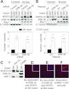
A and B: GSK-3β does not affect NFAT5 nuclear localization. A: HEK293 cells were transfected with siRNA against GSK-3β or control siRNAs and treated as in Fig. 1 except that osmolality was increased for only 2 h. Proteins were extracted separately from cytoplasm and nuclei and then analyzed by Western blotting, and the nuclear-to-cytoplasmic ratio of NFAT5 was calculated. Note that the subcellular distributions of β-tubulin (cytoplasmic marker) and Brg-1 (nuclear marker) are unaffected by NaCl concentration. B: as in A except that HEK293FT cells were transiently cotransfected with plasmids coding for GSK-3β-HA and NFAT5-V5. C–E: lack of evidence for direct association of GSK-3β with NFAT5. C: NFAT5-V5 was immunoprecipitated from HEK293 cells that stably express it (32) after the medium was replaced for 2 h either with the same medium at 290 or an otherwise identical medium at 500 mosmol/kgH2O (NaCl added). SHP-1 coimmunoprecipitates with NFAT5-V5, but GSK-3β does not. D and E: after HeLa cells were transfected with NFAT5-V5, they were transferred for 24 h to Lab-Tek chamber slides for an additional 22 h before the medium was replaced for 2 h either with the same medium at 290 or an otherwise identical medium at 500 mosmol/kgH2O (NaCl added). Then, Duolink in situ assay was performed. No association (red staining) is detected between NFAT5-V5 and GSK-3β. F: positive control for the Duolink assay confirming in HeLa cells the previously observed (73) direct association of p38 with MKP-1-GST in mouse inner medullary collecting duct cells (mIMCD3) cells.
Fig. 4.
Fig. 4.
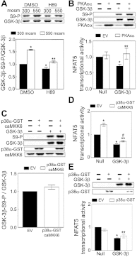
High NaCl increases phosphorylation of GSK-3β-S9 and decreases GSK-3β activity. A: HEK293 cells: medium was replaced for 30 min with the same medium at 290 or an otherwise identical medium at 500 mosmol/kgH2O (NaCl added); mIMCD3 cells: medium was replaced for 30 min either with the same medium at 300 or an otherwise identical medium at 550 mosmol/kgH2O (NaCl added). GSK-3β-S9 phosphorylation was measured by Western blot analysis of the ratio of phospho-GSK-3β/total GSK-3β. The activity of GSK-3β immunoprecipitated from HEK293 cells was measured with the ADP Glo Kinase Assay system (*P < 0.05 compared with respective control, paired t-test, n = 3 for phosphorylation assay and n = 4 for activity assay). B: overexpression of wild-type GSK-3β-HA reduces NFAT5 transcriptional activity in GSK-3β-null MEF cells, and overexpression of GSK-3β-S9A reduces NFAT5 transcriptional activity even more. GSK-3β−/− mouse embryonic fibroblasts (MEFs) were cotransfected with a luciferase reporter of NFAT5 transcriptional activity (ORE-X) together either with the empty vector (EV) pcDNA3, pcDNA3-GSK-3β-HA wild-type (GSK-3β-HA), or pcDNA3-GSK-3β-S9/A-HA (S9A-HA) for 24 h, and then osmolality was increased to 500 mosmol/kgH2O (NaCl added) or left at 300 mosmol/kgH2O for an additional 24 h before ORE-X reporter activity was measured. *P < 0.05 vs. EV at 300 mosmol/kgH2O. **P < 0.05 vs. EV at 500 mosmol/kgH2O. ***P < 0.05 vs. GSK-3β-HA at 500 mosmol/kgH2O, repeated-measures ANOVA, n = 4.
Fig. 5.
Fig. 5.
A: PKA contributes to high NaCl-induced phosphorylation of GSK-3β-S9. mIMCD3 cells were preincubated with 0.1% DMSO (control) or 10 μM PKA inhibitor H89 in DMSO for 60 min at 300 mosmol/kgH2O, and then the medium was changed for 30 min to an identical one or an otherwise identical one in which osmolality was increased to 500 mosmol/kgH2O (NaCl added). Phospho-GSK-3β-S9 and total GSK-3β were measured by Western blot analysis. *P < 0.05 compared with DMSO at 300 mosmol/kgH2O. **P < 0.05, compared with DMSO at 550 mosmol/kgH2O, repeated-measures ANOVA, n = 3. B: PKA increases phosphorylation of GSK-3β-S9 which contributes to high NaCl-induced increase of NFAT5 transcriptional activity. GSK-3β-null MEFs were cotransfected at 300 mosmol/kgH2O with a luciferase reporter of NFAT5 transcriptional activity (ORE-X), GSK-3β-HA (GSK-3β), or EV (Null), and catalytically active PKA (PKAcα) or EV for 24 h, and then the medium was refreshed for an additional 24 h before luciferase activity was measured. *P < 0.05 vs. EV in the null cells. **P < 0.05 vs. EV in the GSK-3β reconstituted group, GSK-3β, repeated-measures ANOVA, n = 4. Expression of PKAcα and GSK-3β-HA was identified by anti-PKA and anti-GSK-3β antibodies, respectively (top). Reconstitution of the null cells with GSK-3β inhibits NFAT5 transcriptional activity, but PKA activity increases phosphorylation of GSK-3β and reverses the inhibition. (C to E). The stimulatory effect of p38α on NFAT5 activity is not mediated by inhibitory phosphorylation of GSK-3β-S9. C and D: GSK-3β-null MEFs were cotransfected at 300 mosmol/kgH2O with a luciferase reporter of NFAT5 transcriptional activity (ORE-X), GSK-3β-HA (GSK-3β), or EV (Null), and p38α-GST plus catalytically active MKK6 (caMKK6) or their EVs as in B, before measurement of luciferase activity and Western blot analysis. *P < 0.05 vs. EV in the null cells. **P < 0.05 vs. EV in the GSK-3β reconstituted group, GSK-3β. #P < 0.05 vs. p38α-GST and caMKK6 in the null cells, repeated-measures ANOVA, n = 3. E: GSK-3β-null MEFs were cotransfected at 300 mosmol/kgH2O with a luciferase reporter of NFAT5 transcriptional activity (ORE-X), GSK-3β-HA (GSK-3β) or EV (Null), and p38α-GST or its EV as in B, and then luciferase activity was measured. *P < 0.05 vs. EV in the null cells. **P < 0.05 vs. p38α-GST in the null cells, repeated-measures ANOVA, n = 3.
Fig. 6.
Fig. 6.
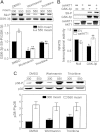
A: PI3 kinase and AKT1 contribute to high NaCl-induced phosphorylation of GSK-3β-S9. mIMCD3 cells were preincubated with 0.1% DMSO (control), 200 nM wortmannin (PI3K inhibitor), or 10 μM triciribine (AKT1 inhibitor) for 60 min at 300 mosmol/kgH2O, and then the medium was changed for 30 min to the identical one or to an otherwise identical one at 550 mosmol/kgH2O (NaCl added). Phospho-GSK-3β-S9 and total GSK-3β were measured by Western blot analysis. *P < 0.05 vs. DMSO at 300 mosmol/kgH2O. **P < 0.05 vs. DMSO at 550 mosmol/kgH2O, repeated-measures ANOVA, n = 3. Inhibition of PI3K or AKT1 prevents the high NaCl-induced increase of phosphorylation of GSK-3β-S9. B: overexpression of caAKT1 eliminates the inhibitory effect of GSK-3β on NFAT5 transcriptional activity accompanied by increased phosphorylation of GSK-3β-S9. GSK-3β-null MEFs were transfected at 300 mosmol/kgH2O with luciferase reporter of NFAT5 transcriptional activity (ORE-X), GSK-3β-HA (GSK-3β), or EV and caAKT1 or EVs for 24 h, and then the medium was replaced with one still at 300 mosmol/kgH2O for an additional 24 h, before measuring luciferase activity. Lysate was immunoblotted to verify expression of caAKT1, GSK-3β, and phosphorylation of GSK-3β-S9 (top). *P < 0.05 vs. EV in null cells. **P < 0.05 vs. EV in GSK-3β reconstituted cells, repeated-measures ANOVA, n = 3. C: wortmannin or triciribine has no significant effect on phosphorylation of p38. mIMCD3 cells were treated as in A (n = 3).
Fig. 7.
Fig. 7.
Summary of kinases and phosphatases known to regulate NFAT5 and of proteins that interact with them. All the depicted interactions are in the context of hypertonicity. Bold lines indicate direct interaction with another component of a pathway or with NFAT5 itself. Dashed lines indicate that the effect is not known to be direct.

Similar articles

Cited by

References

    1. Anastassiadis T, Deacon SW, Devarajan K, Ma H, Peterson JR. Comprehensive assay of kinase catalytic activity reveals features of kinase inhibitor selectivity. Nat Biotechnol 29: 1039–1045, 2011 - PMC - PubMed
    1. Beals CR, Clipstone NA, Ho SN, Crabtree GR. Nuclear localization of NF-ATc by a calcineurin-dependent, cyclosporin-sensitive intramolecular interaction. Genes Dev 11: 824–834, 1997 - PubMed
    1. Beals CR, Sheridan CM, Turck CW, Gardner P, Crabtree GR. Nuclear export of NF-ATc enhanced by glycogen synthase kinase-3. Science 275: 1930–1934, 1997 - PubMed
    1. Burg MB, Ferraris JD, Dmitrieva NI. Cellular response to hyperosmotic stresses. Physiol Rev 87: 1441–1474, 2007 - PubMed
    1. Buxade M, Lunazzi G, Minguillon J, Iborra S, Berga-Bolanos R, Del VM, Aramburu J, Lopez-Rodriguez C. Gene expression induced by Toll-like receptors in macrophages requires the transcription factor NFAT5. J Exp Med 209: 379–393, 2012 - PMC - PubMed

Publication types

MeSH terms

Substances