. 2013 Feb 1;12(3):511-21.
doi: 10.4161/cc.23406. Epub 2013 Jan 16.

TLR2 enhances ovarian cancer stem cell self-renewal and promotes tumor repair and recurrence

Affiliations

TLR2 enhances ovarian cancer stem cell self-renewal and promotes tumor repair and recurrence

Ilana Chefetz et al. Cell Cycle. .

Abstract

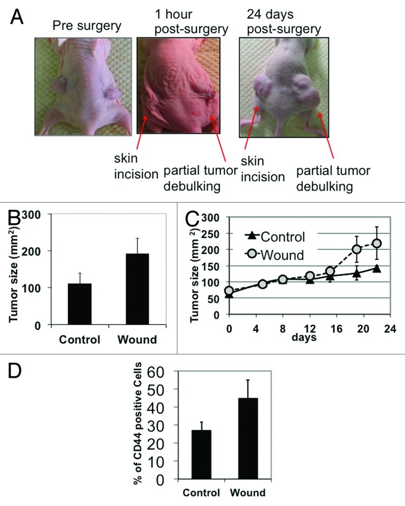
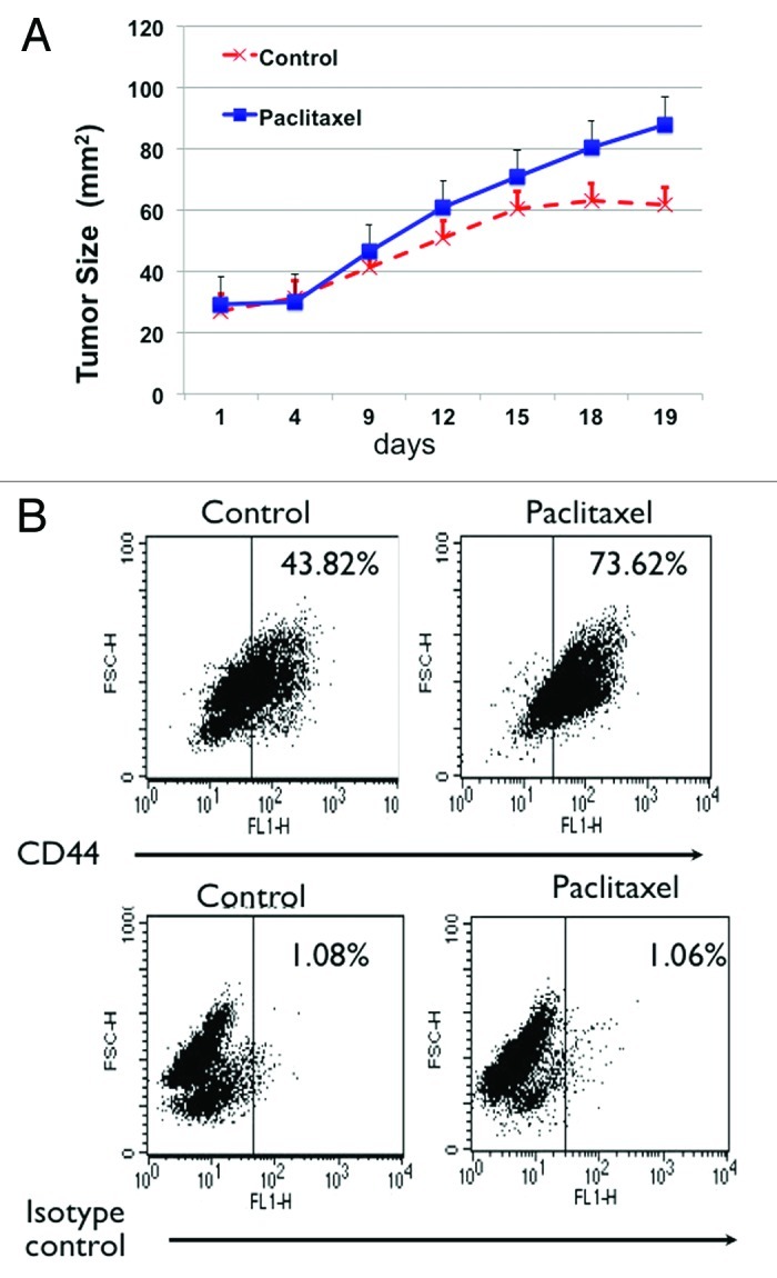
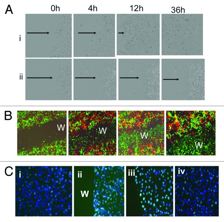
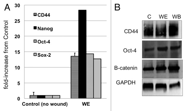
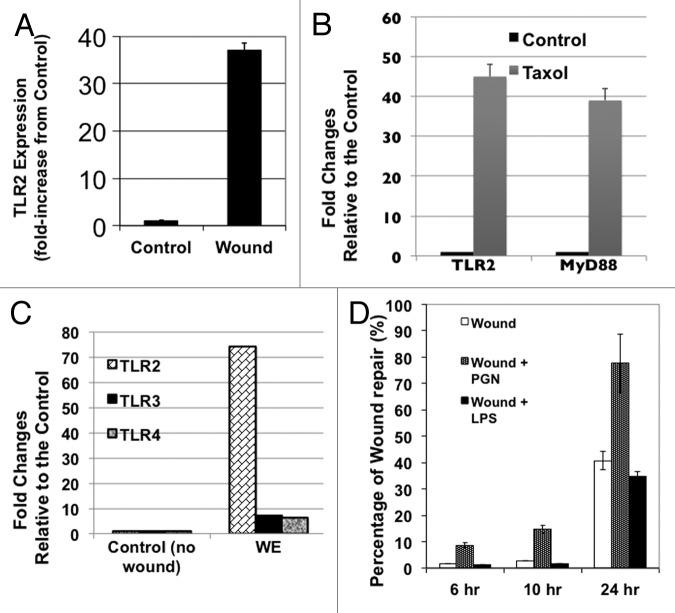
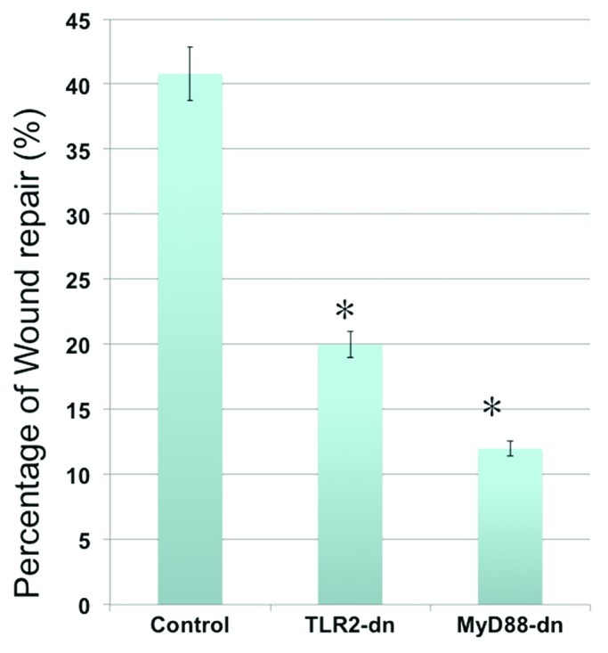
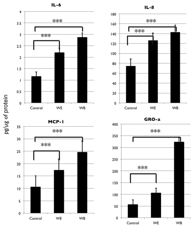
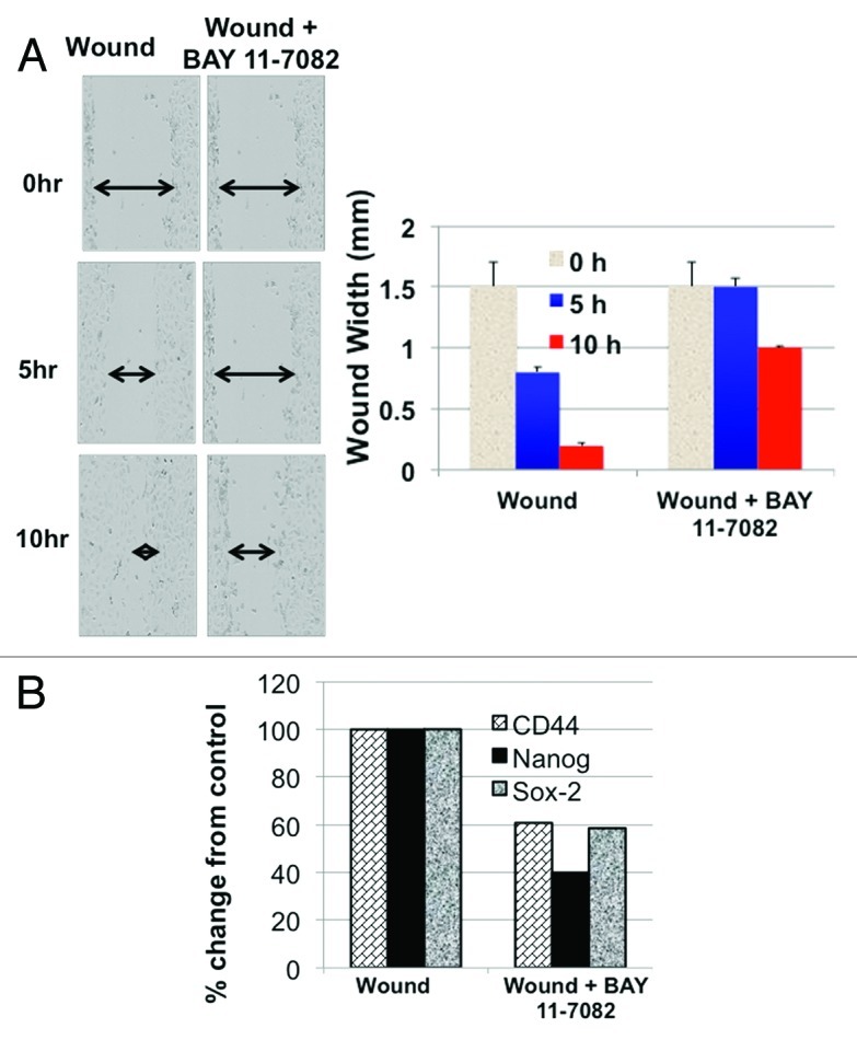
Primary ovarian cancer is responsive to treatment, but chemoresistant recurrent disease ensues in majority of patients. Recent compelling evidence demonstrates that a specific population of cancer cells, the cancer stem cells, initiates and sustains tumors. It is therefore possible that this cell population is also responsible for recurrence. We have shown previously that CD44+/MyD88+ epithelial ovarian cancer stem cells (CD44+/MyD88+ EOC stem cells) are responsible for tumor initiation. In this study, we demonstrate that this population drives tumor repair following surgery- and chemotherapy-induced tumor injury. Using in vivo and in vitro models, we also demonstrate that during the process of tumor repair, CD44+/MyD88+ EOC stem cells undergo self-renewal as evidenced by upregulation of stemness-associated genes. More importantly, we show that a pro-inflammatory microenvironment created by the TLR2-MyD88-NFκB pathway supports EOC stem cell-driven repair and self-renewal. Overall, our findings point to a specific cancer cell population, the CD44+/MyD88+ EOC stem cells and a specific pro-inflammatory pathway, the TLR2-MyD88-NFκB pathway, as two of the required players promoting tumor repair, which is associated with enhanced cancer stem cell load. Identification of these key players is the first step in elucidating the steps necessary to prevent recurrence in EOC patients.

Keywords: TLR2; ovarian cancer stem cells; recurrence; self-renewal; tumor repair.


Figures

None
Figure 1. Tumor injury accelerates tumor growth in vivo. (A) Subcutaneous ovarian cancer xenografts were established on both sides of nude mice. Partial tumor debulking was performed on the right tumor, and skin was incised on the left side but the tumor was left untouched. (B) Size of tumor xenografts was measured ex vivo at day 24 post-surgery. Note that day 24 post-surgery, partially debulked or “wounded” tumor appears larger than the tumor that was not debulked. (C) Graph of tumor kinetics show accelerated growth in “wounded” tumors compared with non-debulked control. Day 0 indicates the measurement immediately preceding wounding. (D) Flow cytomety analysis for CD44 was performed on cancer cells dissociated from the excised tumor xenografts at day 24 post-surgery.
None
Figure 2. Chemotherapy-induced tumor injury enriches for CD44+/MyD88+ EOC stem cells. (A) Paclitaxel treatment (20 mg/kg q3d) enhances growth of tumor implants. (B) Enrichment of CD44 positive cells in tumors from mice treated with Paclitaxel. Tumor implants treated as in (A) were dissociated at the end of the treatment and the number of CD44+ cells was determined by flow cytometry. Note the significant increase in CD44+ cells in the Paclitaxel-treated group.
None
Figure 3. CD44+/MyD88+ EOC stem cells repair in vitro wound. (A) CD44+/MyD88+ EOC stem cells (i) and CD44-/MyD88- EOC cells (ii), were grown to confluence and “wounded” as described in the “Materials and Methods” section. Note that only CD44+/MyD88+ EOC stem cells repair in vitro wound. (B) GFP+/CD44+/MyD88+ EOC stem cells and RFP+/ CD44-/MyD88- EOC cells were grown to confluence and wounded (i). Wound healing was determined by fluorescent microscope at 24 h (ii), 48 h (iii) and 72 h (iv). Note that only GFP+/CD44+/MyD88+ EOC stem cells can repair the wound. (C) Immunohistochemistry for Ki67 (green staining) was determined in (i) no wound control, (ii) wound edge 4 h after wounding, (iii) wound edge 24 h after wounding and (iv) in wound back 24h after wounding. w, wound area. Note: for wound edge and wound back definitions, please see text.
None
Figure 4. In vitro wound/repair process is associated with CD44+/MyD88+ EOC stem cell self-renewal. (A) Levels of CD44, Nanog, Sox2 and Oct4 were determined by qRT-PCR in control cultures (no wound) and in cells from wound edge (WE) 24h after wounding. (B) Levels of CD44, Oct-4, β-catenin and GAPDH were determined by western blot analysis from control cultures (no wound) and from cells isolated from WE and wound back areas. C, control no wound; WE, wound edge; WB, wound back.
None
Figure 5. TLR2 signaling enhances repair and self-renewal. (A) qRT-PCR analyses for TLR2 expression was performed on cancer cells dissociated from the excised tumor fragments at day 24 post-surgery. *p < 0.05. (B) qRT-PCR analyses for TLR2 expression was performed on cancer cells dissociated from tumors of animals treated with Paclitaxel as described in Figure 2A. (C) Levels of TLR2, TLR3 and TLR4 were determined by qRT-PCR from control cultures (no wound) and from cells isolated from wound edge (WE) area 8 h post-wounding. Data represent the average from three independent experiments, measured in triplicate and normalized to GAPDH mRNA. (D) Wounded CD44+/MyD88+ EOC stem cultures were left untreated or treated with 2 µg/ml of PGN or 10 μg/ml of LPS and effect on wound repair was determined. (E) Levels of CD44 and Nanog were determined by qRT-PCR from wounded CD44+/MyD88+ EOC stem cells in the presence or absence of PGN.
None
Figure 6. Inhibition of TLR2 or MyD88 activity inhibits repair. CD44+/MyD88+ EOC stem cells were transfected with a dominant-negative form of TLR2 or a dominant-negative form MyD88 prior to wounding. The effect of blocking TLR2 or MyD88 function on repair is then determined. Note the significant decrease on wound repair in cells transfected with TLR2 or MyD88 dominant-negative plasmids. TLR2-dn, TLR2-dominant-negative; MyD88-dn, MyD88-dominant-negative. *p < 0.005.
None
Figure 7. In vitro wound/repair process upregulates NFκB-mediated pro-inflammatory cytokines. Levels of IL-6, IL-8, MCP-1 and GROa were determined from cell lysates obtained from control cultures (no wound) and from cells isolated from wound edge (WE) and wound back (WB) areas using xMAP technology.
None
Figure 8. Inhibition of NFκB inhibits repair and self-renewal. (A) CD44+/MyD88+ EOC stem cells were wounded and repair process determined in the presence of absence of the NFκB inhibitor, BAY 11-7082 (2.5 μM). (B) CD44, Sox2, Nanog expression levels were determined using qRT-PCR from wounded CD44+/MyD88+ EOC stem cells in the presence or absence of BAY 11-7082. *p < 0.05.

References

    1. Silasi DA, Illuzzi JL, Kelly MG, Rutherford TJ, Mor G, Azodi M, et al. Carcinosarcoma of the ovary. Int J Gynecol Cancer. 2008;18:22–9. doi: 10.1111/j.1525-1438.2007.00948.x. - DOI - PubMed
    1. Jemal A, Bray F, Center MM, Ferlay J, Ward E, Forman D. Global cancer statistics. CA Cancer J Clin. 2011;61:69–90. doi: 10.3322/caac.20107. - DOI - PubMed
    1. Baker TR, Piver MS, Hempling RE. Response to Taxol chemotherapy in resistant ovarian carcinoma. Eur J Gynaecol Oncol. 1993;14:449–54. - PubMed
    1. Armstrong DK. Relapsed ovarian cancer: challenges and management strategies for a chronic disease. Oncologist. 2002;7(Suppl 5):20–8. doi: 10.1634/theoncologist.7-suppl_5-20. - DOI - PubMed
    1. Bast RC, Jr., Hennessy B, Mills GB. The biology of ovarian cancer: new opportunities for translation. Nat Rev Cancer. 2009;9:415–28. doi: 10.1038/nrc2644. - DOI - PMC - PubMed

Publication types

MeSH terms