. 2013 Feb 15;12(4):596-605.
doi: 10.4161/cc.23409. Epub 2013 Jan 16.

Differences in degradation lead to asynchronous expression of cyclin E1 and cyclin E2 in cancer cells

Affiliations

Differences in degradation lead to asynchronous expression of cyclin E1 and cyclin E2 in cancer cells

C Elizabeth Caldon et al. Cell Cycle. .

Abstract

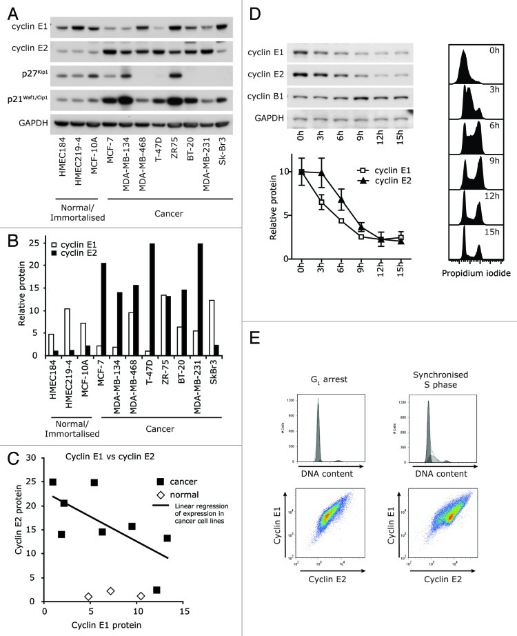
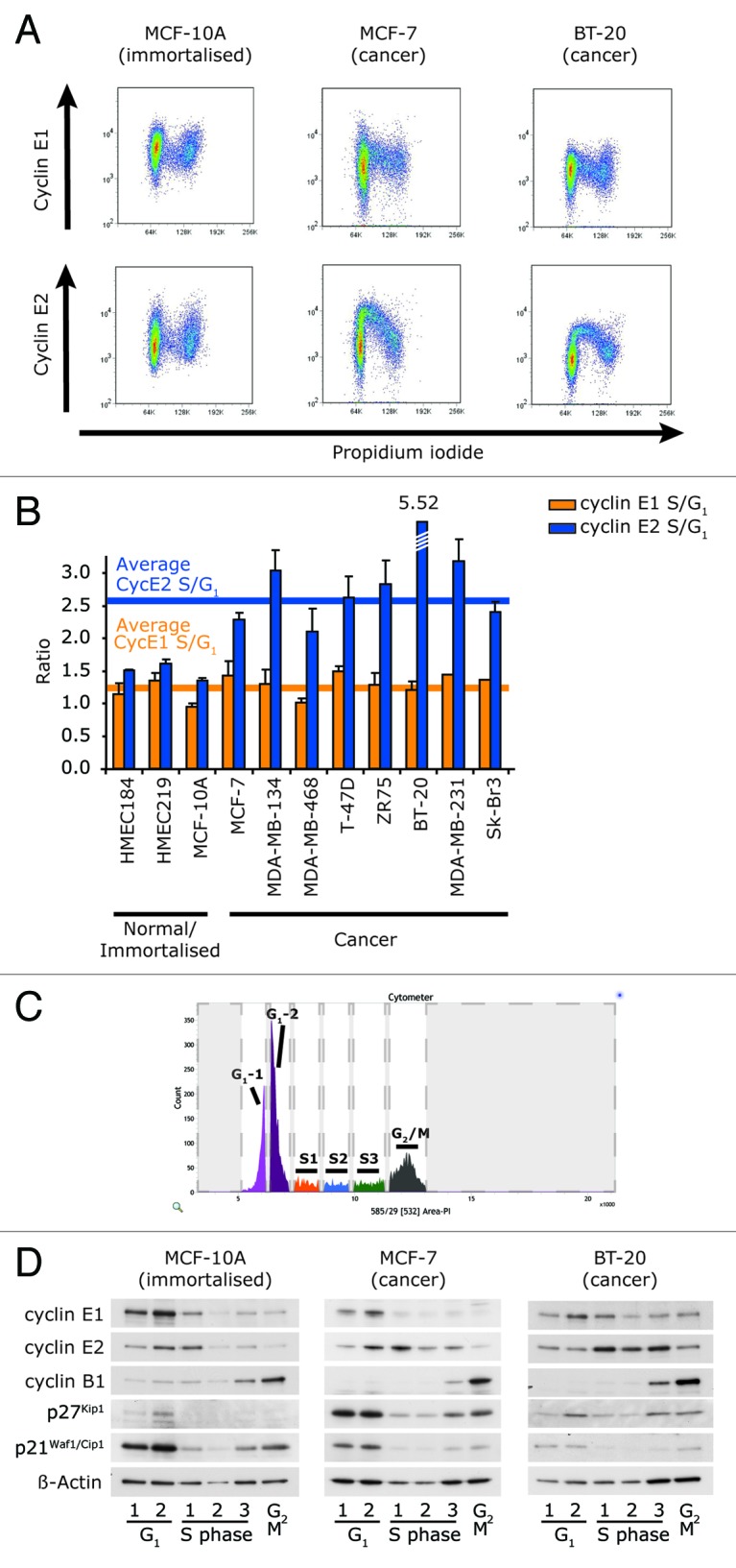
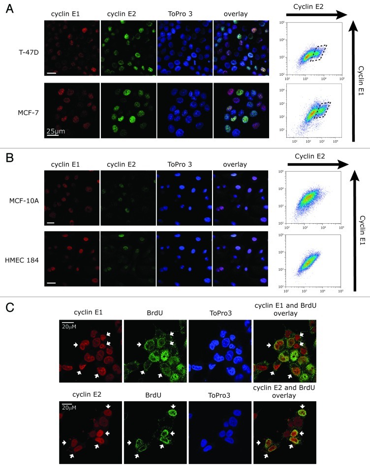
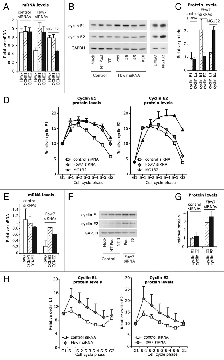
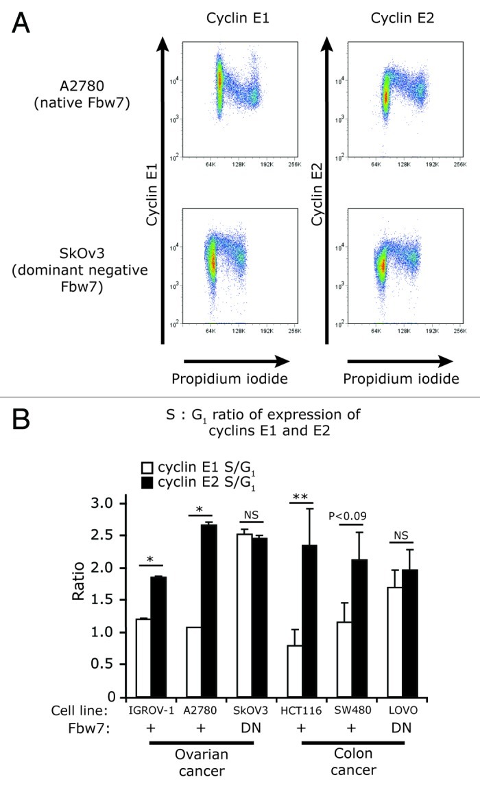
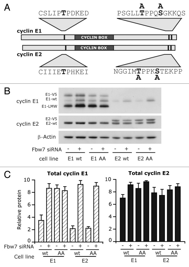
Cyclin E1 is expressed at the G 1/S phase transition of the cell cycle to drive the initiation of DNA replication and is degraded during S/G2M. Deregulation of its periodic degradation is observed in cancer and is associated with increased proliferation and genomic instability. We identify that in cancer cells, unlike normal cells, the closely related protein cyclin E2 is expressed predominantly in S phase, concurrent with DNA replication. This occurs at least in part because the ubiquitin ligase component that is responsible for cyclin E1 downregulation in S phase, Fbw7, fails to effectively target cyclin E2 for proteosomal degradation. The distinct cell cycle expression of the two E-type cyclins in cancer cells has implications for their roles in genomic instability and proliferation and may explain their associations with different signatures of disease.

Keywords: Fbw7; cell cycle; cyclin E1; cyclin E2; proliferation.


Figures

None
Figure 1. Cyclin E1 and E2 have discordant cell cycle expression in cancer cells. (A) Breast cancer and normal/immortalized cell line lysates were immunoblotted for cyclins E1 and E2, p21Waf1/Cip1, p27Kip1 and GAPDH. (B) Densitometry was performed on the levels of cyclins E1 and E2, and normalized to expression of GAPDH. (C) Linear regression identified a negative correlation between the expression of cyclin E1 and cyclin E2 (y = -1.0426x + 22.999, R2 = 0.4591; p < 0.0648). (D) T-47D cells were synchronized with HU, released into the cell cycle, and matched lysates and flow cytometry samples collected 0–15h post release. Lysates were immunoblotted as indicated, and densitometry performed on cyclin E1 and E2 expression normalized to GAPDH expression. Cell cycle distribution was analyzed by propidium iodide staining. Data are from duplicate experiments and error bars represent range between replicates. (E) MCF-7 cells in G0/G1 phase (ICI 182780 arrest) and early S phase (estrogen stimulated) were analyzed for DNA content by propidium iodide staining (PI) and expression of cyclins E1 and E2 by flow cytometry.
None
Figure 2. Cyclin E2 is expressed in S phase in a panel of cancer cell lines, but not normal cell lines. (A) Fixed cells were immunoprobed for either cyclin E1 or E2, counterstained with PI and analyzed by flow cytometry. Representative examples of three cell lines are shown. (B) S/G1 phase ratio of expression of cyclin E1 and E2 was quantitated by calculating the ratio of intensity of the geometric mean of expression of each cyclin in S phase to the geometric mean of intensity of expression in G1 phase and determined across a panel of immortalized and breast cancer cell lines. Error bars represent range between replicates. Data are pooled from, or are representative examples of, duplicate data sets. (C) Exponentially growing cells were stained with PI and sorted into six contiguous fractions of the cell cycle according to DNA content, representative example of BT-20 cells shown. (D) MCF-10A, MCF-7 and BT-20 cells were sorted as in (C), and 25,000 cells from each fraction was prepared in sample buffer and separated by SDS-PAGE, followed by western blotting with the antibodies shown.
None
Figure 3. Cyclin E1 and E2 are expressed in different cell subsets in breast cancer cells. (A and B) Confocal images of (A) breast cancer cells (MCF-7 and T-47D) and (B) immortalized (MCF-10A) or normal (HMEC184) immunoprobed with cyclin E1 (red) or cyclin E2 (green), and counterstained with ToPro3 (blue, nuclei). Fixed cells were analyzed in parallel by flow cytometry for cyclin E1 and E2 levels (right). (C) T-47D cells were pulsed with 20μM BrdU, immunostained for BrdU, cyclin E1 and cyclin E2, and counterstained with ToPro3. White arrows indicate cyclin E1 or cyclin E2 high cells, where high cyclin E2 expression coincides with high BrdU, but high cyclin E1 expression coincides with low BrdU. Scale bars are 20 μm.
None
Figure 4. Cyclin E1 and E2 are differentially regulated by Fbw7 in cancer cell lines, but not normal cell lines. (A) T-47D cells were transfected with Fbw7 and control siRNAs for 48 h, or treated with 8 μM MG132. Lysates were analyzed for Fbw7, CCNE1 and CCNE2 mRNA expression, and (B and C) immunoblotted for cyclin E1 and E2 expression and quantitated by densitometry with normalization to GAPDH. (D) Fixed cells were further immunoprobed for either cyclin E1 or E2, counterstained with PI, and analyzed by flow cytometry. The geometric mean of intensity of expression of cyclin E1 and E2 was quantitated at distinct positions in S phase as identified by PI staining, as indicated in Figure 2C. Data were pooled for control (Mock, NT Pool, NT1) and Fbw7 siRNA (Pool, #8, #9, #10) from quadruplicate experiments and error bars represent S.E.M.. (E–H) HMEC184 cells were transfected with pooled Fbw7 siRNA, Fbw7 siRNA #8, and control treatments (Mock, Non-targeting Pool, Non-targeting #1). (E) Lysates were analyzed by qRT-PCR for Fbw7, CCNE1 or CCNE2 mRNA. (F and G) Lysates were immunoblotted for cyclin E1 and E2 expression and quantitated by densitometry with normalization to GAPDH. (H) Fixed cells were further immunoprobed with antibodies to either cyclin E1 or cyclin E2, counterstained with PI and analyzed by flow cytometry. The geometric mean of intensity of expression of cyclin E1 and E2 was quantitated at distinct positions in S phase as identified by PI staining. Data are pooled from duplicate experiments and error bars represent range between replicates.
None
Figure 5. Cyclin E2 stability is not affected by mutation of Fbw7 in ovarian and colon cancer cell lines. (A) Fixed cells from ovarian cancer cell lines A2780 (Fbw7 +/+) and SkOV3 (Fbw7 dominant-negative - DN) were immunoprobed for either cyclin E1 or E2, counterstained with PI, and analyzed by flow cytometry. (B) S/G1 phase ratio of expression of cyclin E1 and E2 in cell lines with wild-type (+) or dominant-negative (DN) Fbw7. SkOv3 (ovarian) and LoVo (colon) cell lines were compared with other ovarian (A2780, IGROV-1) and colon (HCT116, SW480) cancer cell lines. Data are pooled from duplicate experiments and error bars represent range between replicates. Statistical analysis was performed using two-tailed t-tests, ** = p < 0.005, * = p < 0.05.
None
Figure 6. Cyclin E2 stability is not affected by mutation of Fbw7-recognition sites in cancer cell lines. (A) Schematic of cyclin E1 and cyclin E2, and position of phosphorylation sites and serine/threonine to alanine substitutions. (B) T-47D cells overexpressing cyclin E1 (wt), cyclin E1 T395A/S399A (AA), cyclin E2 (wt) or cyclin E2 T392A/S396A (AA) were transfected with Fbw7 and control siRNAs. Lysates collected at 72 h were immunoblotted for cyclin E1 and E2, and β-actin. (C) Levels of cyclins E1 and E2 were quantitated by densitometry normalized to β-actin.

References

    1. Hwang HC, Clurman BE. Cyclin E in normal and neoplastic cell cycles. Oncogene. 2005;24:2776–86. doi: 10.1038/sj.onc.1208613. - DOI - PubMed
    1. Welcker M, Singer J, Loeb KR, Grim J, Bloecher A, Gurien-West M, et al. Multisite phosphorylation by Cdk2 and GSK3 controls cyclin E degradation. Mol Cell. 2003;12:381–92. doi: 10.1016/S1097-2765(03)00287-9. - DOI - PubMed
    1. Koepp DM, Schaefer LK, Ye X, Keyomarsi K, Chu C, Harper JW, et al. Phosphorylation-dependent ubiquitination of cyclin E by the SCFFbw7 ubiquitin ligase. Science. 2001;294:173–7. doi: 10.1126/science.1065203. - DOI - PubMed
    1. Juan G, Cordon-Cardo C. Intranuclear compartmentalization of cyclin E during the cell cycle: disruption of the nucleoplasm-nucleolar shuttling of cyclin E in bladder cancer. Cancer Res. 2001;61:1220–6. - PubMed
    1. Ekholm-Reed S, Méndez J, Tedesco D, Zetterberg A, Stillman B, Reed SI. Deregulation of cyclin E in human cells interferes with prereplication complex assembly. J Cell Biol. 2004;165:789–800. doi: 10.1083/jcb.200404092. - DOI - PMC - PubMed

Publication types

MeSH terms

LinkOut - more resources