. 2013 Jan 16;15(1):7.
doi: 10.1186/1532-429X-15-7.

Functionalization of gadolinium metallofullerenes for detecting atherosclerotic plaque lesions by cardiovascular magnetic resonance

Affiliations

Functionalization of gadolinium metallofullerenes for detecting atherosclerotic plaque lesions by cardiovascular magnetic resonance

Anthony Dellinger et al. J Cardiovasc Magn Reson. .

Abstract

Background: The hallmark of atherosclerosis is the accumulation of plaque in vessel walls. This process is initiated when monocytic cells differentiate into macrophage foam cells under conditions with high levels of atherogenic lipoproteins. Vulnerable plaque can dislodge, enter the blood stream, and result in acute myocardial infarction and stroke. Imaging techniques such as cardiovascular magnetic resonance (CMR) provides one strategy to identify patients with plaque accumulation.

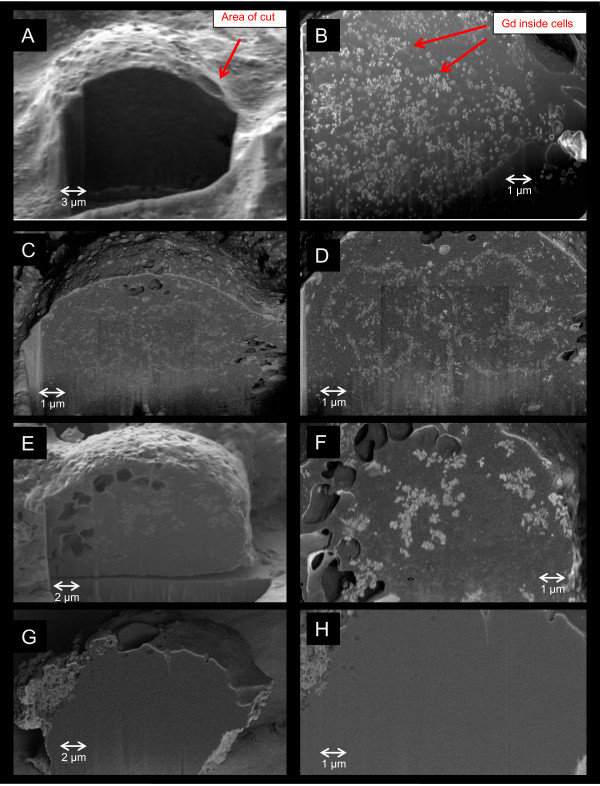
Methods: We synthesized an atherosclerotic-targeting contrast agent (ATCA) in which gadolinium (Gd)-containing endohedrals were functionalized and formulated into liposomes with CD36 ligands intercalated into the lipid bilayer. In vitro assays were used to assess the specificity of the ATCA for foam cells. The ability of ATCA to detect atherosclerotic plaque lesions in vivo was assessed using CMR.

Results: The ATCA was able to detect scavenger receptor (CD36)-expressing foam cells in vitro and were specifically internalized via the CD36 receptor as determined by focused ion beam/scanning electron microscopy (FIB-SEM) and Western blotting analysis of CD36 receptor-specific signaling pathways. The ATCA exhibited time-dependent accumulation in atherosclerotic plaque lesions of ApoE -/- mice as determined using CMR. No ATCA accumulation was observed in vessels of wild type (C57/b6) controls. Non-targeted control compounds, without the plaque-targeting moieties, were not taken up by foam cells in vitro and did not bind plaque in vivo. Importantly, the ATCA injection was well tolerated, did not demonstrate toxicity in vitro or in vivo, and no accumulation was observed in the major organs.

Conclusions: The ATCA is specifically internalized by CD36 receptors on atherosclerotic plaque providing enhanced visualization of lesions under physiological conditions. These ATCA may provide new tools for physicians to non-invasively detect atherosclerotic disease.


Figures

Figure 1
Figure 1
ATCA can detect inflammatory plaque in vivo. ApoE−/− mice (23 wks; n=5/grp) were injected i.v. (100 μg/100 μl) with ATCA (top and middle) or non-targeted control (bottom) and images acquired at the indicated times. Bar = 1.0 mm. Arrows indicate contrast agent binding to the plaque. A. Representative images from two (ATCA) or one (control) different mice injected with ATCA. B, C. Quantification of signal enhancement of ATCA. In B, the brightest voxels (approximately >10 spots/section) in the aorta wall from 5 separate animals were measured excluding artifact. The ratio of the Signal Intensity (SI) of the brightest voxel in the aorta wall to the SI of the (non-affected) myocardium for each time point was calculated. In C, the ratio of SI of the whole region lining the aorta wall to the SI of the (non-affected) myocardium was measured and the average signal intensity was recorded for 5 separate slices from each time-point. The SI-enhanced/SI-myo ratio from each time point to its pre scan SI-enhanced/SI-myo ratio were normalized (prescan values = 1). Data is expressed as mean of 5 CMR slices ± SD (n = 5; animals/grp; * indicates significance (p<0.05). D. Plaque lesions express macrophages and CD36 receptors. Aortas were stained with H&E (left), rat anti-mouse CD36 (middle), rat anti-mouse CD68 (bottom) or non-specific rat IgG (nsIgG). Peroxidase-conjugated anti-rat Abs were added and developed using AEC. The brown staining represents CD36- or CD68-positive cells located in the plaque lesions. Results are representative of at least three separate aortas.
Figure 2
Figure 2
A. Upregulation of CD36 receptors on foam cells. U937 cells (in duplicate) were treated as described in Methods, washed and incubated with antibodies to CD36 or an isotype matched control. The graph shows the average mean fluorescent intensity (MFI) of the signal (±SD) and is representative of three separate experiments. B. Testing ATCA for foam cell binding. U937 cells (non-foam) were converted into foam cells (foam) using oxLDL and PMA as described. Cells were washed and ATCA (10 μg/ml) added overnight in a CO2 incubator. The next day cells were washed with media and the supernatants (S) and pellets (P) subjected to ICP for Gd detection. The percentage of Gd in the pellet and supernatant was calculated based on the total amount added to the cells.
Figure 3
Figure 3
A-H. ATCA are taken up by foam cells. Foam cells were incubated with ATCA (10 μg/ml; A-F) or non-targeted control (G,H) for 16 (A,B), 48 (C,D) or 72 (E,F) hrs, washed, fixed, and subjected to FIB-SEM. Left columns (A,C,E,G) shows the morphology images taken by secondary electron detector after focused ion beam cut and the right column (B,D,F,H) shows the corresponding material contrast images taken by energy and angle selective backscattered electron (EsB) detector for the same region. The foam cell-targeted ATCA are clearly revealed inside the cells but not in the non-targeted controls. In parallel, an aliquot of cells were used to measure cell viability by trypan blue exclusion and revealed no cell death induced by ATCA addition compared to non-treated controls (not shown).
Figure 4
Figure 4
A,B. ATCA activate CD36-specific signaling intermediates. Foam cells were incubated with or without varying concentrations of ATCA (A; dose response) for 10 min or with 10 μg/ml ATCA (B; time course) for varying times. Cells were lysed and Western blotted using phospho-specific antibodies. Band intensities were quantified and the numbers represent the fold increase of each band compared to the housekeeping gene β-actin which did not demonstrate any change upon ATCA challenge. Results are representative of three separate experiments.
Figure 5
Figure 5
A. Plaque-targeting contrast agents do not accumulate in major organs. A representative plaque-targeting contrast agent and non-targeted control was injected as above and the mouse placed supine for whole body scan. Results are representative of four separate mice. B. C. ATCA does not accumulate in liver or induce toxicity. Mice were treated with or without 1000 μg/100 μl of ATCA via tail vein injection. Two and six days later the serum was used for ALT and AST levels. Data is presented as an average of four (untreated) or four (treated) mice ± SD. No increase in activity was observed between the untreated and treated samples. Activity is measure by Units/L obtained using linear regression from a standard curve.

Similar articles

Cited by

References

    1. Buxton DB, Antman M, Danthi N. et al.Report of the national heart, lung, and blood institute working group on the translation of cardiovascular molecular imaging. Circulation. 2011;123(19):2157–2163. doi: 10.1161/CIRCULATIONAHA.110.000943. - DOI - PMC - PubMed
    1. Underhill HR, Hatsukami TS, Fayad ZA, Fuster V, Yuan C. MRI of carotid atherosclerosis: clinical implications and future directions. Nat Rev Cardiol. 2010;7(3):165–173. doi: 10.1038/nrcardio.2009.246. - DOI - PubMed
    1. Stevenson S, Fowler PW, Heine T. et al.A stable non-classical metallofullerene family. Nature. 2000;408(6811):427–428. doi: 10.1038/35044199. - DOI - PubMed
    1. MacFarland DK, Walker KL, Lenk RP. et al.Hydrochalarones: a novel endohedral metallofullerene platform for enhancing magnetic resonance imaging contrast. J Med Chem. 2008;51(13):3681–3683. doi: 10.1021/jm800521j. - DOI - PMC - PubMed
    1. Podrez EA, Poliakov E, Shen Z. et al.Identification of a novel family of oxidized phospholipids that serve as ligands for the macrophage scavenger receptor CD36. J Biol Chem. 2002;277(41):38503–38516. doi: 10.1074/jbc.M203318200. - DOI - PubMed

Publication types

MeSH terms