. 2013 Jan-Feb;4(1):61-73.
doi: 10.4161/nucl.23388. Epub 2013 Jan 1.

Soft substrates normalize nuclear morphology and prevent nuclear rupture in fibroblasts from a laminopathy patient with compound heterozygous LMNA mutations

Affiliations

Soft substrates normalize nuclear morphology and prevent nuclear rupture in fibroblasts from a laminopathy patient with compound heterozygous LMNA mutations

Chiara Tamiello et al. Nucleus. 2013 Jan-Feb.

Abstract

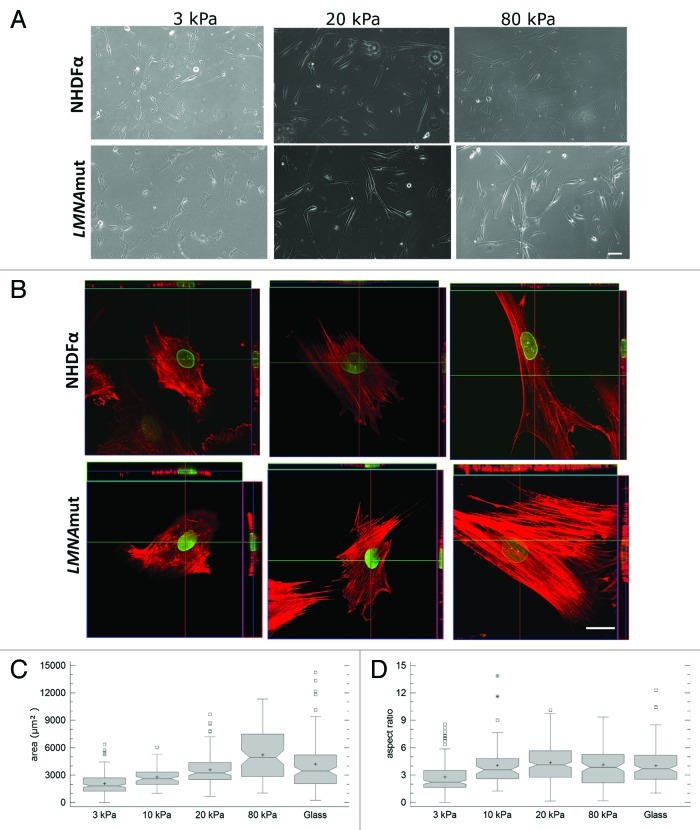
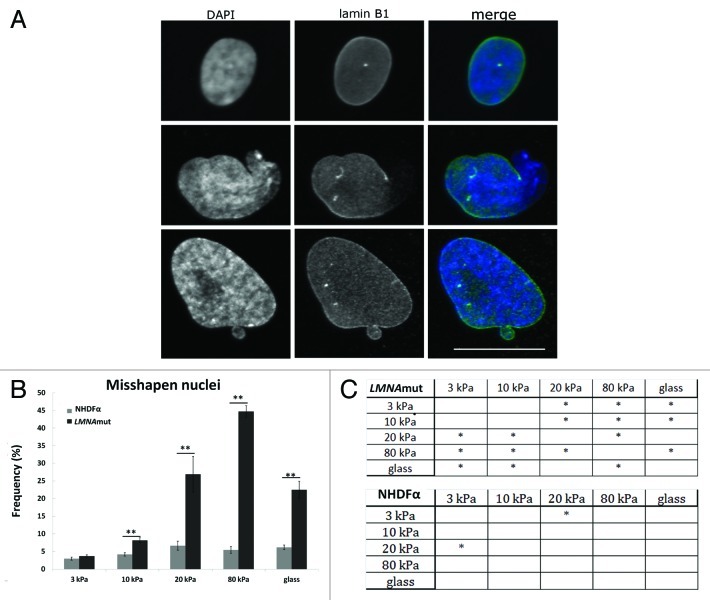
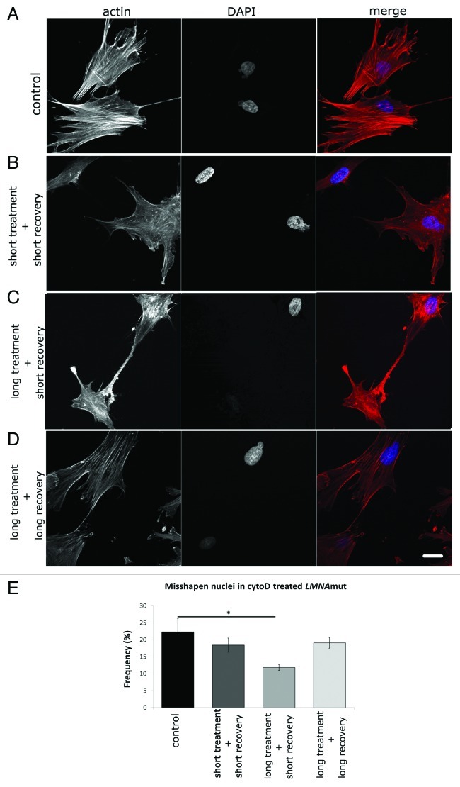
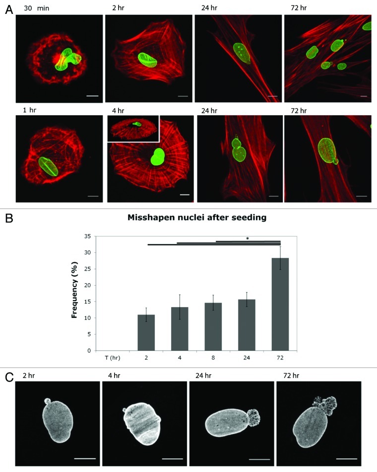
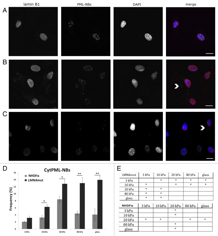
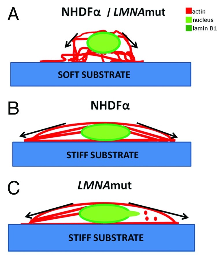
Laminopathies, mainly caused by mutations in the LMNA gene, are a group of inherited diseases with a highly variable penetrance; i.e., the disease spectrum in persons with identical LMNA mutations range from symptom-free conditions to severe cardiomyopathy and progeria, leading to early death. LMNA mutations cause nuclear abnormalities and cellular fragility in response to cellular mechanical stress, but the genotype/phenotype correlations in these diseases remain unclear. Consequently, tools such as mutation analysis are not adequate for predicting the course of the disease. Here, we employ growth substrate stiffness to probe nuclear fragility in cultured dermal fibroblasts from a laminopathy patient with compound progeroid syndrome. We show that culturing of these cells on substrates with stiffness higher than 10 kPa results in malformations and even rupture of the nuclei, while culture on a soft substrate (3 kPa) protects the nuclei from morphological alterations and ruptures. No malformations were seen in healthy control cells at any substrate stiffness. In addition, analysis of the actin cytoskeleton organization in this laminopathy cells demonstrates that the onset of nuclear abnormalities correlates to an increase in cytoskeletal tension. Together, these data indicate that culturing of these LMNA mutated cells on substrates with a range of different stiffnesses can be used to probe the degree of nuclear fragility. This assay may be useful in predicting patient-specific phenotypic development and in investigations on the underlying mechanisms of nuclear and cellular fragility in laminopathies.

Keywords: lamina; laminopathies; nuclear rupture; nuclear shape alteration; substrate stiffness.


Figures

None
Figure 1. Effect of substrate stiffness on cell morphology and actin cytoskeleton organization. (A) Representative bright field images of NHDFα and LMNAmut cells seeded on polyacrylamide gels with stiffness ranging from 3 kPa to 80 kPa taken 48 h after seeding. Fewer and less spread cells were present on 3 kPa polyacrylamide gels than on stiffer substrates for both cell types. Scale bar: 100 μm. (B) Actin organization in NHDFα and LMNAmut (phalloidin-TRITC, red) showed increased organization and tension on substrates stiffer than 3 kPa. Green color is given by lamin B1 staining. Scale bar: 20 μm. (C and D) Cell area and aspect ratio presented as box-and-whisker plots. The measurements of NHDFα and LMNAmut did not show significant difference, thus the values were considered as a group.
None
Figure 2. Nuclear morphological abnormality regulation by substrate stiffness. (A) Immunofluorescent labeling of cell nuclei with DAPI (blue), lamin B1 (green) and overlay of the two in the most right panels allowed to distinguish between normally (upper row) and abnormally shaped nuclei (second and third row). In particular, the nucleus in the second row shows a protrusion and in the third row a bleb can be observed. Scale bars: 10 μm. (B) Frequency of abnormally shaped nuclei on increasing PA gel stiffness for LMNAmut and control NHDFα. Values represent means from at least 300 cells from two experiments. Bars represent SEM * p < 0.05, ** p < 0.01 vs NHDFα on the same substrate stiffness (C) Statistical analyses of differences in frequency of misshapen nuclei for LMNAmut and NHDFα on the different substrate stiffness’s. *, p < 0.05; no star, p > 0.05.
None
Figure 3. Influence of substrate stiffness on cytoskeletal actin organization and aberrations. Confocal z-series taken from half height of the whole cell and relative orthogonal cross sections of NHDFα and LMNAmut immunocytochemical stained for F-actin in red (phalloidin-TRITC) and Lamin B1 in green at 48 h after seeding. (A) Representative fibroblast seeded on 3 kPa PA gels. It shows short and not tensed actin fibers, which are missing in the perinuclear region. An actin cap is running above the nucleus (white arrowhead). No differences could be noticed between LMNAmut and NHDFα. Thus no aberrations could be detected in the actin cytoskeleton of cells plated on soft substrates. (B) Control NHDFα on PA gel stiffer than 3 kPa, precisely on the 20 kPa PA gel. Actin stress fibers are tensed and well-structured also in the perinuclear region. The actin cap made of thick stress fibers run above the nucleus the (white arrowhead) nucleus. (C and D) Representative aberrations found in LMNAmut seeded on 20 (C) and 80 kPa (D) PA gels. Cells have a misshapen nucleus. Yellow arrowhead indicates the lack of actin fibers in the perinuclear region (D and E) and a speckled distribution of actin (E). The actin cap is running above the nucleus (white arrowhead). Scale bars: 20 μm. (E) Representative images of cells on three substrate stiffnesses. NHDFα (left panel) and LMNAmut (right panel) on 3kPa (F), 20 kPa (G) and 80 kPa (H) PA gels. Scale bar: 50 μm.
None
Figure 4. Effects of transient cytoD treatment on LMNAmut nuclei. Representative confocal sections of LMNAmut seeded on collagen I coated glass substrates incubated with cytoD 1μM and recovered in normal growth medium. After fixation, cells were stained with DAPI (blue) to check for nuclear abnormalities and phalloidin-TRITC (red) to check for actin organization. (A) Untreated control LMNAmut. (B) Short treatment + short recovery: LMNAmut treated for 30 min with cytoD and recovered for 1 h. (C) Long treatment + short recovery: LMNAmut treated for 3 h with cytoD and recovered for 1 h. (D) Long treatment + long recovery LMNAmut treated for 3 h with cytoD and recovered overnight. Scale bar: 20 μm. (E) Frequency of misshapen nuclei in LMNAmut upon treatment with cytoD. At least 600 cells were assessed per each group.*, p < 0.05; no star, p > 0.05
None
Figure 5. Alterations in nuclear shape and actin organization upon attachment of cells after trypsin treatment. (A) Representative confocal sections of LMNAmut seeded on collagen I coated glass substrates at 30 min, 1, 2, 4, 24 and 72 h after seeding. Cells were immunocytochemical stained for F-actin in red (phalloidin-TR) and lamin A/C in green. Inset at 4 h: 3D view (generated by ImageJ 3D-viewer, showing the position of the nucleus (green)at the upper region of the cell, with very few tense actin stress fibers (red) surrounding the nucleus). Scale bar: 10 μm. (B) Frequency of misshapen nuclei after seeding. .*, p < 0.05; no star, p > 0.05. (C) Changes in nuclear bleb size upon attachment, visualized by immunofluorescence using the Jol2 lamin A/C antibody. Note the increase in size as well as the aberrant shape of the nuclear blebs. Note also that in most blebs a typical honeycomb structure of the lamina staining can be seen. Scale bar: 10 μm.
None
Figure 6. PML-NBs localization as a marker for cellular compartmentalization. (A−C) Confocal sections representative of cell nuclei were immunolabeled with Lamin B1 (red), DAPI (blue) and PML-NBs (green) to investigate the localization of PML-NBs. Nuclei were counterstained with DAPI (blue). The most right panel shows the triple overlay. Scale bars: 10 μm. (A) Nuclei showing normal morphology and internal localization of PML-NBs. Cellular compartmentalization is intact. (B) Cytoplasmic localization of PML-NBs (cytPML-NBs) around a nucleus showing an abnormal morphology (white arrowhead). Loss of cellular compartmentalization is indicated by the exit of PML-NBs to the cytoplasm. (C) CytPML-NBs could be found also in normally shaped nuclei (white arrowhead) indicating that loss of cellular compartmentalization is not directly related to nuclear morphology abnormalities. (D) Relative frequency of NHDFα and LMNAmut showing cytPML-NBs. Values represent means from at least 600 cells from 2 experiments. Bars represent SEM * p < 0.05, ** p < 0.01 vs NHDFα on the same substrate stiffness (E) Statistical analyses of differences in frequency of cytPML-NBs for LMNAmut and NHDFα on the different substrate stiffness’s. *, p < 0.05; no star, p > 0.05.
None
Figure 7. Spontaneous ruptures of the nuclear membrane do not occur on soft substrates. (A) Montage of selected images from a time-lapse recording of LMNAmut cell cultured on 10 kPa PA gel and transfected with EYFP-NLS, sampled at 1 min intervals for 2 h (Video S1). Nuclear membrane rupture causes the decrease in intranuclear EYFP signal and increase in cytoplasmic EYFP signal (at 0:36, yellow arrowhead). Subsequently, the nuclear signal is gradually up taken by the nucleus and the rupture appears to be restored (at 0:51, orange arrowhead) (B) Evolution in time of EYFP-NLS (mean) intensity in the nucleus of the cell shown in (A). (C) Frequency of spontaneous nuclear membrane ruptures on the different substrates for LMNAmut. Error bars represent the square root of the number of recording.
None
Figure 8. Proposed mechanism for actin cytoskeleton organization and effects on nuclear abnormalities on soft and stiff substrates. Schematic representation of the cross section of a cell seeded on substrates with different stiffness. Red represents the actin cytoskeleton while green the nucleus (dark green nuclear lamina). (A) When seeded on a soft substrate NHDFα and LMNAmut cells have the same response. They develop not tensed actin stress fibers which are not directly connected to the nucleus in the perinuclear region. Fibers run below and on top of the nucleus. Since low forces are exerted on the nucleus, the onset of nuclear abnormalities is prevented. (B) NHDFα cells seeded on a stiff substrate (stiffer than 3 kPa) develop tensed actin stress fibers which are well-organized in the whole cytoplasm. Stress fibers connect to the nucleus and form also the actin cap running on top of the nucleus. (C)LMNAmut cells on top of a stiff substrate develop tensed stress fibers. These stress fibers appear to be lacking in the perinuclear region and aggregates of actin are often visible. The pushing action of the actin cap (stress fibers running on top of the nucleus) together with the uneven distribution of pushing actin in the area around the nucleus lead to the development of nuclear abnormalities, further resulting in nuclear damage.

Similar articles

Cited by

References

    1. Gilbert PM, Havenstrite KL, Magnusson KEG, Sacco A, Leonardi NA, Kraft P, et al. Substrate elasticity regulates skeletal muscle stem cell self-renewal in culture. Science. 2010;329:1078–81. doi: 10.1126/science.1191035. - DOI - PMC - PubMed
    1. Discher DE, Janmey P, Wang YL. Tissue cells feel and respond to the stiffness of their substrate. Science. 2005;310:1139–43. doi: 10.1126/science.1116995. - DOI - PubMed
    1. Wilson KL, Foisner R. Lamin-binding Proteins. Cold Spring Harb Perspect Biol. 2010;2:a000554. doi: 10.1101/cshperspect.a000554. - DOI - PMC - PubMed
    1. Crisp M, Liu Q, Roux K, Rattner JB, Shanahan C, Burke B, et al. Coupling of the nucleus and cytoplasm: role of the LINC complex. J Cell Biol. 2006;172:41–53. doi: 10.1083/jcb.200509124. - DOI - PMC - PubMed
    1. Huang H, Kamm RD, Lee RT. Cell mechanics and mechanotransduction: pathways, probes, and physiology. Am J Physiol Cell Physiol. 2004;287:C1–11. doi: 10.1152/ajpcell.00559.2003. - DOI - PubMed

LinkOut - more resources