Review
. 2013 Jan-Feb;7(1):66-75.
doi: 10.4161/pri.23499. Epub 2013 Jan 1.

Grey matter damage in multiple sclerosis: a pathology perspective

Affiliations
Review

Grey matter damage in multiple sclerosis: a pathology perspective

Roel Klaver et al. Prion. 2013 Jan-Feb.

Abstract

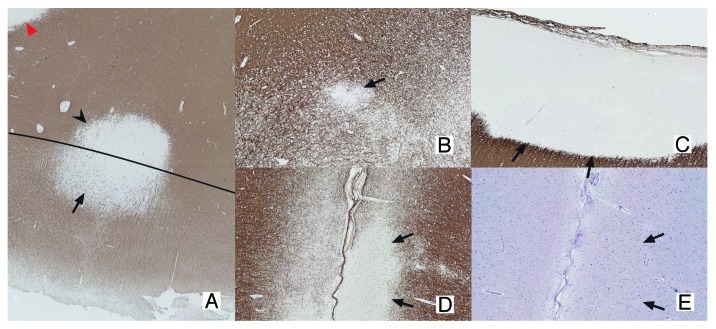
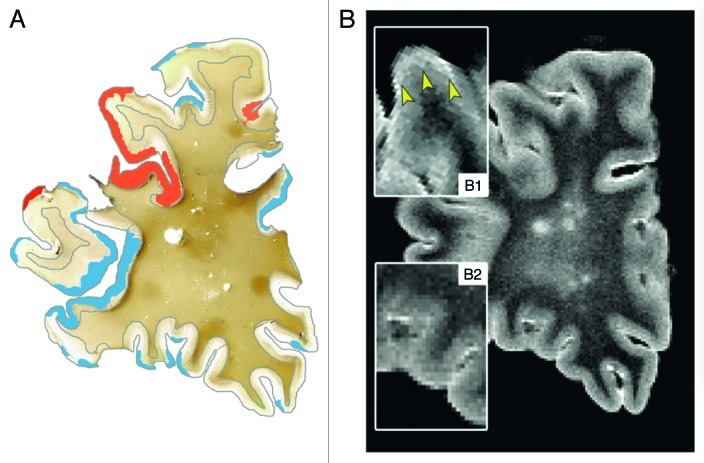
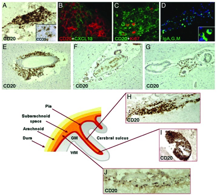
Over the past decade, immunohistochemical studies have provided compelling evidence that gray matter (GM) pathology in multiple sclerosis (MS) is extensive. Until recently, this GM pathology was difficult to visualize using standard magnetic resonance imaging (MRI) techniques. However, with newly developed MRI sequences, it has become clear that GM damage is present from the earliest stages of the disease and accrues with disease progression. GM pathology is clinically relevant, as GM lesions and/or GM atrophy were shown to be associated with MS motor deficits and cognitive impairment. Recent autopsy studies demonstrated significant GM demyelination and microglia activation. However, extensive immune cell influx, complement activation and blood-brain barrier leakage, like in WM pathology, are far less prominent in the GM. Hence, so far, the cause of GM damage in MS remains unknown, although several plausible underlying pathogenic mechanisms have been proposed. This paper provides an overview of GM damage in MS with a focus on its topology and histopathology.

Keywords: clinical relevance; grey matter pathology; imaging; multiple sclerosis; topology.


Figures

None
Figure 1.Classification of GM lesions as described by Bö et al. Type I lesions (A) are GM/WM mixed lesions (original magnification 2.5x). The black line indicates the border between WM and GM. The GM and WM parts of the lesion are indicated with an arrow and arrowhead, respectively. The red arrowhead indicates a WM lesion. Type II lesions (B) are located completely intracortically, while type III and IV lesions run from the pial surface toward deeper cortical layers (D and E) or all the way to the GM/WM border, respectively (C; original magnification 5x). Both D en E are sections showing the same type III lesion. The lesion is visible in the MBP staining (D), showing a clearly demyelinated area (arrows indicate lesion borders), while the same area is invisible in a consecutive slide stained with a more conventional histochemical staining (E; Luxol Fast-Blue). The original magnification is 5x. (A-D), sections stained using myelin basic protein (MBP) antibodies. (E) Section stained using LFB.
None
Figure 2. Post-mortem MRI and accompanying PLP immunohistochemical staining of a coronal section. T2 weighted MRI scans (B) were compared with PLP stainings (A) of the same coronal sections. This figure clearly shows the difficulty to detect cortical lesions with conventional MRI sequences. (A): MRI-visible cortical lesions are shown in red, while MRI-invisible cortical lesions are shown in blue (original magnification 0.7x); (B1): MRI-visible lesions are indicated with arrowheads. MRI-visible cortical lesions show only very subtle increase of signal intensity; (B2) MRI-invisible lesions. Reproduced from Seewann et al., with permission from SAGE Publishers.
None
Figure 3. Meningeal inflammation in MS. Immunostainings of MS brain sections show intrameningeal ectopic B-cell follicle like structures in a cerebral sulcus containing CD20+ B cells (A), ramified stromal cells/FCD expressing CD35 (inset A) and expressing CXCLI3 (B), Ki67+ proliferating B cells (C), and plasmablast/plasma cells stained with an anti- Ig-G, -A, -M polyclonal antibody (D; the inset shows two intrafollicular plasma cells at high magnification). CD20+ B cells are also present in periventricular WM lesions (E) and in scarcely inflamed meninges entering a cerebral sulcus in a SPMS (F) and PPMS case (G). The lower schematic picture illustrates the localization of ectopic B-cell follicle like structures in the MS brain. These follicle like structures are present along (H) and in the depth (I) of the cerebral sulci, whereas scattered B lymphocytes (J) are detected in the meninges covering the external brain surface. Original magnifications: E-G = 100x; A, D, H-J = 200x, B, C and insets in A and D = 400x. Reproduced from Magliozzi et al. with permission from Oxford University Press.

Similar articles

Cited by

References

    1. Lublin FD, Reingold SC, National Multiple Sclerosis Society (USA) Advisory Committee on Clinical Trials of New Agents in Multiple Sclerosis Defining the clinical course of multiple sclerosis: results of an international survey. Neurology. 1996;46:907–11. doi: 10.1212/WNL.46.4.907. - DOI - PubMed
    1. Rovaris M, Confavreux C, Furlan R, Kappos L, Comi G, Filippi M. Secondary progressive multiple sclerosis: current knowledge and future challenges. Lancet Neurol. 2006;5:343–54. doi: 10.1016/S1474-4422(06)70410-0. - DOI - PubMed
    1. Miller DH, Leary SM. Primary-progressive multiple sclerosis. Lancet Neurol. 2007;6:903–12. doi: 10.1016/S1474-4422(07)70243-0. - DOI - PubMed
    1. Cottrell DA, Kremenchutzky M, Rice GP, Koopman WJ, Hader W, Baskerville J, et al. The natural history of multiple sclerosis: a geographically based study. 5. The clinical features and natural history of primary progressive multiple sclerosis. Brain. 1999;122:625–39. doi: 10.1093/brain/122.4.625. - DOI - PubMed
    1. Bö L, Geurts JJG, van der Valk P, Polman C, Barkhof F. Lack of correlation between cortical demyelination and white matter pathologic changes in multiple sclerosis. Arch Neurol. 2007;64:76–80. doi: 10.1001/archneur.64.1.76. - DOI - PubMed

Publication types

LinkOut - more resources