. 2013 Feb 12;110(7):E602-10.
doi: 10.1073/pnas.1208524110. Epub 2013 Jan 16.

Wnt5a cooperates with canonical Wnts to generate midbrain dopaminergic neurons in vivo and in stem cells

Affiliations

Wnt5a cooperates with canonical Wnts to generate midbrain dopaminergic neurons in vivo and in stem cells

Emma R Andersson et al. Proc Natl Acad Sci U S A. .

Abstract

Wnts are a family of secreted proteins that regulate multiple steps of neural development and stem cell differentiation. Two of them, Wnt1 and Wnt5a, activate distinct branches of Wnt signaling and individually regulate different aspects of midbrain dopaminergic (DA) neuron development. However, several of their functions and interactions remain to be elucidated. Here, we report that loss of Wnt1 results in loss of Lmx1a and Ngn2 expression, as well as agenesis of DA neurons in the midbrain floor plate. Remarkably, a few ectopic DA neurons still emerge in the basal plate of Wnt1(-/-) mice, where Lmx1a is ectopically expressed. These results indicate that Wnt1 orchestrates DA specification and neurogenesis in vivo. Analysis of Wnt1(-/-);Wnt5a(-/-) mice revealed a greater loss of Nurr1(+) cells and DA neurons than in single mutants, indicating that Wnt1 and Wnt5a interact genetically and cooperate to promote midbrain DA neuron development in vivo. Our results unravel a functional interaction between Wnt1 and Wnt5a resulting in enhanced DA neurogenesis. Taking advantage of these findings, we have developed an application of Wnts to improve the generation of midbrain DA neurons from neural and embryonic stem cells. We thus show that coordinated Wnt actions promote DA neuron development in vivo and in stem cells and suggest that coordinated Wnt administration can be used to improve DA differentiation of stem cells and the development of stem cell-based therapies for Parkinson's disease.


Conflict of interest statement

The authors declare no conflict of interest.

Figures
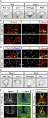
Fig. 1.
Fig. 1.
Wnt1 is required for ventral midbrain FP neurogenesis. (A) Shh and Foxa2 mRNA expression in the VM of Wnt1−/− mice is delayed compared with WT mice at E11.5; their expression is lost in lateral positions (*), and the medial down-regulation of Shh in WT mice (arrow) is not detected in Wnt1−/− mice. (B) Foxa2 protein and TH are found throughout the VM of WT mice at E12, whereas TH+ DA neurons are severely reduced in number and are found solely in the lateral BP in Wnt1−/− mice. The boxed regions in the left panels demarcate the regions that are magnified in the panels at the far right. V = ventricle. (C) TH+ cells are found only in Lmx1a+ domains, which are laterally displaced from the FP to the BP in Wnt1−/− mice. The boxed regions in the left panels demarcate the regions that are magnified in the panels at the far right. (D) The expression of the proneural factors Mash1 and Ngn2 is lost from the FP and is displaced laterally in Wnt1−/− mice at E11.5. (E) At E12.5, the FP of Wnt1−/− mice shows a reduced ventricular zone and contains no neuronal somas (Tuj+Topro3+ cells). (F) Although the WT FP contains Sox2+ progenitors and Nurr1+ postmitotic cells, the Wnt1−/− FP shows fewer Sox2+ progenitors, and all Nurr1+ cells are found in the BP.
Fig. 2.
Fig. 2.
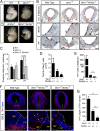
Wnt1/Wnt5a double-mutant mice reveal redundant and nonredundant functions during ventral midbrain development. (A) Wnt1+/−;Wnt5a+/− mice were mated to produce WT, Wnt1−/−;Wnt5a+/+ (labeled Wnt1−/−), Wnt1+/+;Wnt5a−/− (labeled Wnt5a−/−), and Wnt1−/−;Wnt5a−/− mice. At E10.5, Wnt1−/− mice display the previously described phenotypes of midbrain deletion and a less distinct isthmus (yellow arrowhead), whereas Wnt5a−/− mice display A–P shortening which is most obvious in the tail region (white arrow). Wnt1−/−;Wnt5a−/− double-mutant mice resemble a combination of the two single knockouts, with a shortened A–P axis and a significantly shorter midbrain region (dashed lines). (B and C) Proliferation, assessed by staining for the mitotic marker PH3, is specifically reduced in the FP but not the BP of Wnt1−/− mice. Panels show whole midbrain (Top Row) and magnifications of the BP and FP (Middle Row). The areas demarcated by red dotted boxes are magnified in the bottom row. The reduction in the numbers of PH3+ cells in the FP of Wnt1−/− mice is partially rescued by loss of Wnt5a. (D) The total number of Nurr1+ cells decreases to a greater extent in double mutants than in single mutants or WT littermates. (E and F) TH+ cells are dramatically reduced in number and are laterally displaced (white arrowheads in F) from the FP to the BP in Wnt1−/−;Wnt5a+/+ mice. These defects are worsened in Wnt1−/−;Wnt5a−/− mice, in which even fewer cells are found (E), and they are positioned further dorsolaterally in the BP (white arrowheads in F). The VM hinge point (invagination of the ventricle; green arrow in F) is elongated in Wnt1−/− Wnt5a+/+ mice and flattened in Wnt1−/−;Wnt5a−/− mice. (G) The decrease of the A–P length of the TH-expressing midbrain domain observed in Wnt1−/− mice is exacerbated by loss of Wnt5a. *P < 0.05, **P < 0.01, ***P < 0.001.
Fig. 3.
Fig. 3.
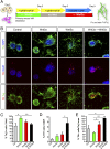
Sequential Wnt3a and Wnt5a treatment of primary ventral midbrain neurospheres improves the yield of DA neurons. (A) Mouse E11.5 VM cultures were grown for the first 6 d in the presence of FGF8b (25 ng/mL), Shh (300 ng/mL), and bFGF (20 ng/mL). Wnt3a was added during the first 3 d, and Wnt5a was added during days 6–9, in the presence of BDNF (20 ng/mL) and GDNF (10 ng/mL). (B) Immunocytochemistry for TUJ1/DAPI, TH/DAPI, and TH/TUJ1 revealed that Wnt3a increased Tuj1+ neurons and that sequential Wnt3a and Wnt5a treatment increased the number of TH+ neurons in VM neurospheres. (C) Wnt3a alone increased the percentage of neurons (Tuj1+ cells per field), but Wnt5a alone or in combination with Wnt3a had no effect, and the effect of Wnt3a was reversed by sequential administration of Wnt5a. (D) Although neither Wnt3a nor Wnt5a significantly increased the percentage of TH+ neurons out of the total cells in the culture, sequential administration of Wnt3a and Wnt5a greatly increased the percentage of TH+/DAPI cells. (E) Although Wnt3a had no effect, Wnt5a increased the percentage of TH+ cells per Tuj1+ area, and sequential Wnt3a and Wnt5a treatment increase this percentage to a greater extent. *P < 0.05 and **P < 0.01 (n = 3–4).
Fig. 4.
Fig. 4.
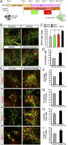
Sequential Wnt3a and Wnt5a treatment improves the midbrain DA differentiation of mES cells. (A) ES cells were cultured according to the following scheme: ES cells were neuralized by Noggin (5 d) in SRM and then were patterned with Shh (200 ng/mL) and Fgf8b (25 ng/mL) for 2 d. From days 7–13, cells were grown in N2 containing bFGF (10 ng/mL), Shh, and Fgf8b. From days 13–15/16, cells were differentiated further with ascorbic acid (0.2 mM), BDNF (20 ng/mL), and GDNF (10 ng/mL) before fixation and staining. The effect of Wnt3a was tested by treatment from days 7–9, and the effect of Wnt5a was tested from days 11–13. (BD) Differentiated cells were stained for TH and Tuj1, and the number of TH+ cells was counted. Sequential treatment with Wnt3a and Wnt5a significantly improved the yield of DA neurons by 60% (%TH+ cells per area, C) and the proportion of neurons that become TH+ neurons by 80% (%TH/TUJ1+ cells, D). (EI) Immunohistochemistry for typical midbrain DA neuron markers such as Foxa2, Lmx1a, Pitx3, and Nurr1 revealed that sequential Wnt3a and Wnt5a treatment induced a tendency for TH+ cells to express Pitx3 (H) and a significant increase in the number of TH+ cells that express Foxa2 (F), Lmx1a, (G), and Nurr1 (I). These results indicate that sequential Wnt3a and Wnt5a treatment improves not only the number of TH+ DA neurons but also the acquisition of a midbrain DA neuron phenotype. *P < 0.05, ***P < 0.001, n = 3–4.
Fig. P1.
Fig. P1.
Wnts cooperate to generate midbrain DA neurons in vivo and in stem cells. Wnt1 and Wnt5a interact at multiple stages of DA neuron development in a cooperative or competitive manner. Wnt1 and Wnt5a antagonize each other’s effects on proliferation, but they co-operate to regulate morphogenesis, neurogenesis, and differentiation. Taking advantage of these results, we developed a protocol to improve the generation of midbrain DA neurons from neural or embryonic stem cells. Sequential administration of Wnt3a, to activate Wnt/β-catenin and induce specification and proliferation, followed by Wnt5a, to activate Wnt/PCP/Rac1 and induce DA differentiation, improved the generation of midbrain DA neurons derived from neural and embryonic stem cells.

Similar articles

Cited by

References

    1. Chien AJ, Conrad WH, Moon RT. A Wnt survival guide: From flies to human disease. J Invest Dermatol. 2009;129(7):1614–1627. - PMC - PubMed
    1. Sugimura R, Li L. Noncanonical Wnt signaling in vertebrate development, stem cells, and diseases. Birth Defects Res C Embryo Today. 2010;90(4):243–256. - PubMed
    1. Inestrosa NC, Arenas E. Emerging roles of Wnts in the adult nervous system. Nat Rev Neurosci. 2010;11(2):77–86. - PubMed
    1. Grigoryan T, Wend P, Klaus A, Birchmeier W. Deciphering the function of canonical Wnt signals in development and disease: Conditional loss- and gain-of-function mutations of beta-catenin in mice. Genes Dev. 2008;22(17):2308–2341. - PMC - PubMed
    1. Angers S, Moon RT. Proximal events in Wnt signal transduction. Nat Rev Mol Cell Biol. 2009;10(7):468–477. - PubMed

Publication types

MeSH terms