. 2013 Apr;61(4):283-93.
doi: 10.1369/0022155413477114. Epub 2013 Jan 15.

A computational approach to detect gap junction plaques and associate them with cells in fluorescent images

Affiliations

A computational approach to detect gap junction plaques and associate them with cells in fluorescent images

Joshua S Goldberg et al. J Histochem Cytochem. 2013 Apr.

Abstract

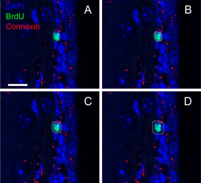
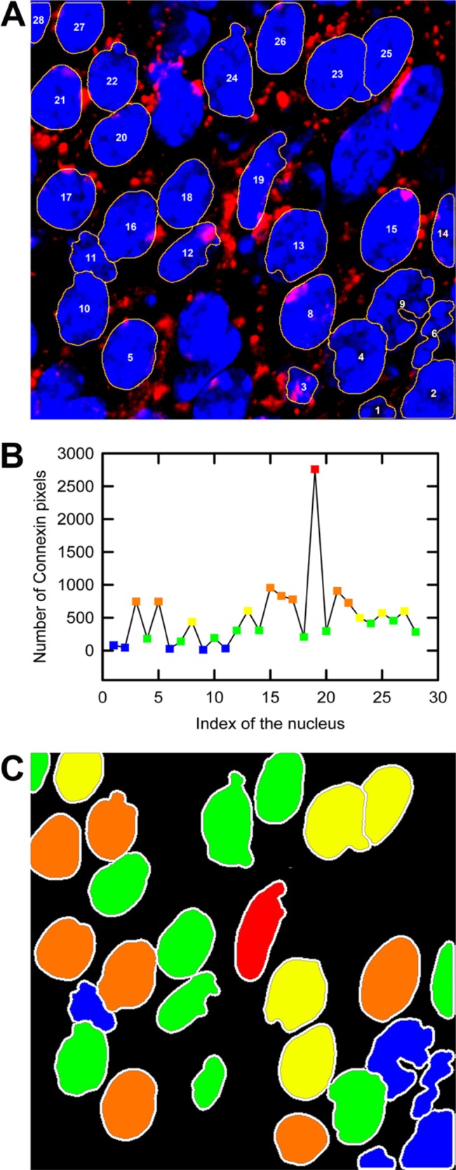
Intercellular signaling is a fundamental requirement for complex biological system function and survival. Communication between adjoining cells is largely achieved via gap junction channels made up of multiple subunits of connexin proteins, each with unique selectivity and regulatory properties. Intercellular communication via gap junction channels facilitates transmission of an array of cellular signals, including ions, macromolecules, and metabolites that coordinate physiological processes throughout tissues and entire organisms. Although current methods used to quantify connexin expression rely on number or area density measurements in a field of view, they lack cellular assignment, distance measurement capabilities (both within the cell and to extracellular structures), and complete automation. We devised an automated computational approach built on a contour expansion algorithm platform that allows connexin protein detection and assignment to specific cells within complex tissues. In addition, parallel implementation of the contour expansion algorithm allows for high-throughput analysis as the complexity of the biological sample increases. This method does not depend specifically on connexin identification and can be applied more widely to the analysis of numerous immunocytochemical markers as well as to identify particles within tissues such as nanoparticles, gene delivery vehicles, or even cellular fragments such as exosomes or microparticles.


Conflict of interest statement

Declaration of Conflicting Interests: The authors declared no potential conflicts of interest with respect to the research, authorship, and/or publication of this article.

Figures

Figure 1.
Figure 1.
A representative confocal microscope image of connexin 43 expression in the murine subventricular zone (SVZ) (coronal view). BrdU (green) marks a subpopulation of quiescent astrocytic cells that are presumed to be neural stem cells (NSCs) (Shen et al. 2008; Tavazoie et al. 2008; Kazanis et al. 2010). The algorithm described herein allows detection and assignment of connexin expression to BrdU+ NSCs (arrows) by performing a nuclear neighborhood search at a pixel-level resolution. Bar = 10 µm.
Figure 2.
Figure 2.
A schematic of the image analysis workflow. Each image in the z-stack is a three-color RGB image. FARSIGHT reads in the confocal z-stack file and segments the nuclear channel of interest (Lin et al. 2003, 2005, 2007; Al-Kofahi et al. 2010). Following segmentation, the confocal z-stack and the FARSIGHT output file, containing the indices of the segmented nuclei, are used as input to the MATLAB programs.
Figure 3.
Figure 3.
Illustration of the nuclear neighborhood search algorithm. (A) A schematic of the contour expansion approach in which the nuclear neighborhood is scanned starting at the surface of the nucleus (black ring) and extending to layers (red, green, and blue layers) beyond the surface layer of pixels. Each layer is one pixel wide in an eight-neighborhood sense. (B) The contour expansion approach is illustrated at the pixel level. Pixels labeled as 1, 2, and 3 correspond to different layers starting from the surface of the nucleus. (C) A nuclear neighborhood filter is then constructed from the contour pixels shown in (B). The filter image of the nucleus has the same dimension as the original fluorescent image. (D) The connexin channel in the original fluorescent image is illustrated here. Each pixel in the connexin channel image is then multiplied with the corresponding pixel in the filter image to extract the pixels in the nuclear neighborhood that are occupied by connexins (E).
Figure 4.
Figure 4.
Testing the performance of the MATLAB program using synthetic data. (A) Program execution times were measured when the number of images in the z-stacks varied. Each image in the z-stack consisted of 512 × 512 pixels. A circular disk with a diameter of 220 pixels was used as the test object in each image of the z-stack. The number of contours used in each image in the z-stack was 10. The execution time scales linearly with the number of planes (R 2 = 0.99, n=3). The error bars are one unit of standard deviation of the mean values and are smaller than the symbols representing the data points. (B) The mean values of the execution times, to scan 10 contours around circular disks of varying diameters in single-plane images of 512 × 512 pixels each (n=3), were recorded. The error bars are one unit of standard deviations of the mean values. (C, solid line) The durations of the program to perform the loop calculations for varying numbers of circular disks of fixed diameter in single-plane images of 512 × 512 pixels each are illustrated here. The diameter of a single circular disk in these images is 45 pixels. The execution time of the program scales linearly with the number of disks (R 2 = 0.98, n=3). (C, dashed line) The dashed line represents the linear scaling of the execution time with the number of disks for the custom-built parallel program (R 2 = 0.99, n=3). The parallel program is significantly faster than the serial program (solid line). The error bars are one unit of standard deviations of the mean values and are smaller than the symbols used to represent the data points. The execution times were calculated using the standard MATLAB commands to measure time.
Figure 5.
Figure 5.
Application of contour expansion algorithm to a representative confocal z-stack image. The z-stack consists of 10 images, and each image consists of 512 × 512 pixels. The x and y pixel scaling is 0.1 µm each, and the axial pixel scaling is 1.0 µm. (B–D) The white loops around the BrdU (green)-stained nucleus (blue) are the contours created by the MATLAB program, labeled as L1, L5, and L10, respectively (Table 1). L1 consists of pixels adjacent to the surface pixels of the nucleus. The surface pixels are obtained following nuclear segmentation using FARSIGHT. The contours sweep the neighborhood of the nucleus starting at the nuclear surface, where each contour is one pixel wide in an eight-neighborhood sense. Similar contours are created with respect to the nuclear surface in all images in the z-stack containing a cross section of the nucleus (Movie 2 [supplemental material]). Bar = 10 µm.
Figure 6.
Figure 6.
Application of FARSIGHT output to a z-stack consisting of 28 DAPI-stained nuclei. (A) The loops (yellow lines around the nuclei) are the surface pixels of each nucleus. The loop expansion algorithm was applied to the FARSIGHT output data to measure the number of connexin pixels. (B) Histogram depicting the number of connexin pixels detected in 10 loops around each nucleus. (C) The nuclei were then grouped based on the number of connexin pixels and color coded corresponding to pixel number. Blue corresponds to nuclei with the least number of connexin pixels detected, whereas red indicates those with maximum pixel detection.

Similar articles

Cited by

References

    1. Al-Kofahi Y, Lassoued W, Lee W, Royam B. 2010. Improved automatic detection and segmentation of cell nuclei in histopathology images. IEEE Trans Biomed Eng. 57:841–852 - PubMed
    1. Beyer EC. 1993. Gap junctions. Int Rev Cytol. 137C:1–37 - PubMed
    1. Brisset AC, Isakson BE, Kwak BR. 2009. Connexins in vascular physiology and pathology. Antioxid Redox Signal. 11:267–282 - PMC - PubMed
    1. Cheng A, Tang H, Cai J, Zhu M, Zhang X, Rao M, Mattson MP. 2004. Gap junctional communication is required to maintain mouse cortical neural progenitor cells in a proliferative state. Dev Biol. 272:203–216 - PubMed
    1. Dang X, Doble BW, Kardami E. 2003. The carboxy-tail of connexin-43 localizes to the nucleus and inhibits cell growth. Mol Cell Biochem. 242(1–2):35–38 - PubMed

Publication types