

Hepatic apoptosis postburn is mediated by c-Jun N-terminal kinase 2
- PMID: 23324888
- PMCID: PMC3552323
- DOI: 10.1097/SHK.0b013e31827f40ab
Hepatic apoptosis postburn is mediated by c-Jun N-terminal kinase 2
Abstract
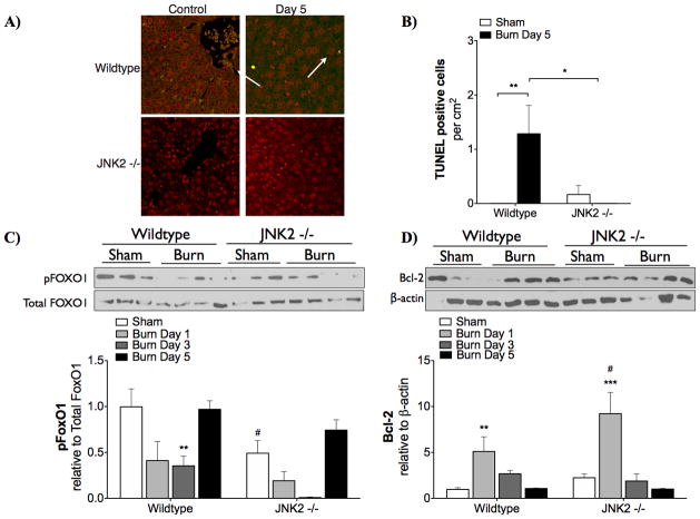
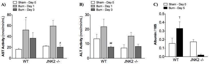
The trauma of a severe burn injury induces a hypermetabolic response that increases morbidity and mortality. Previously, our group showed that insulin resistance after burn injury is associated with endoplasmic reticulum (ER) stress. Evidence suggests that c-Jun N-terminal kinase (JNK) 2 may be involved in ER stress-induced apoptosis. Here, we hypothesized that JNK2 contributes to the apoptotic response after burn injury downstream of ER stress. To test this, we compared JNK2 knockout mice (-/-) with wild-type mice after inducing a 30% total body surface area thermal injury. Animals were killed after 1, 3, and 5 days. Inflammatory cytokines in the blood were measured by multiplex analysis. Hepatic ER stress and insulin signaling were assessed by Western blotting, and insulin resistance was measured by a peritoneal glucose tolerance test. Apoptosis in the liver was quantified by terminal deoxynucleotidyl transferase-mediated dUTP nick end labeling staining. Liver function was quantified by aspartate aminotransferase and alanine aminotransferase activity assays. Endoplasmic reticulum stress increased after burn in both JNK2 and wild-type mice, indicating that JNK2 activation is downstream of ER stress. Knockout of JNK2 did not affect serum inflammatory cytokines; however, the increase in interleukin 6 mRNA expression was prevented in the knockouts. Serum insulin did not significantly increase in the JNK2 group. On the other hand, insulin signaling (PI3K/Akt pathway) and glucose tolerance tests did not improve in JNK2. As expected, apoptosis in the liver increased after burn injury in wild-type mice but not in JNK2. Aspartate aminotransferase/alanine aminotransferase activity revealed that liver function recovered more quickly in JNK2. This study indicates that JNK2 is a central mediator of hepatic apoptosis after a severe burn.
Figures


Similar articles
-
Hepatic autophagy after severe burn in response to endoplasmic reticulum stress.J Surg Res. 2014 Mar;187(1):128-33. doi: 10.1016/j.jss.2013.09.042. Epub 2013 Oct 2. J Surg Res. 2014. PMID: 24209807 Free PMC article.
-
Post-burn hepatic insulin resistance is associated with endoplasmic reticulum (ER) stress.Shock. 2010 Mar;33(3):299-305. doi: 10.1097/SHK.0b013e3181b2f439. Shock. 2010. PMID: 22011639 Free PMC article.
-
Jnk1 Deficiency in Hematopoietic Cells Suppresses Macrophage Apoptosis and Increases Atherosclerosis in Low-Density Lipoprotein Receptor Null Mice.Arterioscler Thromb Vasc Biol. 2016 Jun;36(6):1122-31. doi: 10.1161/ATVBAHA.116.307580. Epub 2016 Apr 21. Arterioscler Thromb Vasc Biol. 2016. PMID: 27102962 Free PMC article.
-
JNK1 but not JNK2 promotes the development of steatohepatitis in mice.Hepatology. 2006 Jan;43(1):163-72. doi: 10.1002/hep.20999. Hepatology. 2006. PMID: 16374858
-
Differential effects of JNK1 and JNK2 inhibition on murine steatohepatitis and insulin resistance.Hepatology. 2009 Jan;49(1):87-96. doi: 10.1002/hep.22578. Hepatology. 2009. PMID: 19053047 Free PMC article.
Cited by
-
Burn-induced mitochondrial dysfunction in hepatocytes: The role of methylation-controlled J protein silencing.J Trauma Acute Care Surg. 2025 Feb 1;98(2):204-211. doi: 10.1097/TA.0000000000004537. Epub 2025 Jan 6. J Trauma Acute Care Surg. 2025. PMID: 39760651
-
Animal models in burn research.Cell Mol Life Sci. 2014 Sep;71(17):3241-55. doi: 10.1007/s00018-014-1612-5. Epub 2014 Apr 9. Cell Mol Life Sci. 2014. PMID: 24714880 Free PMC article. Review.
-
Burn-Induced Apoptosis in the Livers of Aged Mice Is Associated With Caspase Cleavage of Bcl-xL.J Surg Res. 2023 Oct;290:147-155. doi: 10.1016/j.jss.2023.04.020. Epub 2023 May 31. J Surg Res. 2023. PMID: 37267704 Free PMC article.
-
Effects of hydrogen-rich saline on early acute kidney injury in severely burned rats by suppressing oxidative stress induced apoptosis and inflammation.J Transl Med. 2015 Jun 6;13:183. doi: 10.1186/s12967-015-0548-3. J Transl Med. 2015. PMID: 26047940 Free PMC article.
-
Increased proliferation of hepatic periportal ductal progenitor cells contributes to persistent hypermetabolism after trauma.J Cell Mol Med. 2020 Jan;24(2):1578-1587. doi: 10.1111/jcmm.14845. Epub 2019 Dec 3. J Cell Mol Med. 2020. PMID: 31793707 Free PMC article.
References
-
- Finnerty CC, Herndon DN, Przkora R, Pereira CT, Oliveira HM, Queiroz DM, Rocha AM, Jeschke MG. Cytokine expression profile over time in severely burned pediatric patients. Shock. 2006;26:13–19. - PubMed
-
- Przkora R, Barrow RE, Jeschke MG, Suman OE, Celis M, Sanford AP, Chinkes DL, Mlcak RP, Herndon DN. Body composition changes with time in pediatric burn patients. J Trauma. 2006;60:968–971. discussion 971. - PubMed
-
- Biolo G, Fleming RY, Maggi SP, Wolfe RR. Transmembrane transport and intracellular kinetics of amino acids in human skeletal muscle. Am J Physiol. 1995;268:E75–84. - PubMed
Publication types
MeSH terms
Substances
Grants and funding
LinkOut - more resources
Full Text Sources
Other Literature Sources
Medical
Research Materials
Miscellaneous
