. 2013 Jan 27;95(2):293-300.
doi: 10.1097/TP.0b013e3182772244.

Critical role for IL-17A/F in the immunopathogenesis of obliterative airway disease induced by Anti-MHC I antibodies

Affiliations

Critical role for IL-17A/F in the immunopathogenesis of obliterative airway disease induced by Anti-MHC I antibodies

Haseeb Ilias Basha et al. Transplantation. .

Abstract

Background: The IL-17 axis is implicated in pathogenesis of chronic rejection after human lung transplantation. Using a murine model of obliterative airway disease (OAD), we recently demonstrated that Abs to MHC class I antigens can induce immune responses to self-antigens that contributes to immunopathogenesis of chronic rejection. Using a murine model of OAD, we determined the role of IL-17 family members in induction of autoimmunity leading to OAD after ligation of MHC class I.

Methods: Anti-MHC class I or control antibodies (Abs) were administered intrabronchially to wild-type (WT) and IL-17a knock out (IL-17A-/-) C57BL/6.

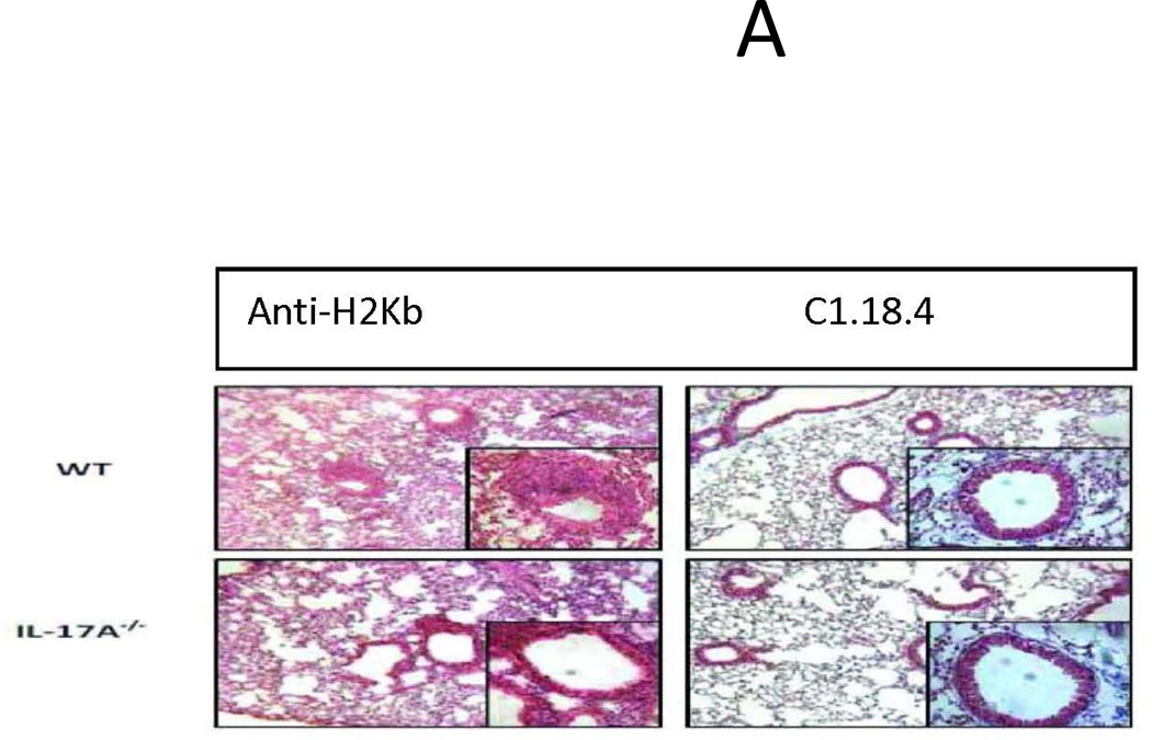
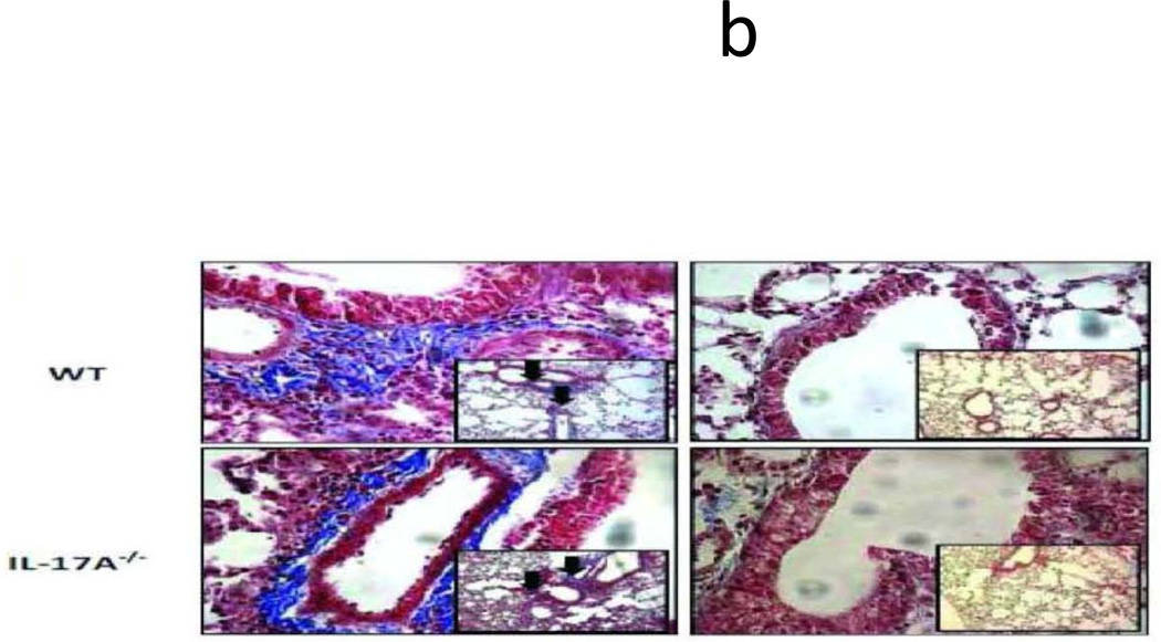
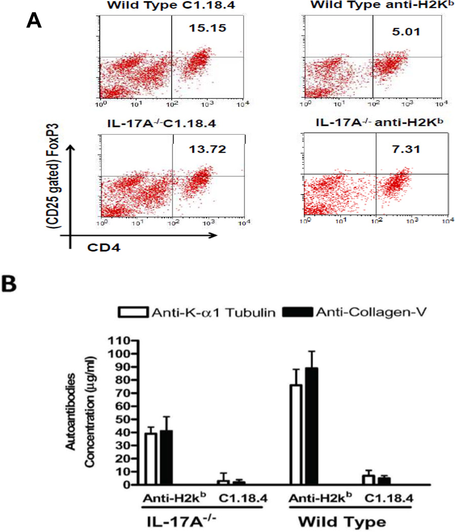
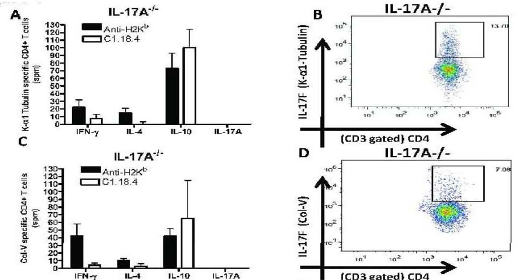
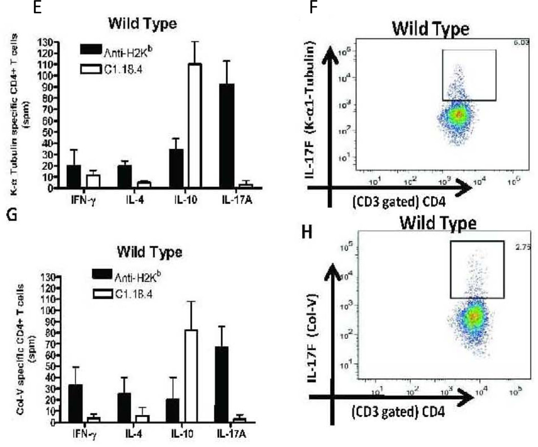
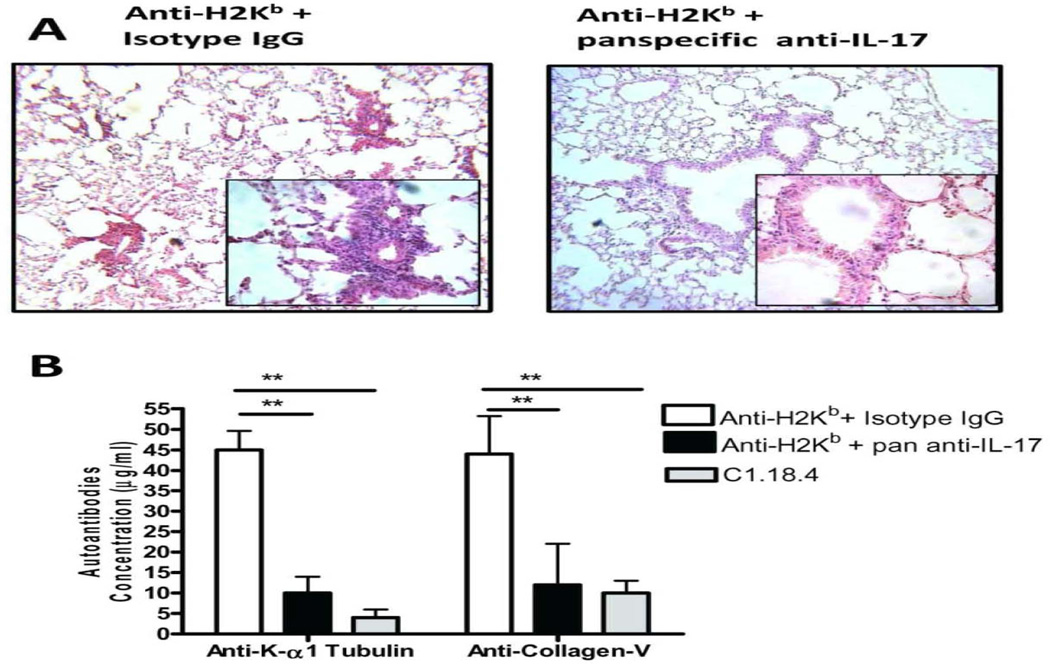
Results: By day 30, anti-MHC I administered endobronchially in IL-17A-/- mice demonstrated significant reduction in cellular infiltration, a 36.8% reduction in CD4 T cells, 62.7% in CD11b macrophages, 37.5% in degree of fibrosis, 1.94 fold and 2.17 fold decrease in anti-KAT and anti-Col-V, respectively, when compared with wild-type mice. Analysis of lung infiltrating cells in anti-MHC I WT revealed increase in IL-17A (KAT:92+21,Col-V:103+19spm) and IL-17F (KAT:5.03%,Col-V:2.75%) secreting CD4+ T cells. However, administration of anti-MHC I in IL-17A-/- demonstrated increase only in IL-17F for KAT (13.70%) and Col-V (7.08%). Anti-IL-17(A-F) mAb administration after anti-MHC I abrogated OAD in both WT and IL-17A-/-.

Conclusion: Our findings indicate that IL-17A and IL-17F secreted by CD4+Th17 cells specific to lung self-antigens are critical mediators of autoimmunity leading to the pathogenesis of OAD.


Conflict of interest statement

The authors have no financial conflicts of interest.

Figures

Figure 1
Figure 1. Significant reduction in induction of cellular infiltration around bronchioles and vessels, epithelial hyperplasia and fibrosis following ligation of anti-MHC class I Abs in IL17A−/− mice
Anti-H2Kb or control C1.18.4 Abs were administered intrabronchially in IL-17A deficient (IL-17A−/−) and WT C57BL/6 mice. A: H&E staining of the lungs. Serial administration of anti-H2Kb Abs resulted in cellular infiltration around bronchioles and vessels, epithelial changes including hyperplasia, fibrosis and luminal occlusion in both IL-17A deficient and WT by day 30 compared to controls treated with C1.18.4 respectively. B: Trichrome staining of the lungs. Collagen deposition around the bronchioles and vessels are stained blue in color. C: Semiquantitative analysis of the cellular infiltrates in the lungs. D: Morphometric analysis of the lungs. Semiquantitative analysis of the slides demonstrated less significant lesions in IL-17A −/− mice compared to WT mice with respect to cellular infiltration, epithelial hyperplasia, fibrosis and luminal occlusion.
Figure 2
Figure 2. Loss of CD4+CD25+Foxp3+ regulatory T cells and Induction of autoAbs to pulmonary self antigens, KAT and Col-V following ligation of anti-MHC class I Abs
(A) Flow cytometric analysis of the frequency of Tregs in the lungs of anti-H2kb treated mice revealed a significant decline in the both IL-17A−/− (anti-H2kb- 7.31; C1.18.4–13.72) and WT mice (anti-H2kb5.01; C1.18.4–15.15) compared to control mice. However, the percentage decline in the frequency of Tregs was higher in WT (10.14%) compared to IL-17A−/− (6.41%) treated with anti-H2kb. (B) Levels of antibodies against the self antigens KAT and Col-V in BAL were measured by ELISA. The titers of the antibodies are presented as mean ± SD (µg/ml). A significant increase in the titer of Abs to both KAT and Col-V was observed in anti-H2kb treated IL-17A−/− and WT mice.
Figure 3
Figure 3. Induction of autoreactive CD4+ T cells which predominantly secrete IL-17A and IL-17F following administration of anti-MHC Class I Abs
Lung infiltrating T cells were isolated from the lungs of IL17A −/− and wild type mice treated with anti-H2Kb or control antibody (n=5 each, the figures shown are a representative of 5 mice). The frequency of T cells secreting IFN-γ, IL-4, IL-17A and IL-10 specific to KAT and Col-V were measured by ELISPOT (Figure 3, A,C,E,G) and presented as means ± SD. The frequency of IL-17F secreting CD4+ T cells specific to self antigens were determined by flow cytometry (Figure 3, B,D,F,H). A significant increase in the frequency of IL-17A secreting CD4+ T cells specific to KAT and Col-V was observed in the lungs of anti-H2kb treated WT mice compared to control animals. An increased frequency of IL17F secreting CD4+ T cells specific to KAT (IL-17A−/−:13.70% vs WT: 5.03%) and Col-V (IL-17A−/:7.08% vs WT: 2.75%) was noted in anti-H2Kb treated IL-17A−/− mice compared to WT mice. No significant difference was seen in the levels of IFN-γ, IL-4 and IL-10 secreting CD4+ T Cells specific to KAT and Col-V was seen between IL-17A−/− and WT administered with anti-H2kb.
Figure 4
Figure 4. Increased induction of IL-17A and IL-17F mediated inflammatory cascades following anti-MHC class I administration
Total RNA from the lungs of anti-H2KB treated WT and IL-17A−/− were extracted using Trizol reagent and used for the expression profile of the signaling cascades intermediates in the autoimmunity pathway using autoimmunity PCR array. The samples were normalized using housekeeping gene expression and the results are expressed as fold expression observed in IL17A −/− mice over expression observed in WT mice. Anti-H2Kb administration in IL-17A−/− resulted in significant increase in levels of IL-17F (7.80 fold) but not IL-17A (−6.4 fold), IL-17B (−0.28 fold), IL-17C (−0.23 fold), IL-17D (0.70 fold), IL-17E (−0.49 fold), IL-6 (−4.01 fold), IL-21(−3.84 fold) and IL-23(−6.18 fold) compared to WT. Anti-H2Kb administered IL-17A−/− demonstrated a significant increase in levels of IL-17RC (3.2 fold) and TRAF-6 (2.3 fold) but not IL-17RA (−2.70 fold). Anti-H2Kb administered IL-17A−/ demonstrated a significant increase in the levels of CXCL1 (5.2 fold), CXCL2 (3.2 fold) and a significant reduction in the levels of CXCL12 (−2.10 fold), CCL7 fold (−4.24 fold) and CX3CL1 (−3.12 fold). Anti-H2Kb administered IL-17A−/− demonstrated differential induction of MMP-3 (6.2 fold), MMP-9 (−3.1 fold) and MMP-13 (−2.8 fold).
Figure 5
Figure 5. Neutralization of IL-17F following administration of anti-MHC class I Abs results in abrogation of OAD lesions and autoimmune responses to lung self antigens, KAT and Col-V in IL17A−/− mice
In order to identify a role for IL-17 cytokine family in development of autoimmunity and OAD, neutralizing pan specific Abs reactive to IL-17A-F or control Abs (50µg i.v) were administered immediately following anti-H2kb in IL-17A−/− mice on days 1,5,12 and 26. (A ,B) administration of pan specific Abs to IL-17A-F following administration in IL-17A−/− resulted in reduction of cell infiltrates around bronchioles and vessels accompanied by (C) marked reduction in the levels of Abs to KAT (4.5 fold) and Col-V (3.6 fold) in the BAL fluid by day 30.

References

    1. Lama VN, Murray S, Lonigro RJ, et al. Course of FEV(1) after onset of bronchiolitis obliterans syndrome in lung transplant recipients. Am J Respir Crit Care Med. 2007;175(11):1192. - PMC - PubMed
    1. Hachem RR, Khalifah AP, Chakinala MM, et al. The significance of a single episode of minimal acute rejection after lung transplantation. Transplantation. 2005;80(10):1406. - PubMed
    1. Jaramillo A, Smith MA, Phelan D, et al. Temporal relationship between the development of anti-HLA antibodies and the development of bronchiolitis obliterans syndrome after lung transplantation. Transplant Proc. 1999;31(1–2):185. - PubMed
    1. Jaramillo A, Smith MA, Phelan D, et al. Development of ELISA-detected anti-HLA antibodies precedes the development of bronchiolitis obliterans syndrome and correlates with progressive decline in pulmonary function after lung transplantation. Transplantation. 1999;67(8):1155. - PubMed
    1. Saini D, Weber J, Ramachandran S, et al. Alloimmunity-induced autoimmunity as a potential mechanism in the pathogenesis of chronic rejection of human lung allografts. The Journal of heart and lung transplantation : the official publication of the International Society for Heart Transplantation. 2011;30(6):624. - PMC - PubMed

Publication types

MeSH terms