

Donor-specific antibodies to class II antigens are associated with accelerated cardiac allograft vasculopathy: a three-dimensional volumetric intravascular ultrasound study
- PMID: 23325007
- PMCID: PMC3552316
- DOI: 10.1097/TP.0b013e318273878c
Donor-specific antibodies to class II antigens are associated with accelerated cardiac allograft vasculopathy: a three-dimensional volumetric intravascular ultrasound study
Abstract
Background: Although a link between donor-specific antibodies against human leukocyte antigens type II (DSA II+) and transplant glomerulopathy has been clearly established, its role in cardiac allograft vasculopathy (CAV) is unclear.
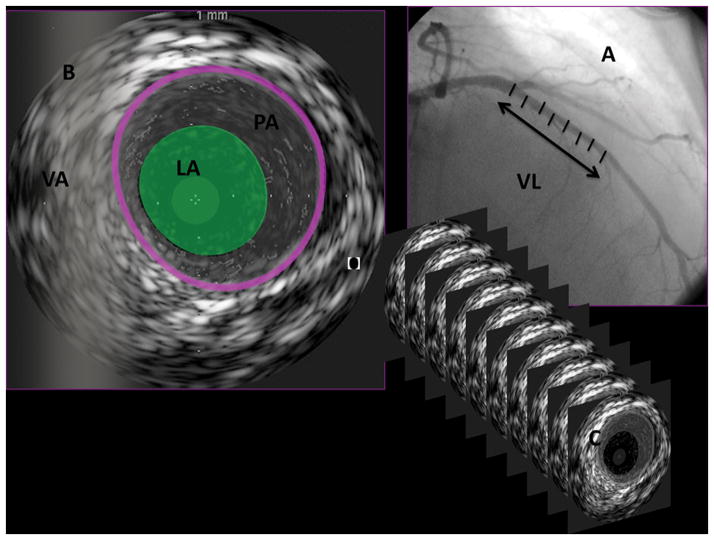
Methods: Donor-specific antibodies were evaluated using solid-phase single-antigen bead assay before transplantation in 51 heart transplant recipients. Coronary angiography and three-dimensional intravascular ultrasound were performed at baseline and approximately 1 year after the baseline examination.
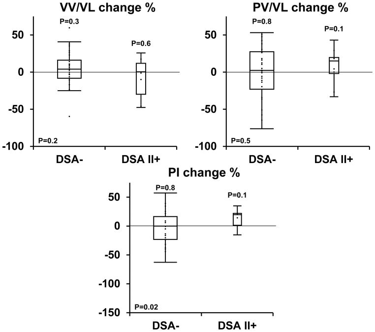
Results: There were 4 (7.8 %), 11 (21.5%), and 2 (3.9%) patients who had DSA against donor class I (DSA I+), DSA II+, or both, respectively. All patients had negative complement-dependent cytotoxic crossmatch. There was accelerated progression of CAV in the DSA II+ group demonstrated by accelerated progression in plaque index (plaque volume/vessel volume) compared to patients with no DSA II+ antibodies (13.8% [12%] vs. -7.9% [37%], P=0.01). The development of any angiographic CAV was also more common in DSA II+ patients as compared to the DSA- patients at 4 years (100% [0%] vs. 64.2% [10%], P=0.05). All other traditional risk factors for CAV or immunosuppression were similar between the groups (P>0.2 for all).
Conclusions: This is the first preliminary study demonstrating that heart transplant recipients with preformed class II DSA may be at an increased risk for accelerated CAV as detected by consecutive volumetric three-dimensional intravascular ultrasound.
Conflict of interest statement
Disclosures
The authors of this manuscript have no conflicts of interest to disclose.
Figures
References
-
- Gebel HM, Bray RA, Nickerson P. Pre-transplant assessment of donor-reactive, HLA-specific antibodies in renal transplantation: contraindication vs. risk. Am J Transplant. 2003 Dec;3(12):1488–500. - PubMed
-
- Tambur AR, Bray RA, Takemoto SK, Mancini M, Costanzo MR, Kobashigawa JA, et al. Flow cytometric detection of HLA-specific antibodies as a predictor of heart allograft rejection. Transplantation. 2000 Oct 15;70(7):1055–9. - PubMed
-
- Przybylowski P, Balogna M, Radovancevic B, Frazier OH, Susskind B, Van Buren C, et al. The role of flow cytometry-detected IgG and IgM anti-donor antibodies in cardiac allograft recipients. Transplantation. 1999 Jan 27;67(2):258–62. - PubMed
-
- Smith JD, Hamour IM, Banner NR, Rose ML. C4d fixing, luminex binding antibodies - a new tool for prediction of graft failure after heart transplantation. Am J Transplant. 2007 Dec;7(12):2809–15. - PubMed
-
- Costanzo MR, Naftel DC, Pritzker MR, Heilman JK, 3rd, Boehmer JP, Brozena SC, et al. Heart transplant coronary artery disease detected by coronary angiography: a multiinstitutional study of preoperative donor and recipient risk factors. Cardiac Transplant Research Database. J Heart Lung Transplant. 1998 Aug;17(8):744–53. - PubMed
Publication types
MeSH terms
Substances
Grants and funding
LinkOut - more resources
Full Text Sources
Other Literature Sources
Medical
