. 2013 Jun;133(6):1655-65.
doi: 10.1038/jid.2013.16. Epub 2013 Jan 16.

Adult zebrafish as a model system for cutaneous wound-healing research

Affiliations

Adult zebrafish as a model system for cutaneous wound-healing research

Rebecca Richardson et al. J Invest Dermatol. 2013 Jun.

Abstract

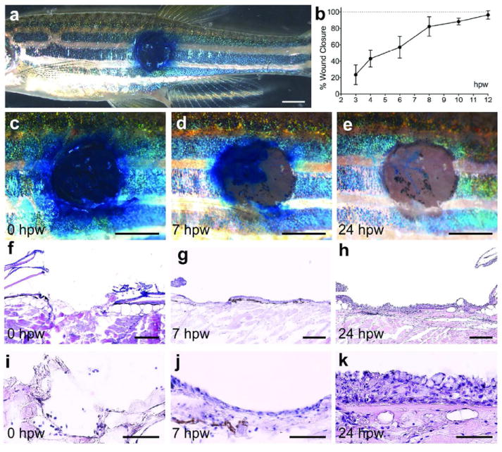
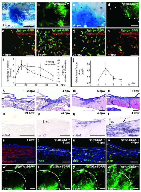
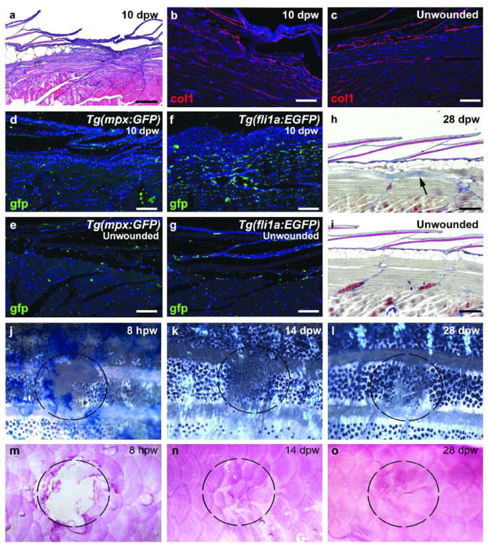
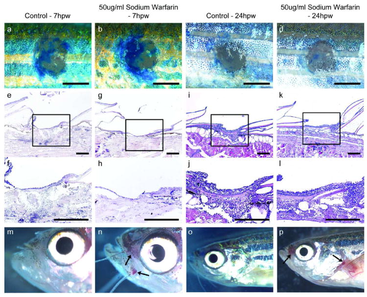
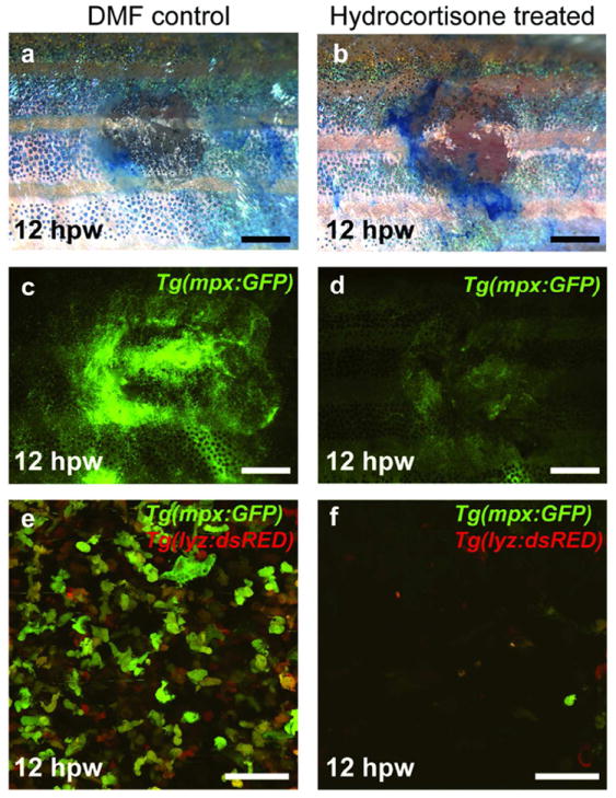
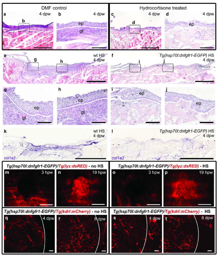
Upon injury, the skin must quickly regenerate to regain its barrier function. In mammals, wound healing is rapid and scar free during embryogenesis, whereas in adults it involves multiple steps including blood clotting, inflammation, re-epithelialization, vascularization, and granulation tissue formation and maturation, resulting in a scar. We have established a rapid and robust method to introduce full-thickness wounds onto the flank of adult zebrafish, and show that apart from external fibrin clot formation, all steps of adult mammalian wound repair also exist in zebrafish. Wound re-epithelialization is extremely rapid and initiates with no apparent lag phase, subsequently followed by the immigration of inflammatory cells and the formation of granulation tissue, consisting of macrophages, fibroblasts, blood vessels, and collagen. The granulation tissue later regresses, resulting in minimal scar formation. Studies after chemical treatment or with transgenic fish further suggest that wound re-epithelialization occurs independently of inflammation and fibroblast growth factor signaling, whereas both are essential for fibroblast recruitment and granulation tissue formation. Together, these results demonstrate that major steps and principles of cutaneous wound healing are conserved among adult mammals and adult zebrafish, making zebrafish a valuable model for studying vertebrate skin repair.


Conflict of interest statement

Conflict of Interest

The authors state no conflict of interest.

Figures

Figure 1
Figure 1. Wounds of adult zebrafish undergo rapid re-epithelialization
(a) Overview of the left flank of an adult zebrafish with an approximately 2 mm circular wound stained with methylene blue. (b) Graphical illustration of time course of wound closure (see Material and Methods); mean values of closure and standard deviations were determined for at least 6 individuals per time point using Excel software. (c-e) Magnified superficial views of the wound. At 0 hpw (a, c), methylene blue penetrates the entire wound, while the epidermal barrier has been partly recovered by 7 hpw (d), and fully recovered by 24 hpw (e). (f-k) H&E staining of longitudinal sections through the wound reveals the removal of epidermis, dermis and scales via the applied wounding protocol (f and I; n=4). At 7 hpw, a thin neo-epidermis is observed on the surface of the wound (g and j; n=6). At 24 hpw, the recovered epidermis appears re-stratified (h and k; n=10). Scale bars: a,c-e = 1 mm; f-h = 200 μm; i-k = 50 μm.
Figure 2
Figure 2. Adult zebrafish exhibit a strong inflammatory response and granulation tissue formation
(a-d) Brightfield (a,c) and fluorescent (b,d) images of a Tg(mpx:GFP) fish at 4 hpw; GFP-positive neutrophils are present in re-epithelialized (methylene-blue excluding) regions of the wound (edges marked by asterisks in c and d; n=6/6). (e-h) Live images of the wound centre of Tg(mpx:GFP)i114/Tg(lyz:DsRED2)117 double transgenic fish reveal inflammatory cells, with a progressive relative increase of macrophages (h). (i,j) Graphical illustrations of time course of wound inflammation (i) and granulation tissue formation (j); mean values and standard deviations of relative fluorescent intensities (i) or granulation tissue areas (j) were determined for at least 6 individuals per time point using Excel software. (k-n) H&E staining reveals formation of granulation tissue beneath the wound from 2-6 dpw, which then regresses (8 dpw, n). (o-r) col1a2 expression beneath the wound is sparse at 24 hpw (o,p; n=4/4), but very prominent at 4 dpw (q,r; n=6/6). In addition to dermal fibroblasts, col1a2 is expressed by basal keratinocytes of the neo-epidermis (indicated by arrow in r). (s-v) Immunofluorescence analysis at 4 dpw reveals Collagen 1 deposition (s; n=4/4), leukocytes (t,u; n=4/4) and blood vessels (v; n=4/4) within the granulation tissue. mpx-positive neutrophils are also present in the neo-epidermis (t; n=4/4). (w-z) Superficial views of a Tg(fli1a:EGFP) fish shows progressive wound vascularization from 24 hpw to 8 dpw (n=4/4). Scale bars: a,b,o,q = 1mm; c,d, w-z = 250 μm; e-h = 50 μm; k-n = 500 μm; p,r,s-v = 100 μm. Abbreviation: ep, epidermis.
Figure 3
Figure 3. Adult zebrafish exhibit minimal scar formation
(a) H&E staining at 10 dpw reveals minimal remaining granulation tissue and newly forming scales. (b,c) At 10 dpw, minimal Collagen1 deposition is observed beneath the regenerating scales (b), similar to an unwounded region (c) (n=6/6). (d-g) Tg(mpx:GFP) (d,e; n=4/4) and Tg(fli1a:EGFP) (f,g; n=4/4) transgenic fish at 10 dpw display normal number of leukocytes (d) and reduced numbers of blood vessels (f) at the regenerating wound site when compared to an unwounded region (e and g). (h,i) AFOG staining at 28 dpw indicates the complete recovery of epidermis, dermis, scales and adipocytes with rarely occurring collagen deposits within the muscle layer beneath (h; n=4/4), compared to unwounded fish (i). (j-o) Superficial views of wounded fish demonstrate the almost complete recovery of stripe pattern (j-l) and scales (m-o; alizarin red) by 28 dpw (n=6/6). Dashed circles mark the position of the wound. Scale bars: a,h,i = 500 μm; b-g = 200 μm.
Figure 4
Figure 4. Blood clot formation plays no role in the re-epithelialization process
Methylene blue penetration assay (a-d) and H&E stained longitudinal sections (e-l) of wounds from control fish (a, c, e, f, i, j) and fish treated with 150 μM Sodium Warfarin (b, d, g, h, k, l), demonstrating no differences in the degree of barrier recovery or wound re-epithelialization at 7 hpw (a, b, e-h) or 24 hpw (c, d, i-l) (n=18/18), even though warfarin-treated fish show localized internal bleeding, particularly around the mouth, gills and pec fins at 7 and 24 hpw (m-p). Scale bars: a-d = 1 mm; e-l = 100 μm.
Figure 5
Figure 5. Reducing the inflammatory response does not affect re-epithelialization
(a-f) Tg(mpx:GFP) (a-d) and Tg(mpx:GFP)/Tg(lyz:DsRED2) fish (e and f) treated with 275 μM Hydrocortisone show no delay in re-epithelialization (a,b; n=19/19), although the number of inflammatory cells at the wound is clearly reduced at 12 hpw when compared to control fish (c-f). Scale bars: a-d = 1 mm; e-f = 50 μm.
Figure 6
Figure 6. Inflammation and FGF signaling are required for granulation tissue formation
(a-d) Hydrocortisone-treated fish at 4 dpw, lacking granulation tissue beneath the wound (c,d; n=9/9) in comparison to DMF-treated controls (a,b). (e-j) H&E staining demonstrates granulation tissue beneath the wound epidermis in heat-shocked wild-type fish (e, g, h; n=6/6), but not in the heat-shocked Tg(hsp70l:dnfgfr1-EGFP) fish (f, i, j; n=6/6) at 4 dpw. (k, l) in situ hybridization analysis reveals normal col1a2 expression in a heat-shocked wild-type fish (k; n=4/4), but strongly reduced expression in a heat-shocked Tg(hsp70l:dnfgfr1-EGFP) fish at 4 dpw (l; n=4/4). (m-t) Analysis of Tg(hsp70l:dnfgfr1-EGFP),Tg(lyz:dsRED) (m-p) or Tg(hsp70l:dnfgfr1-EGFP), Tg(kdrl:HSRAS-mCherry) (q-t) double transgenic fish reveals normal inflammatory responses (m-p) and normal vascularization (q-t) in non-heat-shocked (m,n; n=8/8; q,r; n=8/8) and heat-shocked Tg(hsp70l:dnfgfr1-EGFP) fish (o,p; n=8/8; s,t; n=8/8). Scale bars: a,c,e,f = 500 μm; b,d, g-j, q-t = 100 μm; k-p = 1 mm.

References

    1. Barrientos S, Stojadinovic O, Golinko MS, Brem H, Tomic-Canic M. Growth factors and cytokines in wound healing. Wound Repair Regen. 2008;16:585–601. - PubMed
    1. Broadley KN, Aquino AM, Woodward SC, Buckley-Sturrock A, Sato Y, Rifkin DB, et al. Monospecific antibodies implicate basic fibroblast growth factor in normal wound repair. Lab Invest. 1989;61:571–575. - PubMed
    1. Caddy J, WIlanowski T, Darido C, Dworkin S, Ting SB, Zhao Q, Rank G, Auden A, Srivastava S, Papenfuss TA, Murdoch JN, Humbert PO, Parekh V, Boulos N, Weber T, Zuo J, Cunningham JM, Jane SM. Epidermal wound repair is regulated by the planar cell polarity signaling pathway. Dev Cell. 2010;19:138–47. - PMC - PubMed
    1. Chi NC, Shaw RM, De Val S, Kang G, Jan LY, Black BL, Stainier DY. Foxn4 directly regulates tbx2b expression and atrioventricular canal formation. Genes Dev. 2008;22:734–9. - PMC - PubMed
    1. Dovi JV, He LK, DiPietro LA. Accelerated wound closure in neutrophil-depleted mice. J Leukoc Biol. 2003;73:448–455. - PubMed

Publication types

Substances