. 2013 Jan 16;33(3):950-60.
doi: 10.1523/JNEUROSCI.2949-12.2013.

A corticotropin releasing factor pathway for ethanol regulation of the ventral tegmental area in the bed nucleus of the stria terminalis

Affiliations

A corticotropin releasing factor pathway for ethanol regulation of the ventral tegmental area in the bed nucleus of the stria terminalis

Yuval Silberman et al. J Neurosci. .

Abstract

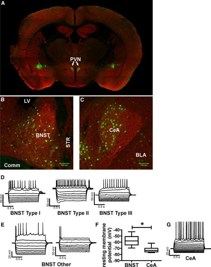
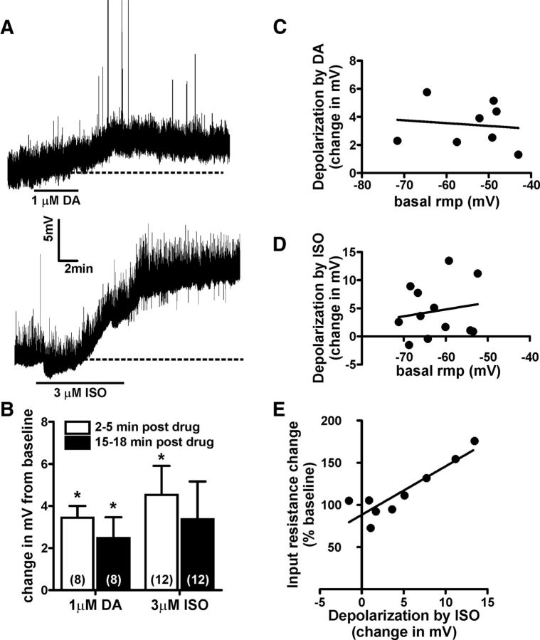
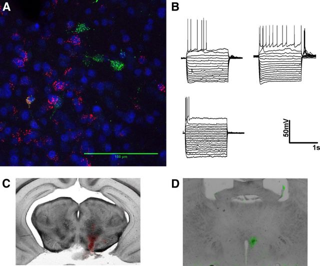
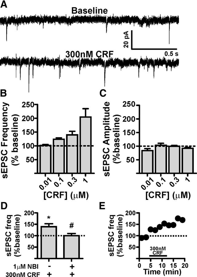
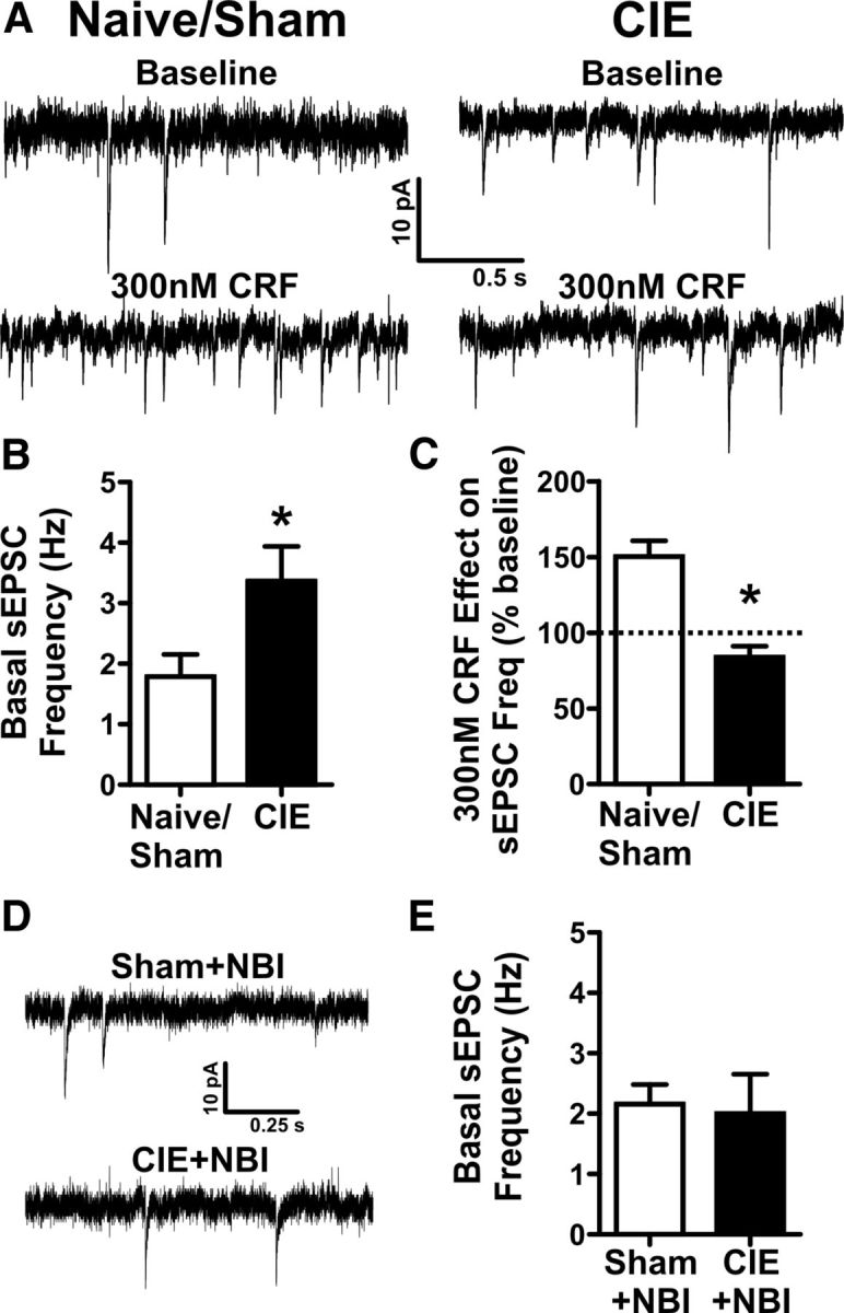
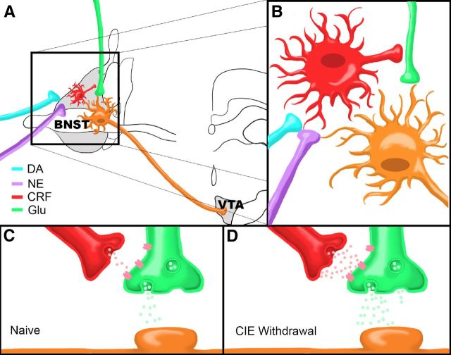
A growing literature suggests that catecholamines and corticotropin-releasing factor (CRF) interact in a serial manner to activate the bed nucleus of the stria terminalis (BNST) to drive stress- or cue-induced drug- and alcohol-seeking behaviors. Data suggest that these behaviors are driven in part by BNST projections to the ventral tegmental area (VTA). Together, these findings suggest the existence of a CRF-signaling pathway within the BNST that is engaged by catecholamines and regulates the activity of BNST neurons projecting to the VTA. Here we test three aspects of this model to determine: (1) whether catecholamines modify CRF neuron activity in the BNST; (2) whether CRF regulates excitatory drive onto VTA-projecting BNST neurons; and (3) whether this system is altered by ethanol exposure and withdrawal. A CRF neuron fluorescent reporter strategy was used to identify BNST CRF neurons for whole-cell patch-clamp analysis in acutely prepared slices. Using this approach, we found that both dopamine and isoproterenol significantly depolarized BNST CRF neurons. Furthermore, using a fluorescent microsphere-based identification strategy we found that CRF enhances the frequency of spontaneous EPSCs onto VTA-projecting BNST neurons in naive mice. This action of CRF was occluded during acute withdrawal from chronic intermittent ethanol exposure. These findings suggest that dopamine and isoproterenol may enhance CRF release from local BNST sources, leading to enhancement of excitatory neurotransmission on VTA-projecting neurons, and that this pathway is engaged by patterns of alcohol exposure and withdrawal known to drive excessive alcohol intake.


Figures

Figure 1.
Figure 1.
Anatomy and electrophysiologic properties of CRF-tomato neurons. AC, Expression pattern of CRF neurons in the PVN (A), BNST (B), and CeA (C). CRF neurons are false colored in green. MAP2 antibody was used as a background label for neurons and false colored in red. D, E, Example traces of membrane voltage responses to positive and negative current injections in BNST CRF neurons. Type I-III neuron classification was based on Hammack and Rainnie (2007). “Other” neurons were BNST CRF neurons that did not fit into previously described cell types. F, Group analysis comparing the average resting membrane potential of CRF neurons in the BNST (n = 17 neurons) and CeA (n = 12 neurons) from eight CRF-tomato mice. Asterisk (*) indicates significant difference between BNST and CeA CRF neurons, p < 0.05. G, Example trace of membrane voltage responses to current injections in a typical CeA CRF neuron.
Figure 2.
Figure 2.
Dopamine and β-adrenergic receptor activation depolarizes BNST CRF neurons. A, Example traces showing the time course of dopamine or the β-adrenergic receptor agonist isoproterenol induced depolarization of BNST CRF neurons. Dashed lines indicate basal resting membrane potential of the neuron being recorded. B, Bar graph summarizing the averaged depolarizing effect of DA and ISO on BNST CRF neurons at peak (2–5 min post-drug removal) and at extended time points (15–18 min post-drug removal). Asterisk (*) indicates significant difference from basal resting membrane potential. Numbers in parenthesis indicates number of neurons per group taken from 8 CRF-tomato mice. C, D, Analysis showing that basal resting membrane potential (rmp) of BNST CRF neurons did not correlate to the magnitude of either DA-induced (C) or ISO-induced (D) depolarization. E, Analysis showing that the magnitude of ISO-induced depolarization was significantly correlated with increases in input resistance (R2 = 0.8, p < 0.05).
Figure 3.
Figure 3.
BNST neurons projecting to the VTA are heterogeneous and largely distinct from neurons projecting to the hypothalamus. A, Magnification (63×) of an example confocal z-stack image used to determine degree of colocalization between different populations of BNST projection neurons in a 50-μm-thick coronal slice. False colored red cells are BNST neurons retrogradely filled with fluorescent tracer microspheres injected into the VTA. False colored green cells are BNST neurons retrogradely filled with Fluorogold injected into the hypothalamus. Blue is pan-neuronal staining with NeuN. B, Example traces of membrane voltage responses to positive and negative current injections in BNST neurons that project to the VTA. C, D, False colored overlaid images showing injection sites of the VTA (red) and hypothalamus (green) from the example animal shown in A.
Figure 4.
Figure 4.
CRF enhances presynaptic glutamatergic transmission onto VTA-projecting BNST neurons. A, Example traces of sEPSCs recorded from a VTA-projecting BNST neuron during baseline and after application of 300 nm CRF. B, Bar graph showing that CRF significantly enhances sEPSC frequency in VTA-projecting BNST neurons in a concentration-dependent manner; n = 3–6 cells per group taken from 13 mice C, Bar graph summarizing the lack of effect of CRF on sEPSC amplitude in VTA-projecting BNST neurons. D, Bar graph summarizing that pretreatment with a selective CRFR1 antagonist NBI 27914 can significantly reduce the potentiating effect of 300 nm CRF on sEPSC frequency; n = 5 neurons from four mice for CRF group; n = 5 neurons from three mice for NBI + CRF group; asterisk (*) indicates significant difference from baseline, p < 0.05; pound sign (#) indicates significant difference between groups, p < 0.05. E, Time course of 300 nm CRF-induced enhancement of sEPSC in the cell shown in A. Dotted lines in BE indicate normalized basal sEPSC frequency.
Figure 5.
Figure 5.
Acute withdrawal from chronic intermittent ethanol, CIE, enhances basal glutamatergic tone onto VTA-projecting BNST neurons and occludes the effect of exogenously applied CRF. A, Example traces of sEPSCs recorded from VTA-projecting BNST neurons from CIE and sham exposed mice during baseline and after exogenous application of 300 nm CRF. B, Bar graph comparing the basal sEPSC frequency in sham/naive controls (n = 26) vs CIE (n = 12) from 24 mice. Asterisk (*) indicates significant difference between groups, p < 0.05. C, Bar graph summarizing the effect of 300 nm CRF on sEPSC frequency in sham/naive (n = 11) vs CIE (n = 5) from 13 mice. D, Example traces of sEPSCs recorded from VTA-projecting BNST neurons from CIE and sham-exposed mice treated with 60 mg/kg NBI 27914 before daily vapor chamber exposure. E, Bar graph showing no differences in basal sEPSC frequency in sham + NBI (n = 6) vs CIE + NBI (n = 6) during acute withdrawal time points in 6 mice. Asterisk (*) indicates significant difference between groups, p < 0.05. Dotted line indicates normalized basal sEPSC frequency.
Figure 6.
Figure 6.
Summary model. A, Dopamine and norepinephrine afferents synapse onto CRF-producing neurons in the BNST that in turn influence neurotransmitter release from glutamatergic afferents (Glu) onto BNST neurons projecting to the VTA. B, Close up view of proposed neurocircuitry described in A. C, D, Model of CRF modulation of glutamatergic transmission onto a VTA-projecting BNST neuron in a drug-naive state (C) or during acute ethanol withdrawal following CIE (D). Note that there are higher levels of CRF and glutamate release during withdrawal compared to the drug-naive state.

Similar articles

Cited by

References

    1. Becker HC, Lopez MF. Increased ethanol drinking after repeated chronic ethanol exposure and withdrawal experience in C57BL/6 mice. Alcohol Clin Exp Res. 2004;28:1829–1838. - PubMed
    1. Berglind WJ, Case JM, Parker MP, Fuchs RA, See RE. Dopamine D1 or D2 receptor antagonism within the basolateral amygdala differentially alters the acquisition of cocaine-cue associations necessary for cue-induced reinstatement of cocaine-seeking. Neuroscience. 2006;137:699–706. - PubMed
    1. Briand LA, Vassoler FM, Pierce RC, Valentino RJ, Blendy JA. Ventral tegmental afferents in stress-induced reinstatement: the role of cAMP response element-binding protein. J Neurosci. 2010;30:16149–16159. - PMC - PubMed
    1. Brown ZJ, Tribe E, D'souza NA, Erb S. Interaction between noradrenaline and corticotrophin-releasing factor in the reinstatement of cocaine seeking in the rat. Psychopharmacology (Berl) 2009;203:121–130. - PubMed
    1. Brown ZJ, Nobrega JN, Erb S. Central injections of noradrenaline induce reinstatement of cocaine seeking and increase c-fos mRNA expression in the extended amygdala. Behav Brain Res. 2011;217:472–476. - PubMed

Publication types

LinkOut - more resources