. 2013 Jan 16;33(3):1002-13.
doi: 10.1523/JNEUROSCI.3891-12.2013.

Distinct pathways for rule-based retrieval and spatial mapping of memory representations in hippocampal neurons

Affiliations

Distinct pathways for rule-based retrieval and spatial mapping of memory representations in hippocampal neurons

Rapeechai Navawongse et al. J Neurosci. .

Abstract

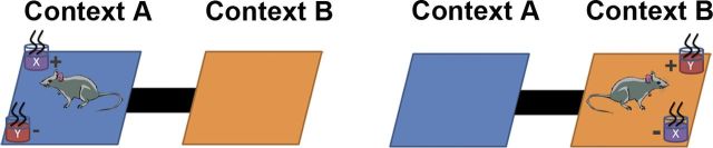
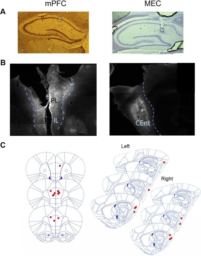
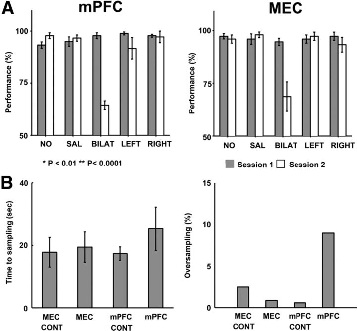
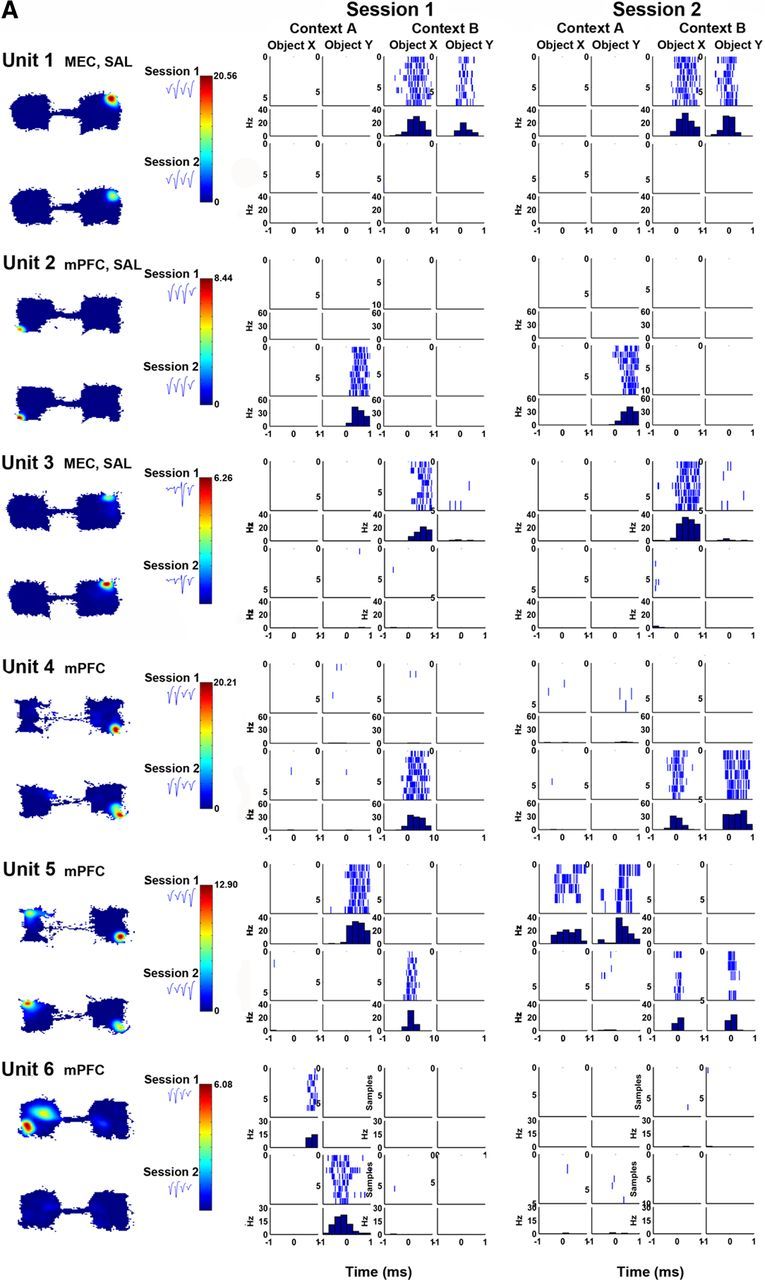
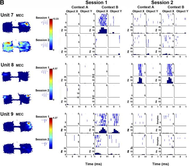
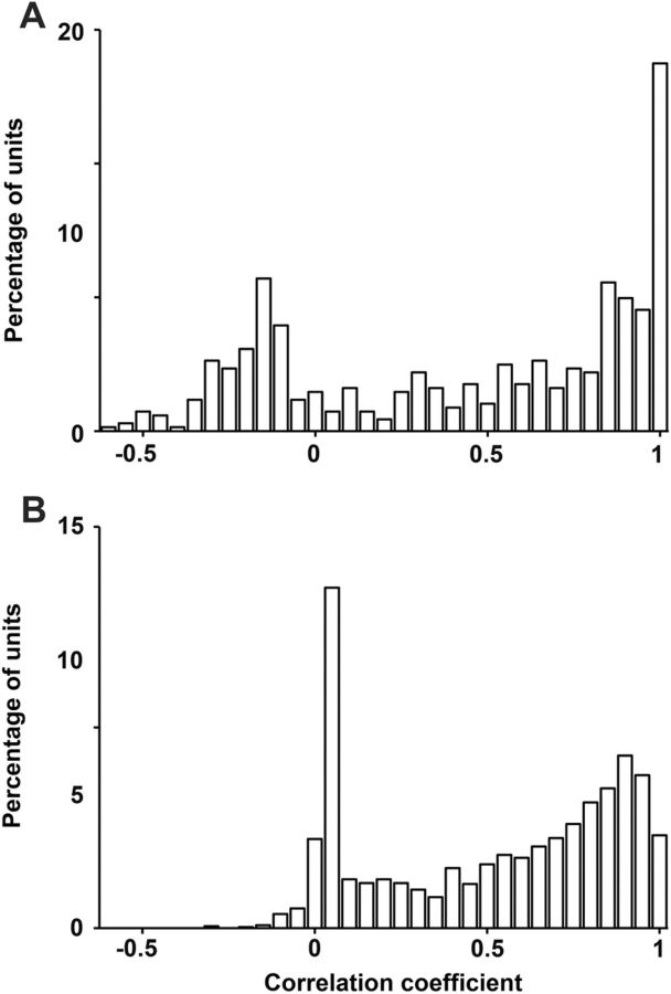
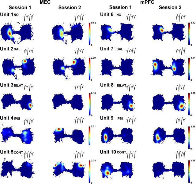
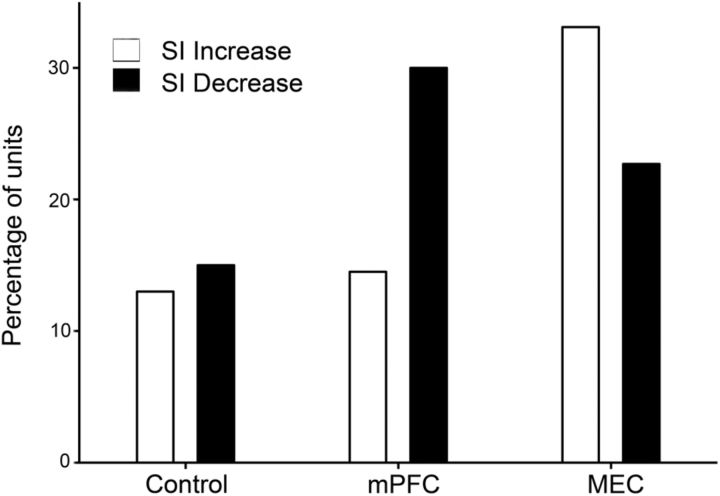
Hippocampal neurons encode events within the context in which they occurred, a fundamental feature of episodic memory. Here we explored the sources of event and context information represented by hippocampal neurons during the retrieval of object associations in rats. Temporary inactivation of the medial prefrontal cortex differentially reduced the selectivity of rule-based object associations represented by hippocampal neuronal firing patterns but did not affect spatial firing patterns. In contrast, inactivation of the medial entorhinal cortex resulted in a pervasive reorganization of hippocampal mappings of spatial context and events. These results suggest distinct and cooperative prefrontal and medial temporal mechanisms in memory representation.


Figures

Figure 1.
Figure 1.
Context-guided object discrimination task. Rats initially move into one of two environmental contexts (A or B) and explore without the presence of object stimuli for 20 s. Then two objects (X, Y) are placed in two corners of the context; the left–right positions of X and Y are pseudorandomized across trials. In Context A, object X is baited (+) and Y is not (−), whereas in Context B, object Y is baited and X is not.
Figure 2.
Figure 2.
A, Lesion marks made at tips of tetrodes located in CA1 of one rat from the mPFC group and one from the MEC group. The slices obtained from mPFC group were in the coronal plane and those from the MEC group were in the sagittal plane. B, Fluorescent muscimol deposition at injection sites. The left image shows cannula track and deposition in mPFC, while the right shows cannula tracks and deposition in MEC. The bright area localizes the muscimol deposition in each area. C, Histological plates showing locations of bilateral cannula in both mPFC (n = 6) and MEC (n = 5) groups. Histological plates were adopted from Paxinos and Watson (2010). CEnt, caudal medial entorhinal cortex; PL, prelimbic; IL, infralimbic.
Figure 3.
Figure 3.
A, Comparison of the mean (±SE) performance accuracy on the context-guided object association task between session 1 and session 2 of all five treatment conditions from both MEC and mPFC groups. NO, no injection; SAL, saline injection; BILAT, bilateral muscimol injection; LEFT, unilateral muscimol injection on the left hemisphere; RIGHT, unilateral muscimol injection on the right hemisphere. B, Comparisons of behavior during object sampling by control and bilateral mPFC and MEC inactivation groups. Left, Mean ± SE of time of object sampling before choice. CONT, control group with no treatment and saline infusion combined. Right, Number of trials with four or more object samplings before the choice response.
Figure 4.
Figure 4.
Spatial firing rate maps and averaged tetrode waveforms during the entire session (A) and rasters and peri-event histograms plotted for object-sampling events (B) for each object (X or Y) at each position (shown separately in upper and bottom) within each context (A or B) for example neurons that fired during the object-sampling period in each group. See descriptions of firing patterns in text.
Figure 4.
Figure 4.
Spatial firing rate maps and averaged tetrode waveforms during the entire session (A) and rasters and peri-event histograms plotted for object-sampling events (B) for each object (X or Y) at each position (shown separately in upper and bottom) within each context (A or B) for example neurons that fired during the object-sampling period in each group. See descriptions of firing patterns in text.
Figure 5.
Figure 5.
A, The distribution of correlation coefficients between object-sampling firing patterns in the two sessions for control (no treatment plus saline) MEC and mPFC groups. B, The distribution of correlation coefficients between spatial firing rate maps in the two sessions for control MEC and mPFC groups.
Figure 6.
Figure 6.
Example spatial firing maps and tetrode waveforms of CA1 place cells in MEC and mPFC groups. Color bars on the right represent firing rates (in Hz). Units 1 and 6 are examples where no treatment was performed and Units 2 and 7 are examples where saline was infused between two sessions. Units 1, 2, and 6 of neurons had stable spatial firing maps (correlation coefficients > 0.58) between sessions, whereas Unit 7 is an example where the cell developed place fields in the second session. Units 3–5 are examples of neurons that remapped (correlation coefficients < 0.58) after bilateral (unit 3), ipsilateral (unit 4), or contralateral (unit 5) MEC inactivation. Units 8 and 9 are examples of neurons that were stable after mPFC bilateral or ipsilateral inactivation, respectively, whereas Unit 10 is an example of the occasional loss of spatial firing after a contralateral mPFC inactivation.
Figure 7.
Figure 7.
Comparison of the percentage of object-sampling neurons that increased or decreased in object selectivity index (SI) in Controls and following mPFC or MEC inactivation.

Similar articles

Cited by

References

    1. Allen TA, Narayanan NS, Kholodar-Smith DB, Zhao Y, Laubach M, Brown TH. Imaging the spread of reversible brain inactivations using fluorescent muscimol. J Neurosci Methods. 2008;171:30–38. - PMC - PubMed
    1. Anderson MI, Killing S, Morris C, O'Donoghue A, Onyiagha D, Stevenson R, Verriotis M, Jeffery KJ. Behavioral correlates of the distributed coding of spatial context. Hippocampus. 2006;16:730–742. - PubMed
    1. Apergis-Schoute J, Pinto A, Paré D. Ultrastructural organization of medial prefrontal inputs to the rhinal cortices. Eur J Neurosci. 2006;24:135–144. - PubMed
    1. Barker GR, Warburton EC. When is the hippocampus involved in recognition memory? J Neurosci. 2011;31:10721–10731. - PMC - PubMed
    1. Barker GR, Bird F, Alexander V, Warburton EC. Recognition memory for objects, place, and temporal order: a disconnection analysis of the role of the medial prefrontal cortex and perirhinal cortex. J Neurosci. 2007;27:2948–2957. - PMC - PubMed

Publication types