. 2013 Jan 16;33(3):1050-61a.
doi: 10.1523/JNEUROSCI.1704-12.2013.

Bone morphogenetic protein 4 mediates estrogen-regulated sensory axon plasticity in the adult female reproductive tract

Affiliations

Bone morphogenetic protein 4 mediates estrogen-regulated sensory axon plasticity in the adult female reproductive tract

Aritra Bhattacherjee et al. J Neurosci. .

Abstract

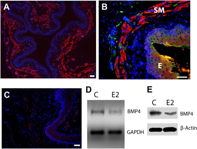
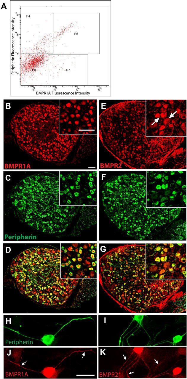
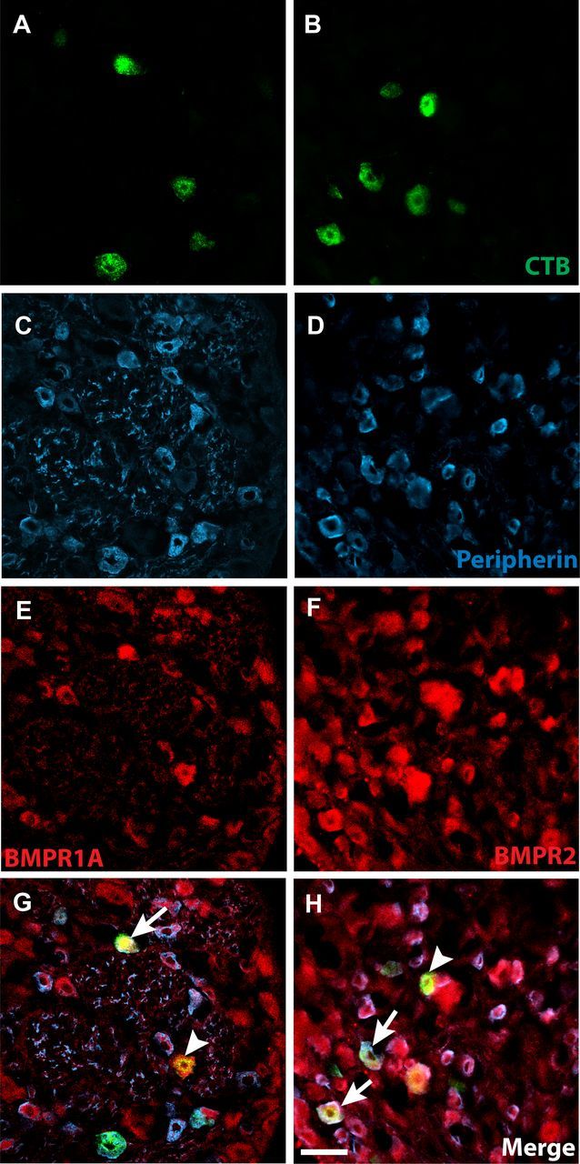
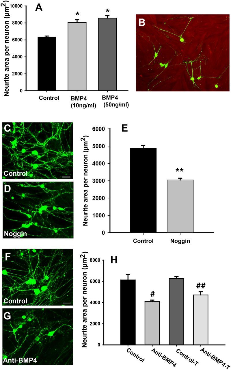
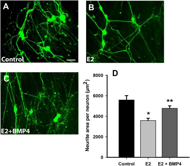
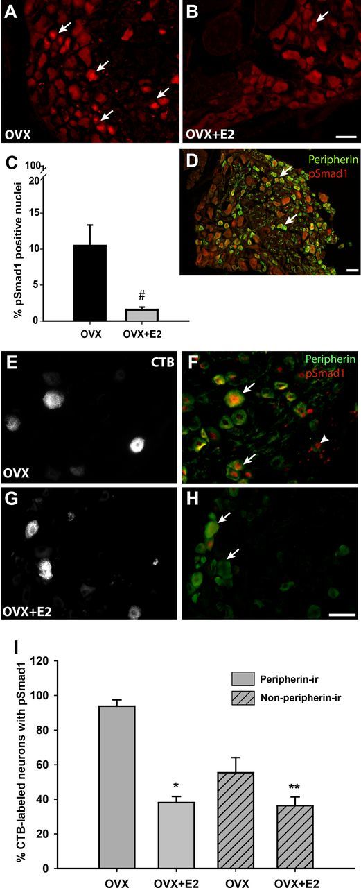
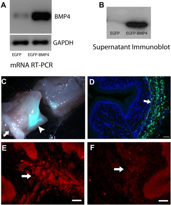
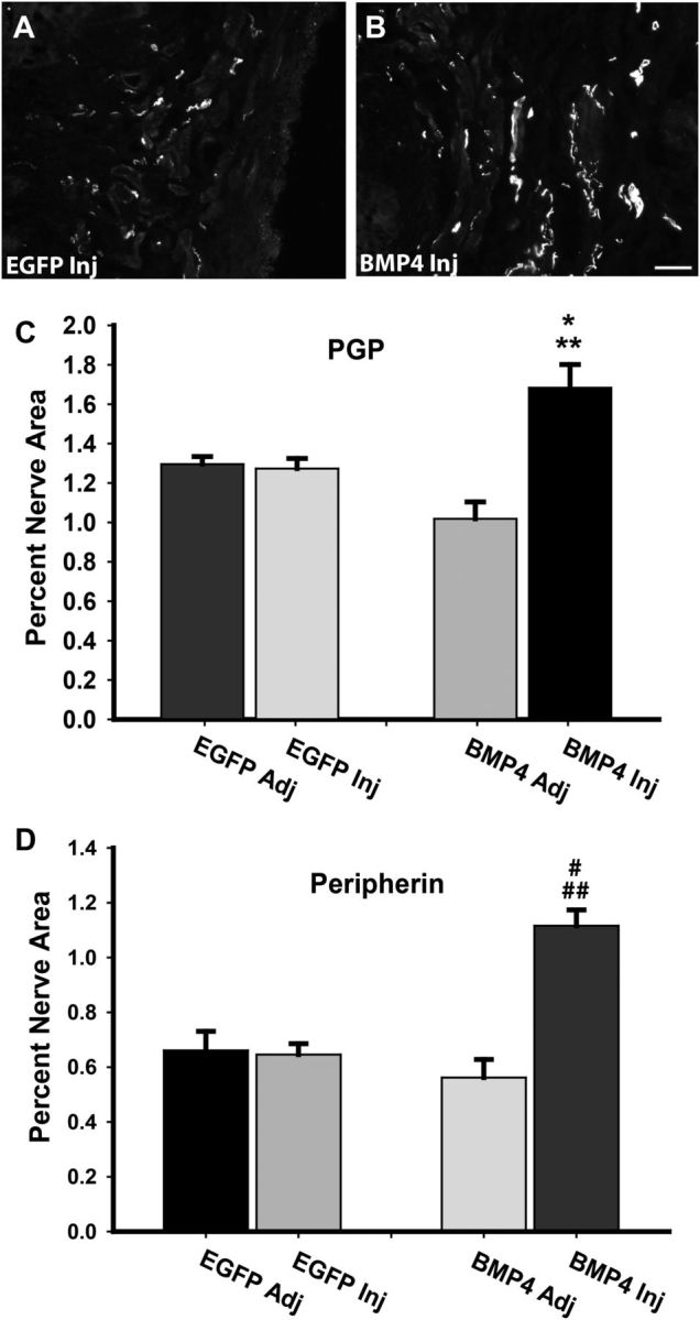
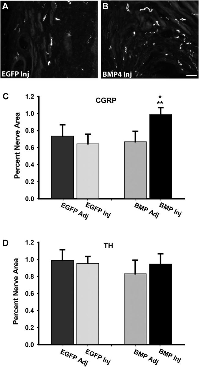
Peripheral axons are structurally plastic even in the adult, and altered axon density is implicated in many disorders and pain syndromes. However, mechanisms responsible for peripheral axon remodeling are poorly understood. Physiological plasticity is characteristic of the female reproductive tract: vaginal sensory innervation density is low under high estrogen conditions, such as term pregnancy, whereas density is high in low-estrogen conditions, such as menopause. We exploited this system in rats to identify factors responsible for adult peripheral neuroplasticity. Calcitonin gene-related peptide-immunoreactive sensory innervation is distributed primarily within the vaginal submucosa. Submucosal smooth muscle cells express bone morphogenetic protein 4 (BMP4). With low estrogen, BMP4 expression was elevated, indicating negative regulation by this hormone. Vaginal smooth muscle cells induced robust neurite outgrowth by cocultured dorsal root ganglion neurons, which was prevented by neutralizing BMP4 with noggin or anti-BMP4. Estrogen also prevented axon outgrowth, and this was reversed by exogenous BMP4. Nuclear accumulation of phosphorylated Smad1, a primary transcription factor for BMP4 signaling, was high in vagina-projecting sensory neurons after ovariectomy and reduced by estrogen. BMP4 regulation of innervation was confirmed in vivo using lentiviral transduction to overexpress BMP4 in an estrogen-independent manner. Submucosal regions with high virally induced BMP4 expression had high innervation density despite elevated estrogen. These findings show that BMP4, an important factor in early nervous system development and regeneration after injury, is a critical mediator of adult physiological plasticity as well. Altered BMP4 expression may therefore contribute to sensory hyperinnervation, a hallmark of several pain disorders, including vulvodynia.


Figures

Figure 1.
Figure 1.
BMP4 expression and regulation in the adult rat vagina. A, Low-magnification image shows gross histology of the vagina in cross-section. α-Smooth muscle actin-IR reveals the submucosal smooth muscle layer (red), which lies beneath the epithelium demarcated by the dense aggregation of DAPI-stained nuclei (blue). B, BMP4-IR (red) is expressed almost exclusively within smooth muscle (SM) lying beneath the epithelium (E), and this corresponds to the location of most peripherin-positive axons (green). C, Preabsorption of the BMP4 antibody with an excess of mouse recombinant BMP4 eliminated staining of vaginal tissues. Scale bars, 50 μm. D, Submucosal vaginal tissue explants cultured for 24 h with 20 nm estrogen (E2) show reduced BMP4 gene expression by RT-PCR relative to GAPDH compared with untreated controls (C). E, In vivo treatment of OVX rats for 7 d with estrogen also reduced BMP4 protein levels relative to β-actin as assessed by immunoblot.
Figure 2.
Figure 2.
BMP receptors are expressed in L6–S2 sensory neurons. A, FACS of dissociated DRG cells immunolabeled for BMPR1A (Cy3) and peripherin (Alexa Fluor 488) reveal high levels of BMPR1A fluorescence intensity in cells displaying high peripherin fluorescence intensity (P6). Other populations include peripherin-negative cells with high BMPR1A expression (P7), peripherin-positive cells with low BMPR1A expression (P4), and cells negative for either marker (P8). B, Immunostaining of an L6 DRG section shows that BMPR1A protein is widely distributed; inset is a higher-power image showing BMPR1A localized predominantly within neurons. C, Distribution of peripherin immunostaining in the section shown in B. D, A merged image shows that BMPR1A-IR is present predominantly in peripherin-positive neurons. E, BMPR2 immunostaining shows wider localization across different neuronal subtypes. F, Immunostaining reveals peripherin-expressing DRG neurons in E. G, Most peripherin-positive neurons show BMPR2-IR. H, I, Peripherin-immunoreactive neurons in dissociated DRG culture. Immunostaining for BMPR1A (J) and BMPR2 (K) show that both receptors are present throughout the axons (arrows) as well as the cell body. Scale bars, 50 μm.
Figure 3.
Figure 3.
Vagina-projecting neurons express BMP4 receptors. A, B, Neurons projecting specifically to the vaginal submucosa were labeled after Alexa Fluor-488-conjugated recombinant CTB injection, as shown here in an S1 DRG. C, D, Retrogradely labeled neurons frequently display peripherin-IR. E, F, Vagina-projecting neurons exhibit IR for both BMPR1A and BMPR2. G, H, Arrows indicate CTB-labeled, peripherin-positive neurons expressing BMPR. Arrowheads indicate CTB-labeled, peripherin-negative neurons expressing BMPR. Scale bar, 50 μm (for all panels).
Figure 4.
Figure 4.
Target-derived BMP4 promotes sensory neurite outgrowth. A, Dissociated DRG neurons grown on collagen-coated plates showed increased neurite outgrowth in the presence of 10 or 50 ng/ml BMP4 (*p ≤ 0.007). B, Vaginal smooth muscle cells immunostained for α-smooth muscle actin (red) were grown on collagen-coated plastic. DRG neurons immunostained for peripherin (green) were plated on smooth muscle cells, and most extended neurites that made contacts with muscle cells at 48 h. C, Vehicle-treated cocultures immunostained for peripherin showed many neurites. D, Cocultures treated with noggin (250 ng/ml) had fewer neurites. E, Quantitative analysis confirmed a reduction in neurite outgrowth per neuron in noggin-treated cocultures (**p = 0.001). F, Neurites elaborated by neurons grown directly on smooth muscle cocultures (Control) are reduced by the addition of a BMP4 function-neutralizing antibody (#p = 0.005; G, H). Anti-BMP4 was equally effective in reducing axon outgrowth when smooth muscle cells were grown in transwell inserts (H, Control-T, Anti-BMP4-T; ##p = 0.004). Scale bars, 50 μm.
Figure 5.
Figure 5.
Neurite outgrowth in cocultures is downregulated by estrogen and rescued by exogenous BMP4. A, Smooth muscle cells (unstained) were grown for 72 h, on which dissociated sensory neurons (green) were cultured for 48 h. Scale bar: A–C, 50 μm. B, When smooth muscle cells were grown in the presence of 2 × 10⁻⁸ m 17β-estradiol, neurite outgrowth appeared to be reduced. C, Addition of 10 ng/ml recombinant BMP4 appeared to increase numbers of neurites. D, Quantitative analysis showed that neurite outgrowth per neuron was reduced by estrogen (E2) pretreatment (*p = 0.001 vs control), and the decrease was abrogated by BMP4 (#p = 0.004 vs E2). There was no significant difference between control and E2 + BMP4.
Figure 6.
Figure 6.
pSmad1 expression in DRG neurons is reduced after estrogen administration. A, In vehicle-injected OVX rats, many nuclei show high levels of pSmad1-IR (arrows). B, At 24 h after estrogen administration (OVX+E2), pSmad1-positive nuclei were observed infrequently. C, Quantitative analysis confirms that numbers of pSmad1-positive nuclei are markedly reduced by estrogen administration (*p = 0.038). D, Double immunostaining for peripherin (green) and pSmad1 (red) show that positive nuclei are frequently present in peripherin-positive neurons in the OVX rat. E, Vaginal submucosal injection of CTB resulted in retrograde labeling of DRG neurons of OVX rats. F, Several CTB-labeled neurons in this low-estrogen condition (E) display nuclear pSmad1. Arrows points to peripherin-positive CTB-labeled neurons, and arrowhead shows a peripherin-negative CTB neuron. G, Vaginal CTB injection also labeled DRG neurons in OVX rats receiving estrogen. H, In the presence of estrogen, CTB-labeled neurons (G) in both peripherin-positive (arrow) and peripherin-negative (arrowhead) groups were frequently pSmad1 negative. I, In CTB-labeled neurons, pSmad1-immunoreactive nuclei occurred more frequently in peripherin-positive compared with peripherin-negative neurons (p = 0.004 by 2-way ANOVA). Both populations responded to estrogen with a reduction in the percentage of pSmad1-expressing nuclei (**p < 0.001, ***p = 0.034 by Student–Newman–Kuels tests). However, the decline was much stronger in the peripherin-positive population (p = 0.007). All images from the L6 and S1 DRG. Scale bars, 50 μm.
Figure 7.
Figure 7.
Lentiviral injection regulates BMP4 expression in vaginal smooth muscle. A, HEK293T cells overexpress BMP4 as assessed by RT-PCR 48 h after transduction with viral particles containing vectors for EGFP–BMP4 expression compared with those with only EGFP expressing control. GAPDH was used as a housekeeping control gene. B, Immunoblot showing abundant amounts of secreted BMP4 protein in conditioned media of HEK293T cells transduced with EGFP–BMP4 compared with EGFP alone. C, Bright-field and epifluorescence overlay of the whole vagina (cut transversely) shows intense EFGP fluorescence at the injection site (arrowhead) at 7 d. Arrow indicates the vaginal orifice. D, Overlay of DAPI staining and EGFP in a section of the specimen shown in C. Arrow indicates abundant EGFP expression in the smooth muscle layer. Scale bar, 200 μm. E, Immunostaining for BMP4 shows robust fluorescence in vaginal smooth muscle (arrow) of an estrogen-treated OVX rat transduced with EGFP–BMP4 at 7 d. F, Estrogen-treated rats receiving control virus with EGFP alone showed very low levels of BMP4 immunostaining. Scale bars: E, F, 50 μm.
Figure 8.
Figure 8.
BMP4 overexpression preserves vaginal innervation at high estrogen. A, Immunostaining for PGP9.5 at the site of injection 7 d after injection of lentivirus driving expression of only EGFP (EGFP Inj) showed low innervation density typical of OVX rats receiving estrogen supplementation. B, PGP-positive innervation density at sites injected with lentivirus driving BMP4 expression (BMP4 Inj) was considerably higher, characteristic of low-estrogen OVX rats. Scale bar, 50 μm. C, Injection of EGFP lentivirus did not alter innervation density relative to uninjected adjacent tissues (EGFP Adj), or to innervation at sites adjacent to injection with the BMP4 lentivirus (BMP4 Adj). Innervation density at sites receiving BMP4 lentivirus was significantly greater than that of other sites (*p = 0.019 for BMP4 Inj vs EGFP Inj and **p = 0.026 for BMP4 Inj vs BMP4 Adj). D, Staining for peripherin confirmed that changes noted with PGP9.5 also occur within this axonal population (#p ≤ 0.001 for BMP4 Inj vs EGFP Inj, and ##p ≤ 0.001 for BMP4 Inj vs BMP4 Adj).
Figure 9.
Figure 9.
BMP4 overexpression selectively affects sensory innervation density. A, In OVX rats receiving estrogen and injected with EGFP lentivirus (EGFP Inj), sensory axons immunoreactive for CGRP were depleted. B, Sites injected with the BMP4 lentivirus (BMP4 Inj) showed higher levels of CGRP-immunoreactive innervation. Scale bar, 50 μm. C, Quantitative analysis confirmed that CGRP axon density in tissue receiving BMP4 lentiviral injections was greater than that of tissue adjacent to the injection site (BMP Adj; *p = 0.012) and to tissue receiving EGFP lentivirus (EGFP Inj; **p = 0.049). D, In contrast, injection with BMP4 lentivirus did not affect densities of axons expressing the sympathetic axon marker TH.

Similar articles

Cited by

References

    1. Abir R, Ben-Haroush A, Melamed N, Felz C, Krissi H, Fisch B. Expression of bone morphogenetic proteins 4 and 7 and their receptors IA, IB, and II in human ovaries from fetuses and adults. Fertil Steril. 2008;89:1430–1440. - PubMed
    1. Ai X, Cappuzzello J, Hall AK. Activin and bone morphogenetic proteins induce calcitonin gene-related peptide in embryonic sensory neurons in vitro. Mol Cell Neurosci. 1999;14:506–518. - PubMed
    1. Baker JC, Beddington RS, Harland RM. Wnt signaling in Xenopus embryos inhibits bmp4 expression and activates neural development. Genes Dev. 1999;13:3149–3159. - PMC - PubMed
    1. Baleato RM, Aitken RJ, Roman SD. Vitamin A regulation of BMP4 expression in the male germ line. Dev Biol. 2005;286:78–90. - PubMed
    1. Berkley KJ, Robbins A, Sato Y. Functional differences between afferent fibers in the hypogastric and pelvic nerves innervating female reproductive organs in the rat. J Neurophysiol. 1993;69:533–544. - PubMed

Publication types

MeSH terms

LinkOut - more resources