

Moderate prenatal alcohol exposure reduces plasticity and alters NMDA receptor subunit composition in the dentate gyrus
- PMID: 23325244
- PMCID: PMC3563269
- DOI: 10.1523/JNEUROSCI.1217-12.2013
Moderate prenatal alcohol exposure reduces plasticity and alters NMDA receptor subunit composition in the dentate gyrus
Abstract
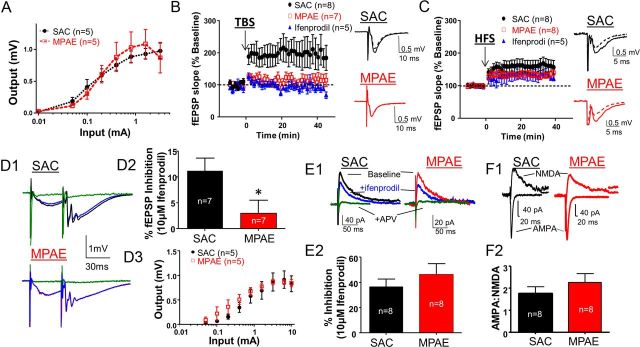
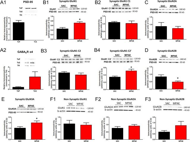
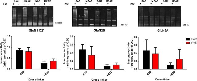
Although it is well documented that heavy consumption of alcohol during pregnancy impairs brain development, it remains controversial whether moderate consumption causes significant damage. Using a limited access, voluntary consumption paradigm, we recently demonstrated that moderate prenatal alcohol exposure (MPAE) is associated with dentate gyrus-dependent learning and memory deficits that are manifested in adulthood. Here, we identified a novel mechanism that may underlie this effect of MPAE. We found that MPAE mice exhibit deficits in NMDA receptor (NMDAR)-dependent long-term potentiation (LTP) in the dentate gyrus. Further, using semiquantitative immunoblotting techniques, we found that the levels of GluN2B subunits were decreased in the synaptic membrane, while levels of C2'-containing GluN1 and GluN3A subunits were increased, in the dentate gyrus of MPAE mice. These data suggest that MPAE alters the subunit composition of synaptic NMDARs, leading to impaired NMDAR-dependent LTP in the dentate gyrus.
Figures
Similar articles
-
Prenatal ethanol exposure persistently impairs NMDA receptor-dependent activation of extracellular signal-regulated kinase in the mouse dentate gyrus.J Neurochem. 2009 Jun;109(5):1311-23. doi: 10.1111/j.1471-4159.2009.06049.x. Epub 2009 Mar 20. J Neurochem. 2009. PMID: 19317851 Free PMC article.
-
Prenatal ethanol (EtOH) exposure alters the sensitivity of the adult dentate gyrus to acute EtOH exposure.Alcohol Clin Exp Res. 2014 Jan;38(1):135-43. doi: 10.1111/acer.12227. Epub 2013 Aug 5. Alcohol Clin Exp Res. 2014. PMID: 23915337
-
Sex-specific effect of prenatal alcohol exposure on N-methyl-D-aspartate receptor function in orbitofrontal cortex pyramidal neurons of mice.Alcohol Clin Exp Res. 2021 Oct;45(10):1994-2005. doi: 10.1111/acer.14697. Epub 2021 Sep 29. Alcohol Clin Exp Res. 2021. PMID: 34523139 Free PMC article.
-
Long-term potentiation and the role of N-methyl-D-aspartate receptors.Brain Res. 2015 Sep 24;1621:5-16. doi: 10.1016/j.brainres.2015.01.016. Epub 2015 Jan 22. Brain Res. 2015. PMID: 25619552 Free PMC article. Review.
-
A Systematic Review of the Effects of Perinatal Alcohol Exposure and Perinatal Marijuana Exposure on Adult Neurogenesis in the Dentate Gyrus.Alcohol Clin Exp Res. 2020 Jun;44(6):1164-1174. doi: 10.1111/acer.14332. Epub 2020 May 14. Alcohol Clin Exp Res. 2020. PMID: 32246781 Free PMC article.
Cited by
-
Prenatal ethanol exposure impairs executive function in mice into adulthood.Alcohol Clin Exp Res. 2014 Dec;38(12):2962-8. doi: 10.1111/acer.12577. Alcohol Clin Exp Res. 2014. PMID: 25581651 Free PMC article.
-
Synaptic Plasticity Abnormalities in Fetal Alcohol Spectrum Disorders.Cells. 2023 Jan 29;12(3):442. doi: 10.3390/cells12030442. Cells. 2023. PMID: 36766783 Free PMC article. Review.
-
Prenatal alcohol exposure is associated with altered subcellular distribution of glucocorticoid and mineralocorticoid receptors in the adolescent mouse hippocampal formation.Alcohol Clin Exp Res. 2014 Feb;38(2):392-400. doi: 10.1111/acer.12236. Epub 2013 Aug 19. Alcohol Clin Exp Res. 2014. PMID: 23992407 Free PMC article.
-
Impact of combined prenatal ethanol and prenatal stress exposures on markers of activity-dependent synaptic plasticity in rat dentate gyrus.Alcohol. 2014 Sep;48(6):523-32. doi: 10.1016/j.alcohol.2014.06.006. Epub 2014 Jul 25. Alcohol. 2014. PMID: 25129673 Free PMC article.
-
Long Term Depression in Rat Hippocampus and the Effect of Ethanol during Fetal Life.Brain Sci. 2017 Nov 28;7(12):157. doi: 10.3390/brainsci7120157. Brain Sci. 2017. PMID: 29182556 Free PMC article. Review.
References
-
- Chandra D, Jia F, Liang J, Peng Z, Suryanarayanan A, Werner DF, Spigelman I, Houser CR, Olsen RW, Harrison NL, Homanics GE. GABAA receptor alpha 4 subunits mediate extrasynaptic inhibition in thalamus and dentate gyrus and the action of gaboxadol. Proc Natl Acad Sci U S A. 2006;103:15230–15235. - PMC - PubMed
-
- Costa ET, Savage DD, Valenzuela CF. A review of the effects of prenatal or early postnatal ethanol exposure on brain ligand-gated ion channels. Alcohol Clin Exp Res. 2000;24:706–715. - PubMed
-
- D'Angelo E, Rossi P, Armano S, Taglietti V. Evidence for NMDA and mGlu receptor-dependent long-term potentiation of mossy fiber-granule cell transmission in rat cerebellum. J Neurophysiol. 1999;81:277–287. - PubMed
Publication types
MeSH terms
Substances
Grants and funding
- R01-AA015614/AA/NIAAA NIH HHS/United States
- R37 AA015614/AA/NIAAA NIH HHS/United States
- R03AA020101/AA/NIAAA NIH HHS/United States
- F31AA020434/AA/NIAAA NIH HHS/United States
- R01 AA014973/AA/NIAAA NIH HHS/United States
- T32 AA014127/AA/NIAAA NIH HHS/United States
- P50 AA022534/AA/NIAAA NIH HHS/United States
- R03 AA020101/AA/NIAAA NIH HHS/United States
- R01-AA014973/AA/NIAAA NIH HHS/United States
- T32AA014127/AA/NIAAA NIH HHS/United States
- R01 AA015614/AA/NIAAA NIH HHS/United States
- R25 GM075149/GM/NIGMS NIH HHS/United States
- F31 AA020434/AA/NIAAA NIH HHS/United States
- P20AA017068/AA/NIAAA NIH HHS/United States
- P20 AA017068/AA/NIAAA NIH HHS/United States
LinkOut - more resources
Full Text Sources
Other Literature Sources
Miscellaneous