Review
. 2013 Apr 15;304(8):C693-714.
doi: 10.1152/ajpcell.00350.2012. Epub 2013 Jan 16.

Physiology of SLC12 transporters: lessons from inherited human genetic mutations and genetically engineered mouse knockouts

Affiliations
Review

Physiology of SLC12 transporters: lessons from inherited human genetic mutations and genetically engineered mouse knockouts

Kenneth B Gagnon et al. Am J Physiol Cell Physiol. .

Abstract

Among the over 300 members of the solute carrier (SLC) group of integral plasma membrane transport proteins are the nine electroneutral cation-chloride cotransporters belonging to the SLC12 gene family. Seven of these transporters have been functionally described as coupling the electrically silent movement of chloride with sodium and/or potassium. Although in silico analysis has identified two additional SLC12 family members, no physiological role has been ascribed to the proteins encoded by either the SLC12A8 or the SLC12A9 genes. Evolutionary conservation of this gene family from protists to humans confirms their importance. A wealth of physiological, immunohistochemical, and biochemical studies have revealed a great deal of information regarding the importance of this gene family to human health and disease. The sequencing of the human genome has provided investigators with the capability to link several human diseases with mutations in the genes encoding these plasma membrane proteins. The availability of bacterial artificial chromosomes, recombination engineering techniques, and the mouse genome sequence has simplified the creation of targeting constructs to manipulate the expression/function of these cation-chloride cotransporters in the mouse in an attempt to recapitulate some of these human pathologies. This review will summarize the three human disorders that have been linked to the mutation/dysfunction of the Na-Cl, Na-K-2Cl, and K-Cl cotransporters (i.e., Bartter's, Gitleman's, and Andermann's syndromes), examine some additional pathologies arising from genetically modified mouse models of these cotransporters including deafness, blood pressure, hyperexcitability, and epithelial transport deficit phenotypes.


Figures
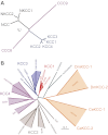
Fig. 1.
Fig. 1.
Evolution of solute carrier 12 (SLC12) proteins. A: cluster dendogram of human SLC12A cotransporters. The two major branches of cation-chloride cotransporters (CCC) separated early during evolution. Functionally uncharacterized CCC9 separated early from the Na+-dependent branch of cation-chloride cotransporters. In a similar fashion, CCC8 separated early from the Na+-independent K-Cl cotransporters. The human Na+-independent cotransporters are more closely related to one another than are the Na+-dependent cotransporters. NKCC2, Na-K-2Cl cotransporter 2. B: cluster dendogram illustrating separation of the Na+-independent cation-chloride cotransporters during vertebrate evolution. Although multiple “K-Cl like” cotransporters are found in roundworms and arthropods, the modern K-Cl cotransporters diverged early during vertebrate evolution. The amino acid sequences used to create the dendograms in both A and B were aligned using Vector Nti Suite software (version 6.0; Invitrogen/Life Technologies), saved as a text file, and then reformatted for use with Promlk, a software component of the Phylogeny Inference Package (PHYLIP; http://evolution.gs.washington.edu/phylip.html). Length of tree branches can be compared with the reference bar, which represents 0.1 amino acid substitutions per site. Dm, Drosophila melanogaster; Ce, Caenorhabditis elegans.
Fig. 2.
Fig. 2.
Storyboard of gene-targeted manipulation. Before the complete sequencing of animal genomes, investigators wishing to target specific genes for knockout in animal models were required to initially map genomic DNA to identify the exons, introns, and unique restriction sites within the gene of interest. Steps involved include 1) endonuclease digestion of genomic DNA; 2) creation of a phage DNA library; 3) screening of phage library; 4) Southern blot analysis with phage DNA probes; 5) rescue of cDNA fragments into bacterial plasmids; 6) sequencing; and 7) design and construction of a targeting vector with upstream and downstream arms of DNA to promote homologous recombination.
Fig. 3.
Fig. 3.
SLC12A cotransporters in the kidney nephron. KCC3 is localized to the basolateral membrane of proximal tubule (PT), NKCC2 on the apical membrane of the thick ascending limb (TAL), and Na-Cl cotransporter (NCC) expression on the apical membrane of the distal convoluted tubule (DCT); KCC4 is found in the DCT and cortical connecting tubule (CNT); and NKCC1 is expressed in the inner medullar collecting duct (IMCD), the afferent arteriole of the glomerulus, and the intra- and extraglomerular mesangium.
Fig. 4.
Fig. 4.
Major SLC12 transporters in mammalian kidneys. A: thick ascending limb epithelial cell of the Loop of Henle. NKCC2 participates in the reabsorption of Na+ and recycles the K+ that exits through the ATP-dependent renal outer medullary potassium channel (ROMK). Additional Na+ ions follow the paracellular pathway driven by an electric gradient generated by ROMK on the apical membrane and CLCK (CLCKA and CLCKB) on the basolateral membrane. B: epithelial cell of the distal convoluted tubule (DCT). NCC expressed on the apical membrane contributes to Na+ reabsorption. The segment also reabsorbs Ca2+ through apical Ca2+ channel (transient receptor potential vanniloid type 5, TRPV5) and basolateral Na+/Ca2+ exchanger (NCX1) and Ca2+ pump. In the DCT cell, Ca2+ is buffered and transported by parvalbumin (Pvlb; DCT1) or calbindin-2 (Calb; DCT2). PMCA1b, plasma membrane Ca2+-ATPase 1b.
Fig. 5.
Fig. 5.
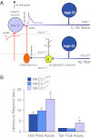
Nociception phenotype in NKCC1 knockout mice. A: model of sensory circuitry in the spinal cord. Nociceptive signals are carried by unmyelinated (C) and thinly myelinated (Aδ) afferent fibers that synapse onto spinal cord lamina I and II neurons. Interneurons (orange), which are activated by projection neurons in deeper spinal cord layers, release GABA at the terminals of the C and Aδ fibers. Because the Cl concentration is high in these afferent neurons, GABA produces a depolarization of the nerve terminal and inhibition of incoming pain signals from the periphery. B: withdrawal latencies for hot plate and tail flick assays in wild-type, heterozygous, and homozygous NKCC1 knockout mice. Both tests were performed at 52°C. Data represent means ± SE. *Significant difference with P < 0.05. Hot plate and tail flick data were taken from references 136 and 74, respectively.
Fig. 6.
Fig. 6.
NKCC1 expression/function alters reproduction and sterility. A: relationship between body weight and testicle weight in heterozygous (red squares) and homozygous (blue circles) NKCC1 knockout mice. Distribution of weight is indicated by the 6 data points per genotype. Inset: control of testosterone secretion. Hypothalamic neurons release gonadotropin releasing hormone (GnRH), which stimulates the pituitary to release luteinizing hormone (LH) and follicle-stimulating hormone (FSH). The two anterior pituitary hormones then stimulate the testis to produce testosterone. B: plasma levels of testosterone and LH determined in homozygous (blue bars) and heterozygous (red bars) NKCC1 knockout mice. C: TUNEL staining in control testis. Note the single layer of large dark cells (spermatogonia) at the base of the seminiferous tubule and large number of spermatids filling the lumen. D: TUNEL staining in homozygous NKCC1-null testis. Note the accumulation and disorganization of spermatogonia. Also note the number of TUNEL-positive (red stained) cells, indicating apoptotic cell death. Sections were counterstained with thionin (unpublished observations from the Delpire laboratory).
Fig. 7.
Fig. 7.
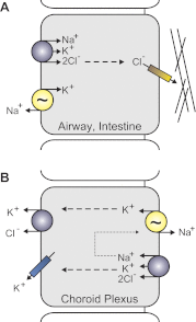
Illustration of differential NKCC1 expression and function. A: prototypical Cl secreting epithelial cells (e.g., salivary, lung, intestine) express NKCC1 on the basolateral membrane. The Na+-K+-ATPase (yellow), coexpressed with NKCC1, recycles Na+ ions thus maintaining the driving force for NKCC1. Chloride ions exit the apical membrane of epithelial through a Cl channel, the cystic fibrosis transmembrane conductance regulator. Lines represent the mucus layer adjacent to the apical membrane. B: apical localization of NKCC1 and the Na+-K+-ATPase in choroid plexus epithelial cells uncouples the reabsorption of K+ from Na+ secretion. Potassium ions exit the basolateral membrane through K-Cl cotransporters and K+ channels.
Fig. 8.
Fig. 8.
Decrease in KCC2 expression leads to seizures and lethality. A: pentylenetetrazole (PTZ)-induced seizures in 6-wk-old wild-type and heterozygote KCC2-null mice. Two groups of mice (21 wild-type and 21 heterozygotes) were injected daily with 60 mg/kg PTZ for a total of 4 days. The number of mice exhibiting seizures scoring >3 within 1 h after the injection are represented. [Modified from Ref. , with permission.] B: Kaplan-Meier plots of KCC2b knockouts and CAMKIIα-driven inducible KCC2 knockouts [where mice were fed with doxycycline chow (200 mg/kg, BioServ, Frenchtown, NJ) since conception until doxycycline withdrawal at postnatal day 40 (P40)]. Lethality occurred between P110 and P130, indicating both slow elimination of the drug and a slow decrease in KCC2 expression. C: Western blot analysis of KCC2 expression at P60 in hindbrain and forebrain of a control and an inducible KCC2 knockout mouse after 2 wk doxycycline withdrawal.
Fig. 9.
Fig. 9.
Axonal defects in KCC3-null mice. A and B: electron micrographs of P8 distal sciatic nerves isolated from wild-type (A) and KCC3-null (B) mice. Scale bar in A, 2 μm. Myelin compaction in mutant fibers (B, inset) is indistinguishable from that of wild-type (A, inset, scale bar, 500 nm). Fibers with periaxonal fluid accumulation appear only in KCC3-null nerves (asterisk, B). CF: varying degrees of the severity of the abnormal fluid accumulation in distal KCC3-null sciatic nerve fibers. Scale bars, 500 nm. Myelin debris (arrows) is observed in the periaxonal space (C and E). a, Axon; pn, paranodes; bv, blood vessel. [Modified from Ref. , with permission.]
Fig. 10.
Fig. 10.
Expression of SLC12 cotransporters in the inner ear. Cartoon depicts the scala vestibule, scala tympani, and scala media of the mammalian inner ear. Illustrated are the organ of Corti, inner and outer hair cells, phalangeal cells, Deiter's cells, and stria vascularis cells. Note the basolateral expression of NKCC1 on the stria vascularis cells that line the scala media. Inner and outer hair cells express KCC3, and supporting phalangeal and Deiter's cells express KCC4.

Similar articles

Cited by

References

    1. Abbas L, Whitfield TT. Nkcc1 (SLC12A2) is required for the regulation of endolymph volume in the otic vesicle and swim bladder volume in the zebrafish larva. Development 136: 2837–2848, 2009 - PMC - PubMed
    1. Adragna NC, Chen Y, Delpire E, Lauf PK, Morris M. Hypertension in K-Cl cotransporter-3 knockout mice. Adv Exp Med Biol 559: 379–385, 2004 - PubMed
    1. Alvarez-Leefmans FJ, Gamiño SM, Giraldez F, Nogueron I. Intracellular chloride regulation in amphibian dorsal root ganglion neurons studied with ion-selective microelectrodes. J Physiol 406: 225–246, 1988 - PMC - PubMed
    1. Alvarez-Leefmans FJ, Leon-Olea M, Mendoza-Sotelo J, Alvarez FJ, Anton B, Garduno R. Immunolocalization of the Na(+)-K(+)-2Cl(-) cotransporter in peripheral nervous tissue of vertebrates. Neuroscience 104: 569–582, 2002 - PubMed
    1. Andermann F, Andermann E, Joubert M, Karpati G, Carpenter S, Melancon D. Familial agenesis of the corpus callosum with anterior horn cell disease: a syndrome of mental retardation, areflexia and paraparesis. Trans Am Neurol Assoc 97: 242–244, 1972

Publication types

Substances