. 2013 Mar;23(3):555-67.
doi: 10.1101/gr.147942.112. Epub 2013 Jan 16.

Dynamic DNA methylation across diverse human cell lines and tissues

Affiliations

Dynamic DNA methylation across diverse human cell lines and tissues

Katherine E Varley et al. Genome Res. 2013 Mar.

Abstract

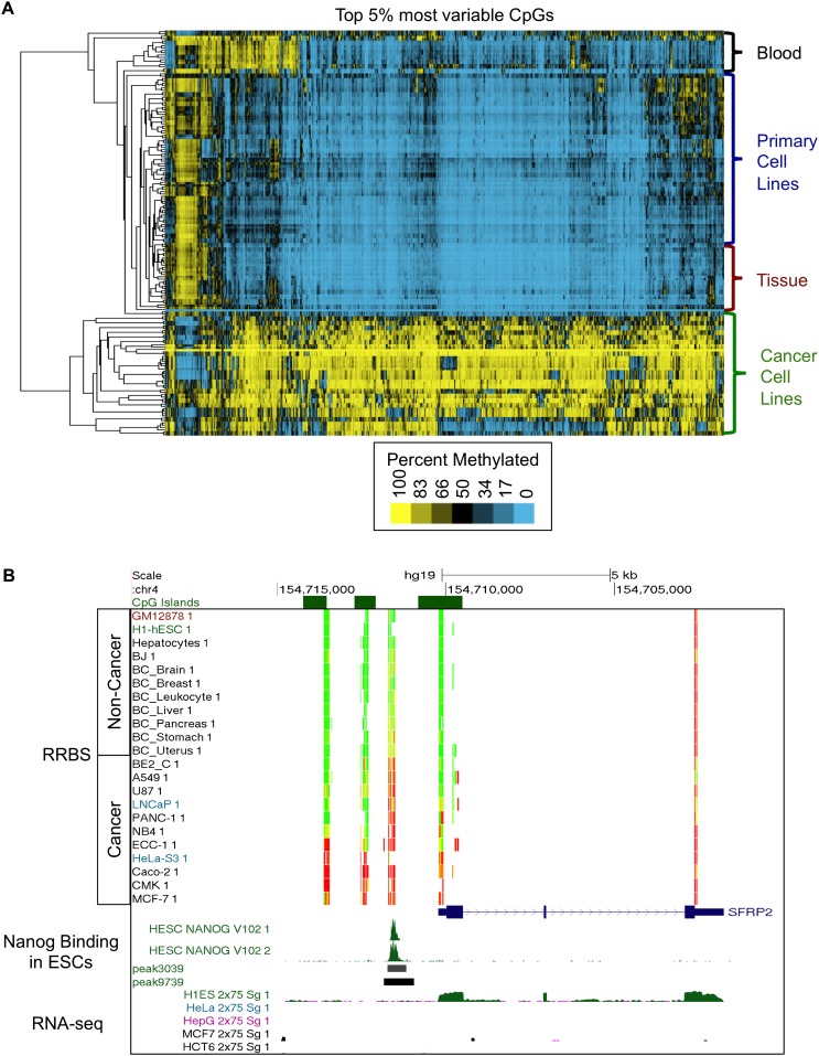
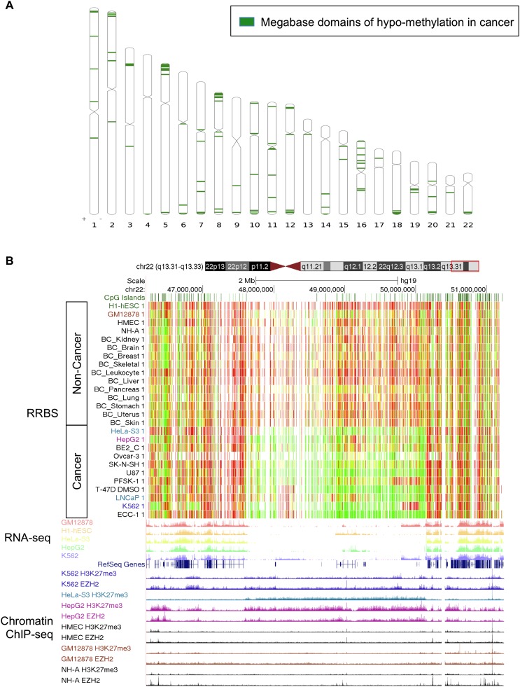
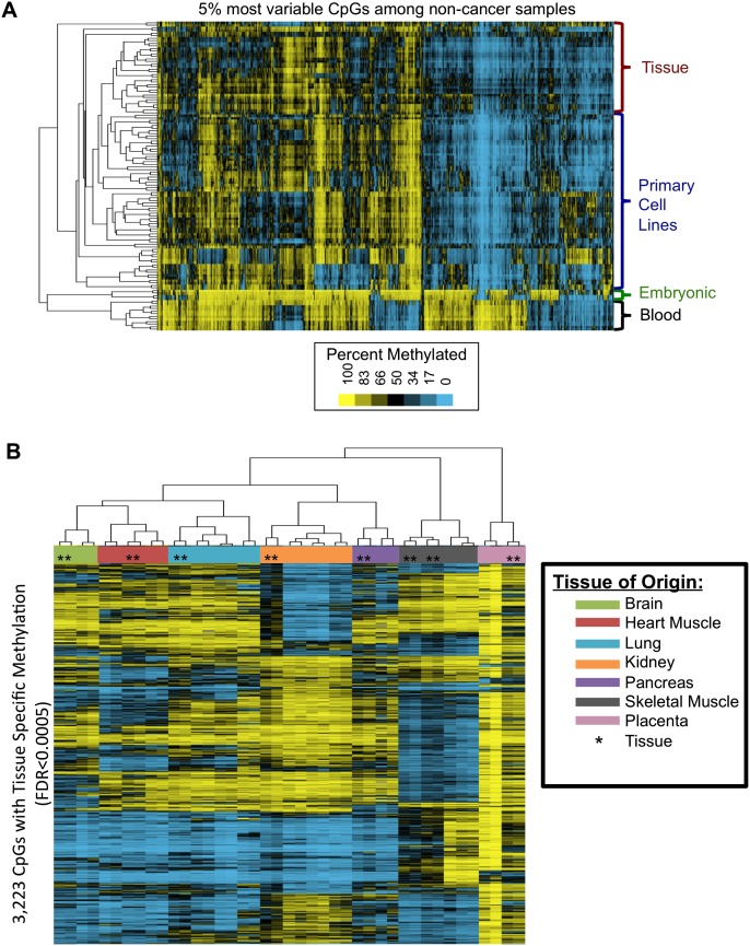
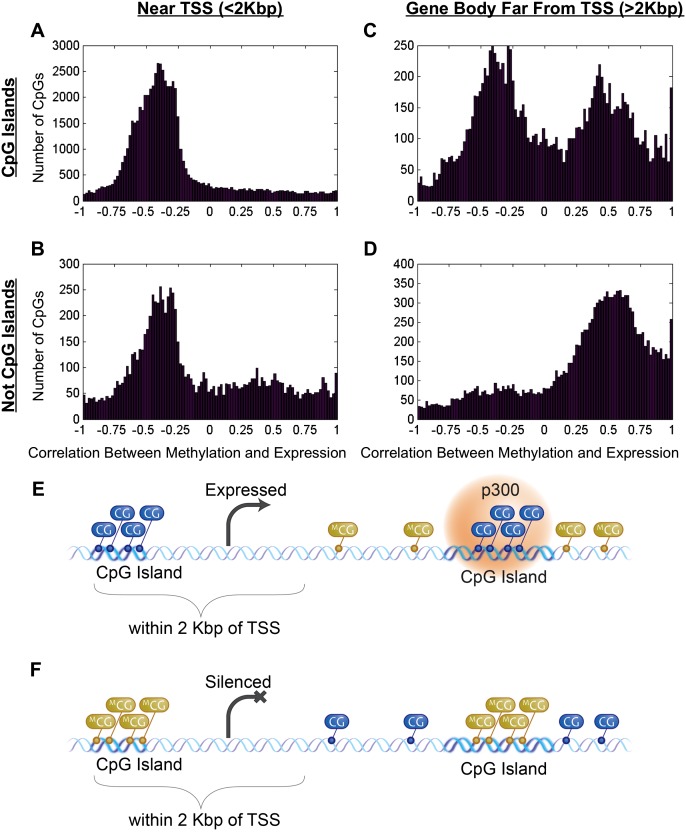
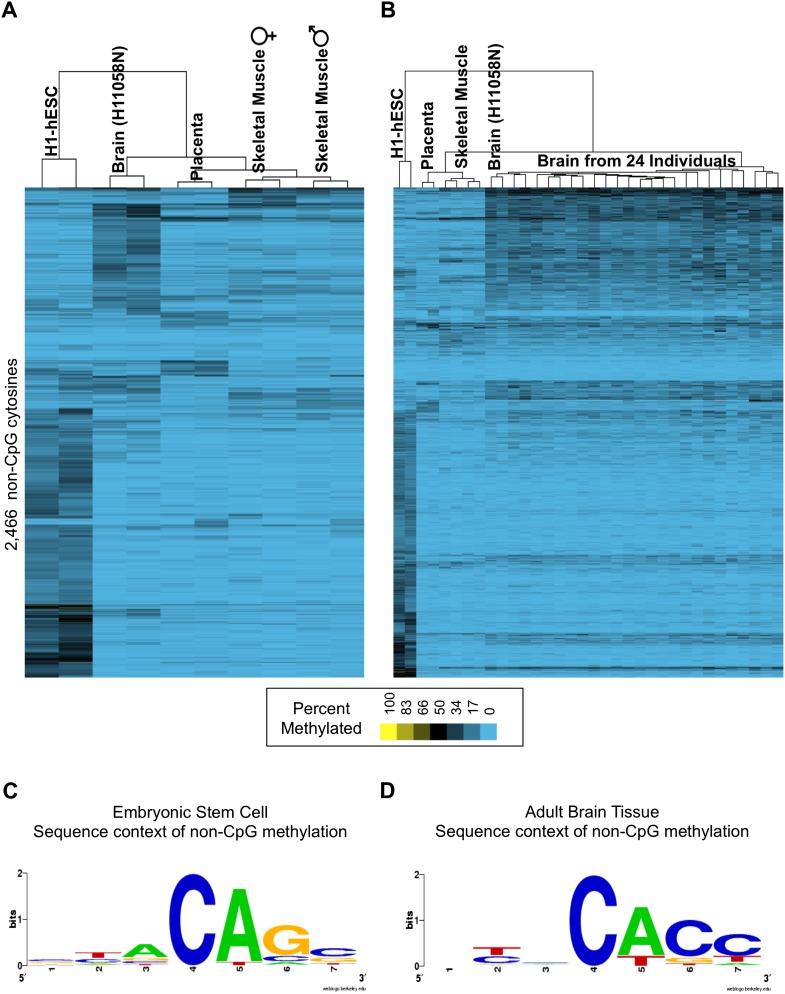
As studies of DNA methylation increase in scope, it has become evident that methylation has a complex relationship with gene expression, plays an important role in defining cell types, and is disrupted in many diseases. We describe large-scale single-base resolution DNA methylation profiling on a diverse collection of 82 human cell lines and tissues using reduced representation bisulfite sequencing (RRBS). Analysis integrating RNA-seq and ChIP-seq data illuminates the functional role of this dynamic mark. Loci that are hypermethylated across cancer types are enriched for sites bound by NANOG in embryonic stem cells, which supports and expands the model of a stem/progenitor cell signature in cancer. CpGs that are hypomethylated across cancer types are concentrated in megabase-scale domains that occur near the telomeres and centromeres of chromosomes, are depleted of genes, and are enriched for cancer-specific EZH2 binding and H3K27me3 (repressive chromatin). In noncancer samples, there are cell-type specific methylation signatures preserved in primary cell lines and tissues as well as methylation differences induced by cell culture. The relationship between methylation and expression is context-dependent, and we find that CpG-rich enhancers bound by EP300 in the bodies of expressed genes are unmethylated despite the dense gene-body methylation surrounding them. Non-CpG cytosine methylation occurs in human somatic tissue, is particularly prevalent in brain tissue, and is reproducible across many individuals. This study provides an atlas of DNA methylation across diverse and well-characterized samples and enables new discoveries about DNA methylation and its role in gene regulation and disease.


Figures

Figure 1.
Figure 1.
Methylation patterns distinguish cell types and reveal aberrant hypermethylation across cancers. (A) Unsupervised hierarchical clustering of the top 5% of CpGs with the most varying methylation across 82 samples distinguishes four major clades, identified as cancer cell lines, tissues, primary cell lines, and blood leukocytes. (B) Loci that are hypermethylated across cancers are significantly enriched for sites that are bound by NANOG in embryonic stem cells. UCSC Genome Browser visualization of the SFRP2 gene showing DNA methylation data, NANOG binding sites in the embryonic stem cell line H1-hESC (H1-hESC), and RNA-seq data. The color in the RRBS track indicates the percent of molecules that are methylated at each CpG position. (Red) 100%, (yellow) 50%, (green) 0%. Hypermethylation across the cancers occurs in the SFRP2 gene promoter where NANOG, a transcription factor, binds in H1-hESC. NANOG binding in H1-hESC is visualized as green peaks in both ChIP-seq replicates, and peak boundaries are depicted as black and gray boxes below the raw signal (darker boxes indicate a more significant peak). The RNA-seq data demonstrate that SFRP2 is expressed in H1-hESC and not expressed in the cancer cell lines (HeLa, HepG2, MCF7, and HCT116).
Figure 2.
Figure 2.
Megabase-size domains are hypomethylated across cancers. (A) We identified 114 megabase windows in the genome that are significantly hypomethylated across cancer cell lines, compared to primary cell lines and tissues. These domains are enriched near the ends and centromeres of chromosomes. (B) UCSC Genome Browser visualization of a 2-Mb hypomethylated domain on the q-arm of chromosome 22. The color in the RRBS track indicates the percent of molecules that are methylated at each CpG position. (Red) 100%, (yellow) 50%, (green) 0%. Hypomethylation across cancers occurs in the 2-Mb gene-depleted region. RNA-seq demonstrates that the methylated regions flanking the cancer-specific hypomethylated domain contain genes that are expressed in both the cancer (HeLa, HepG2, and K562) and noncancer samples (GM12878 and H1-HESC). The chromatin ChIP-seq tracks demonstrate that the hypomethylated region is marked by cancer-specific repressive H3K27me3 and EZH2 binding (cancer = K562, HeLa, HepG2; noncancer = HMEC, GM12878, NH-A).
Figure 3.
Figure 3.
Noncancer samples exhibit methylation differences associated with cell culture, as well as tissue-specific methylation that is preserved between primary cell lines and tissues. (A) Unsupervised hierarchical clustering of the top 5% of CpGs with the most varying methylation across noncancer samples separates clades of samples characterized as tissues, primary cell lines, embryonic cell types, and blood leukocytes. The tissues and primary cell lines are divided into separate clades by a cell culture-associated methylation signature. (B) Seven tissue types were represented by both primary cell lines and tissues in this data set (tissue types listed in legend). ANOVA identified 117,795 CpGs significantly associated with tissue of origin (FDR < 0.05). For this visualization, we performed unsupervised hierarchical clustering on the 3223 significant CpGs with the largest standard deviation of PM values across the samples (SD ≥ 26). Both primary cell lines and primary tissues share a common tissue-specific methylation pattern, and the heat map displays the methylation patterns associated with each tissue of origin. Many CpGs are partially methylated in the tissues (black = 50%) at loci where the cell lines are completely methylated (yellow = 100%), indicating that heterogeneity among the cell types comprising the tissues results in a dampened signal compared to the pure cell population isolated in a cultured cell line (tissues marked by *, cell lines unmarked).
Figure 4.
Figure 4.
Correlation between CpG methylation and gene expression depends on genomic context. (A,B) CpGs <2000 bp away from the transcription start site (TSS) are negatively correlated with expression, regardless of whether they reside in a CpG island. (C) CpGs that are in gene bodies far from the TSS (>2000 bp away) and reside in CpG islands can be either positively or negatively correlated with gene expression. (D) CpGs that are in the gene body far from the TSS (>2000 bp away) and do not reside in CpG islands and are positively correlated with gene expression. (E) Model of relationship between methylation and gene expression. Expressed genes are associated with unmethylated promoters, methylated gene bodies, and unmethylated intragenic CpG island EP300-bound enhancers. (F) Silenced genes are associated with methylated promoters, unmethylated gene bodies, and methylated intragenic CpG island enhancer elements.
Figure 5.
Figure 5.
Non-CpG cytosine methylation. (A) We examined 82 cell lines and tissues and identified 2466 non-CpG cytosines that were methylated in both replicates. The samples with more than 200 methylated non-CpG cytosines are depicted. The human embryonic cell line (H1-hESC) contained 1418 methylated non-CpG cytosines, followed by adult human brain tissue (N = 666), placental tissue (N = 249), and skeletal muscle from two individuals (female N = 261, male N = 235). (B) The non-CpG cytosine methylation identified in the brain tissue was confirmed across post-mortem brain samples from 24 different individuals and occurs at a set of loci distinct from those methylated in the other samples. (C) The non-CpG cytosine methylation found in the embryonic stem cell line occurred primarily at the CAG sequence context, consistent with previous reports (Lister et al. 2009). (D) The non-CpG cytosine methylation discovered in the adult human brain tissue occurred primarily in the CACC sequence context.

Similar articles

Cited by

References

    1. Abel EV, Aplin AE 2010. FOXD3 is a mutant B-RAF-regulated inhibitor of G(1)-S progression in melanoma cells. Cancer Res 70: 2891–2900 - PMC - PubMed
    1. Aran D, Toperoff G, Rosenberg M, Hellman A 2011. Replication timing-related and gene body-specific methylation of active human genes. Hum Mol Genet 20: 670–680 - PubMed
    1. Ball MP, Li JB, Gao Y, Lee JH, LeProust EM, Park IH, Xie B, Daley GQ, Church GM 2009. Targeted and genome-scale strategies reveal gene-body methylation signatures in human cells. Nat Biotechnol 27: 361–368 - PMC - PubMed
    1. Ben-Porath I, Thomson MW, Carey VJ, Ge R, Bell GW, Regev A, Weinberg RA 2008. An embryonic stem cell-like gene expression signature in poorly differentiated aggressive human tumors. Nat Genet 40: 499–507 - PMC - PubMed
    1. Berman BP, Weisenberger DJ, Aman JF, Hinoue T, Ramjan Z, Liu Y, Noushmehr H, Lange CP, van Dijk CM, Tollenaar RA, et al. 2012. Regions of focal DNA hypermethylation and long-range hypomethylation in colorectal cancer coincide with nuclear lamina-associated domains. Nat Genet 44: 40–46 - PMC - PubMed

Publication types