

Stability of citrate-capped silver nanoparticles in exposure media and their effects on the development of embryonic zebrafish (Danio rerio)
- PMID: 23325492
- PMCID: PMC4029426
- DOI: 10.1007/s12272-013-0005-x
Stability of citrate-capped silver nanoparticles in exposure media and their effects on the development of embryonic zebrafish (Danio rerio)
Abstract
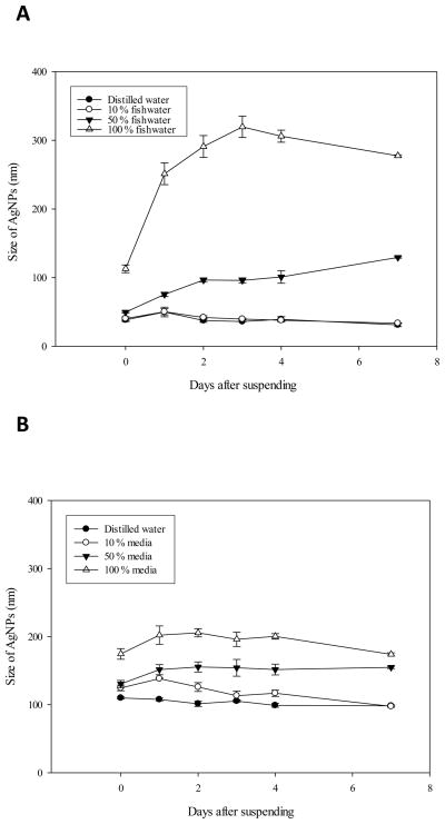
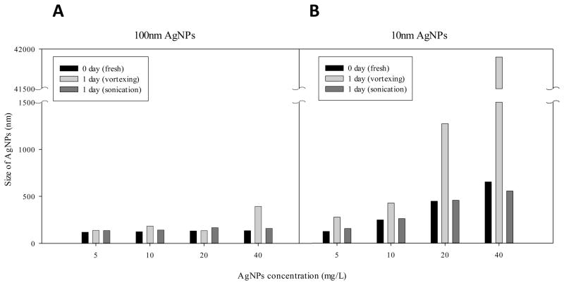
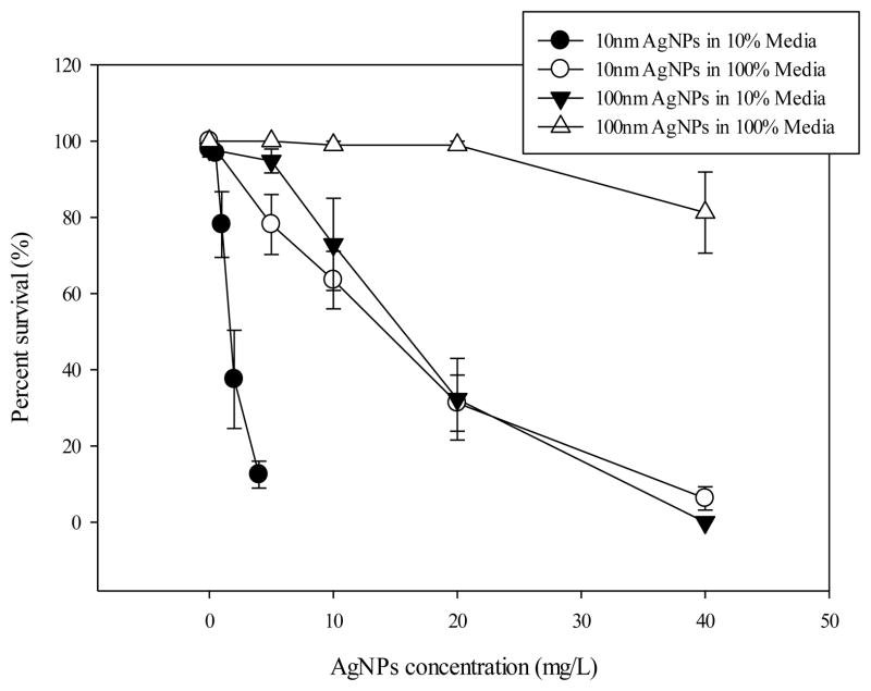
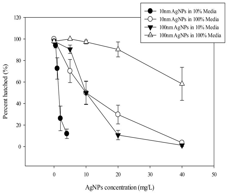
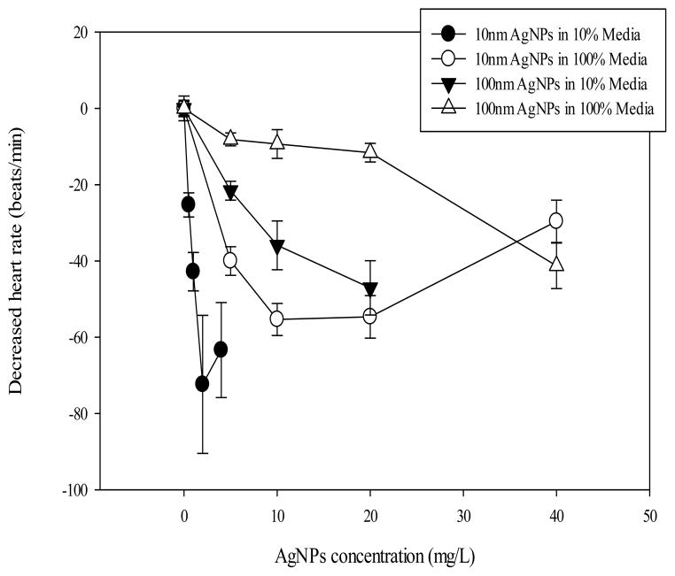
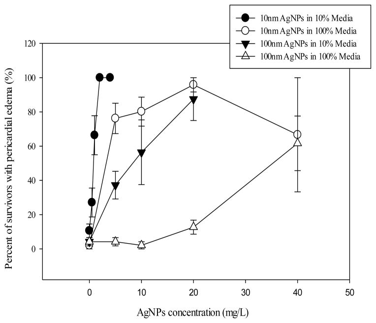
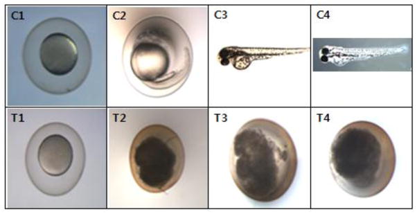
The stability of citrate-capped silver nanoparticles (AgNPs) and the embryonic developmental toxicity were evaluated in the fish test water. Serious aggregation of AgNPs was observed in undiluted fish water (DM-100) in which high concentration of ionic salts exist. However, AgNPs were found to be stable for 7 days in DM-10, prepared by diluting the original fish water (DM-100) with deionized water to 10 %. The normal physiology of zebrafish embryos were evaluated in DM-10 to see if DM-10 can be used as a control vehicle for the embryonic fish toxicity test. As results, DM-10 without AgNPs did not induce any significant adverse effects on embryonic development of zebrafish determined by mortality, hatching, malformations and heart rate. When embryonic toxicity of AgNPs was tested in both DM-10 and in DM-100, AgNPs showed higher toxicity in DM-10 than in DM-100. This means that the big-sized aggregates of AgNPs were low toxic compared to the nano-sized AgNPs. AgNPs induced delayed hatching, decreased heart rate, pericardial edema, and embryo death. Accumulation of AgNPs in the embryo bodies was also observed. Based on this study, citrate-capped AgNPs are not aggregated in DM-10 and it can be used as a control vehicle in the toxicity test of fish embryonic development.
Figures
Similar articles
-
The toxicity of silver nanoparticles to zebrafish embryos increases through sewage treatment processes.Ecotoxicology. 2013 Oct;22(8):1264-77. doi: 10.1007/s10646-013-1114-5. Epub 2013 Aug 22. Ecotoxicology. 2013. PMID: 23975539
-
Toxicity responses of different organs of zebrafish (Danio rerio) to silver nanoparticles with different particle sizes and surface coatings.Environ Pollut. 2019 Mar;246:414-422. doi: 10.1016/j.envpol.2018.12.034. Epub 2018 Dec 13. Environ Pollut. 2019. PMID: 30579210
-
Silver nanoparticles alter zebrafish development and larval behavior: distinct roles for particle size, coating and composition.Neurotoxicol Teratol. 2011 Nov-Dec;33(6):708-14. doi: 10.1016/j.ntt.2011.02.002. Epub 2011 Feb 22. Neurotoxicol Teratol. 2011. PMID: 21315816 Free PMC article.
-
Exploring developmental toxicity of microplastics and nanoplastics (MNPS): Insights from investigations using zebrafish embryos.Sci Total Environ. 2024 Jul 10;933:173012. doi: 10.1016/j.scitotenv.2024.173012. Epub 2024 May 6. Sci Total Environ. 2024. PMID: 38719038 Review.
-
A review on the toxicity of silver nanoparticles against different biosystems.Chemosphere. 2022 Apr;292:133397. doi: 10.1016/j.chemosphere.2021.133397. Epub 2021 Dec 23. Chemosphere. 2022. PMID: 34954197 Review.
Cited by
-
Silver Nanoparticles: Technological Advances, Societal Impacts, and Metrological Challenges.Front Chem. 2017 Feb 21;5:6. doi: 10.3389/fchem.2017.00006. eCollection 2017. Front Chem. 2017. PMID: 28271059 Free PMC article. Review.
-
Injection of ligand-free gold and silver nanoparticles into murine embryos does not impact pre-implantation development.Beilstein J Nanotechnol. 2014 May 21;5:677-88. doi: 10.3762/bjnano.5.80. eCollection 2014. Beilstein J Nanotechnol. 2014. PMID: 24991505 Free PMC article.
-
Mutagenesis and Resistance Development of Bacteria Challenged by Silver Nanoparticles.Antimicrob Agents Chemother. 2022 Oct 18;66(10):e0062822. doi: 10.1128/aac.00628-22. Epub 2022 Sep 12. Antimicrob Agents Chemother. 2022. PMID: 36094196 Free PMC article.
-
The biochemical and toxicological responses of earthworm (Eisenia fetida) following exposure to nanoscale zerovalent iron in a soil system.Environ Sci Pollut Res Int. 2017 Jan;24(3):2507-2514. doi: 10.1007/s11356-016-8001-6. Epub 2016 Nov 7. Environ Sci Pollut Res Int. 2017. PMID: 27822688
-
Oocyte exposure to ZnO nanoparticles inhibits early embryonic development through the γ-H2AX and NF-κB signaling pathways.Oncotarget. 2017 Jun 27;8(26):42673-42692. doi: 10.18632/oncotarget.17349. Oncotarget. 2017. PMID: 28487501 Free PMC article.
References
-
- Ahamed M, Alsalhi MS, Siddiqui MK. Silver nanoparticle applications and human health. Clin Chim Acta. 2010;411:1841–1848. - PubMed
-
- Bar-Ilan O, Albrecht RM, Fako VE, Furgeson DY. Toxicity assessments of multisized gold and silver nanoparticles in zebrafish embryos. Small. 2009;5:1897–1910. - PubMed
-
- Bielmyer GK, Grosell M, Brix KV. Toxicity of silver, zinc, copper, and nickel to the copepod Acartia tonsa exposed via a phytoplankton diet. Environ Sci Technol. 2006;40:2063–2068. - PubMed
-
- Bilberg K, Malte H, Wang T, Baatrup E. Silver nanoparticles and silver nitrate cause respiratory stress in Eurasian perch (Perca fluviatilis) Aquat Toxicol. 2010;96:159–165. - PubMed
Publication types
MeSH terms
Substances
Grants and funding
LinkOut - more resources
Full Text Sources
Other Literature Sources
Research Materials
