. 2013 Mar;87(6):3549-60.
doi: 10.1128/JVI.03219-12. Epub 2013 Jan 16.

Adaptation to the interferon-induced antiviral state by human and simian immunodeficiency viruses

Affiliations

Adaptation to the interferon-induced antiviral state by human and simian immunodeficiency viruses

Julia Bitzegeio et al. J Virol. 2013 Mar.

Erratum in

Abstract

The production of type I interferon (IFN) is an early host response to different infectious agents leading to the induction of hundreds of IFN-stimulated genes (ISGs). The roles of many ISGs in host defense are unknown, but their expression results in the induction of an "antiviral state" that inhibits the replication of many viruses. Here we show that prototype primate lentiviruses human immunodeficiency virus type 1 (HIV-1) and simian immunodeficiency virus of macaques (SIV(MAC) and SIV(MNE)) can replicate in lymphocytes from their usual hosts (humans and macaques, respectively), even when an antiviral state is induced by IFN-α treatment. In contrast, HIV-1 and SIV(MAC)/SIV(MNE) replication was hypersensitive to IFN-α in lymphocytes from unnatural hosts, indicating that the antiviral state can effectively curtail the replication of primate lentiviruses in hosts to which they are not adapted. Most of the members of a panel of naturally occurring HIV-1 and HIV-2 strains behaved like prototype strains and were comparatively insensitive to IFN-α in human lymphocytes. Using chimeric viruses engineered to overcome restriction factors whose antiretroviral specificities vary in a species-dependent manner, we demonstrate that differential HIV-1 and SIV(MAC) sensitivities to IFN-α in lymphocytes from humans and macaques could not be ascribed to TRIM5, APOBEC3, tetherin, or SAMHD1. Single-cycle infection experiments indicated that at least part of this species-specific, IFN-α-induced restriction of primate lentivirus replication occurs early in the retroviral life cycle. Overall, these studies indicate the existence of undiscovered, IFN-α-inducible antiretroviral factors whose spectrum of activity varies in a species-dependent manner and to which at least some HIV/SIV strains have become adapted in their usual hosts.


Figures
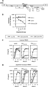
Fig 1
Fig 1
Differential sensitivities of stHIV and SIVMAC239 to IFN-α in huPBMCs and pgtPBMCs. (A) Schematic representation of the stHIV genome. The Vif protein is from SIVMAC239, and the Env protein is from SHIV/KB9. All other sequences are from NL4-3. (B) Equivalent amounts of HIV-1, stHIV, and SIVMAC proviruses expressing GFP were cotransfected with various amounts of rhAPOBEC3G. Supernatants were collected at 48 h posttransfection and assayed for infectivity on CEM cells. (C and D) Activated huPBMCs (B) or pgtPBMCs (C) were infected with stHIV or SIVMAC239/SIVMNE at an MOI of 0.001. At 24 h postinfection, the cells were washed and samples were split into three wells, two of which were treated with 100 or 1,000 U/ml IFN-α. Supernatants were collected every 48 h for the following 15 days, and RT activity was measured with an enzyme-linked immunosorbent assay (Cavidi Tech). LTR, long terminal repeat; p.i., postinfection.
Fig 2
Fig 2
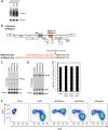
Functional replacement of the stHIV Vpu protein with SIVden Vpu or SIVgsn Vpu. (A) huPBMCs left untreated or treated with 100 or 1,000 U/ml IFN-α for 24 h were lysed and subjected to Western blot analysis with antitetherin and antitubulin antibodies. Note that tetherin is highly and heterogeneously glycosylated and migrates as a smear at 50 to 64 kDa. (B) Schematic representation of the stHIVdenU and stHIVgsnU genomes. The Vpu proteins and Env signal peptides are from SIVden or SIVgsn. All other sequences are from the same stHIV construct described in Fig. 1A. (C) and (D) 293T cells were transfected with stHIV, stHIVΔvpu, stHIVdenU, or stHIVgsnU. At 48 h posttransfection, the virions were harvested, cells were lysed, and both were subjected to Western blot analysis with anti-CA (B) or anti-gp41 (C) antibodies. (E) Virion-containing supernatants were titrated on TZM reporter cells, and infected foci were revealed by X-Gal staining and counted. (F) CEMx174 cells were infected with stHIVΔenvGFP reporter viruses pseudotyped with VSV-G containing the gene for GFP in the place of the gene for Nef. At 48 h postinfection, the cells were fixed and stained for CD4 and analyzed by flow cytometry. LTR, long terminal repeat.
Fig 3
Fig 3
Resistance of stHIVdenU and stHIVgsnU to pigtailed macaque tetherin. (A) Western blot analysis of 293T cells cotransfected with the indicated proviral plasmid and increasing amounts (0, 11, 33, or 100 ng) of a plasmid expressing either human or pigtailed macaque tetherin. Cell and virion lysates were probed with anti-CA monoclonal and/or rabbit anti-HA antibodies, and signals were detected with fluorescent secondary antibodies. (B) Virion-containing supernatants harvested at 48 h postinfection were titrated on TZM reporter cells, and the infectious virion yield was determined with a chemiluminescent β-galactosidase assay. RLU, relative light units.
Fig 4
Fig 4
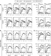
Addition or removal of functional tetherin antagonists does not confer stHIV or SIVMAC239 resistance or sensitivity to IFN-α. (A to D) huPBMCs (A and C) or pgtPBMCs (B and D) were infected with stHIV, stHIVΔvpu, stHIVdenU, stHIVgsnU (A and B), SIVMAC239, or SIVMAC239ΔNef (C and D) at an MOI of 0.001. The next day, cells were washed and divided among three wells that were treated with 0, 100, or 1,000 U/ml IFN-α. Supernatants were collected every 48 h for the following 15 days, and RT activity was measured with an enzyme-linked immunosorbent assay-based assay (Cavidi Tech). Results from two huPBMC and pgtPBMC donors are shown. Note that the virus replication curves in the presence of 100 or 1,000 U/ml IFN-α (gray and open circles) are superimposed in panel B. p.i., postinfection.
Fig 5
Fig 5
Insertion of SIVMAC239 Vpx into the stHIV genome and incorporation into infectious virions. (A) Schematic representation of the stHIVSp6Vpx genome. The Vpx proteins and the Vpx packaging signal within p6 are from SIVMAC239. All other sequences are from the stHIV construct described in Fig. 1A. (B) Western blot analysis of 293T cells and virions transfected with stHIV, stHIVSp6, stHIVVpx, and stHIVSp6Vpx that were harvested at 48 h postinfection. Cell lysates and virions were probed with anti-CA and anti-Vpx monoclonal antibodies, and signals were detected with fluorescent secondary antibodies. (C) Virion-containing supernatants were titrated on TZM reporter cells, and infected foci were revealed by X-Gal staining and counted. LTR, long terminal repeat.
Fig 6
Fig 6
Insertion of Vpx into or its removal from the stHIV or SIVMAC239 genome does not confer resistance or sensitivity to IFN-α. (A to D) huPBMCs (A and C) or pgtPBMCs (B and D) were infected at an MOI of 0.001 with stHIV, stHIVSp6, stHIVVpx, or stHIVSp6Vpx (A and B) or with SIVMAC239 or SIVMAC239ΔVpx (C and D). The next day, cells were washed and divided among three wells that were treated with 0, 100, or 1,000 U/ml IFN-α. Supernatants were collected every 48 h for the following 15 days, and RT activity was measured with an enzyme-linked immunosorbent assay-based assay (Cavidi Tech). Results from two huPBMC and pgtPBMC donors are shown. Note that the virus replication curves in the presence of 100 or 1,000 U/ml of IFN-α (gray and open circles) are superimposed in panels B and C (bottom). p.i., postinfection.
Fig 7
Fig 7
An early, species-dependent but TRIM5-independent block to stHIV and SIVMAC239 infection is induced by IFN-α. (A and B) huPBMCs (A) or pgtPBMCs (B) were treated with the indicated concentrations of IFN-α for 24 h and then infected with SIV-GFP, stHIV-GFP, or stHIV(SCA)-GFP reporter virus in the presence of IFN-α. At 24 h postinfection, the medium was changed and 100 ng/ml 3TC was added. At 48 h postinfection, the cells were fixed and the percentage of GFP-positive cells was determined by flow cytometry. The data represent the mean ± standard deviation of three huPBMC or pgtPBMC donors and are plotted relative to the level of infection in the absence of IFN-α (which was set at 100%).
Fig 8
Fig 8
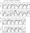
Effects of IFN-α on primary HIV-1 and HIV-2 strain replication in huPBMCs. (A and B) huPBMCs were infected with the indicated HIV-1 (A) or HIV-2 (B) strains at an MOI of 0.001. The next day, cells were washed and divided among three wells that were treated with 0, 100, or 1,000 U/ml IFN-α. Supernatants were collected every 48 h for the following 15 days, and RT activity was measured with an enzyme-linked immunosorbent assay (Cavidi Tech). Results from two huPBMC donors are shown.

Comment in

  • Findings of research misconduct.
    [No authors listed] [No authors listed] NIH Guide Grants Contracts (Bethesda). 2015 Jul 31:NOT-OD-15-131. NIH Guide Grants Contracts (Bethesda). 2015. PMID: 26241998 Free PMC article. No abstract available.
  • Findings of Research Misconduct.
    [No authors listed] [No authors listed] Fed Regist. 2015 Jul 23;80(141):43784. Fed Regist. 2015. PMID: 27737258 Free PMC article. No abstract available.

References

    1. Bowie AG, Unterholzner L. 2008. Viral evasion and subversion of pattern-recognition receptor signalling. Nat. Rev. Immunol. 8:911–922 - PMC - PubMed
    1. Der SD, Zhou A, Williams BR, Silverman RH. 1998. Identification of genes differentially regulated by interferon alpha, beta, or gamma using oligonucleotide arrays. Proc. Natl. Acad. Sci. U. S. A. 95:15623–15628 - PMC - PubMed
    1. Gaines H, von Sydow MA, von Stedingk LV, Biberfeld G, Bottiger B, Hansson LO, Lundbergh P, Sonnerborg AB, Wasserman J, Strannegaard OO. 1990. Immunological changes in primary HIV-1 infection. AIDS 4:995–999 - PubMed
    1. Jacquelin B, Mayau V, Targat B, Liovat AS, Kunkel D, Petitjean G, Dillies MA, Roques P, Butor C, Silvestri G, Giavedoni LD, Lebon P, Barre-Sinoussi F, Benecke A, Muller-Trutwin MC. 2009. Nonpathogenic SIV infection of African green monkeys induces a strong but rapidly controlled type I IFN response. J. Clin. Invest. 119:3544–3555 - PMC - PubMed
    1. Malleret B, Maneglier B, Karlsson I, Lebon P, Nascimbeni M, Perie L, Brochard P, Delache B, Calvo J, Andrieu T, Spreux-Varoquaux O, Hosmalin A, Le Grand R, Vaslin B. 2008. Primary infection with simian immunodeficiency virus: plasmacytoid dendritic cell homing to lymph nodes, type I interferon, and immune suppression. Blood 112:4598–4608 - PubMed

Publication types

Substances

LinkOut - more resources