. 2013 Apr;87(7):3760-73.
doi: 10.1128/JVI.02497-12. Epub 2013 Jan 16.

Simian immunodeficiency virus infects follicular helper CD4 T cells in lymphoid tissues during pathogenic infection of pigtail macaques

Affiliations

Simian immunodeficiency virus infects follicular helper CD4 T cells in lymphoid tissues during pathogenic infection of pigtail macaques

Yin Xu et al. J Virol. 2013 Apr.

Abstract

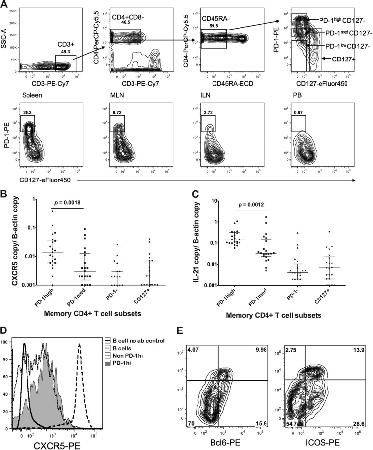
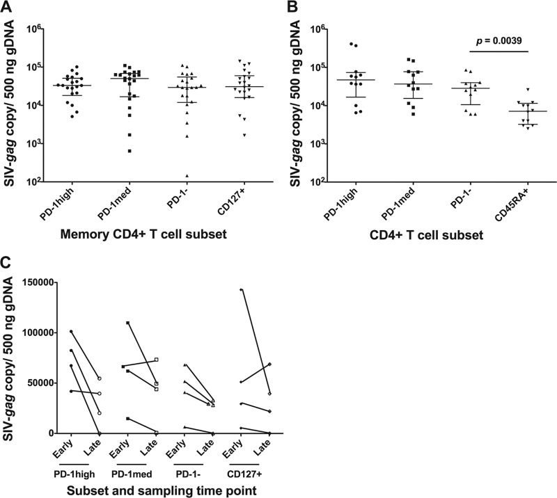
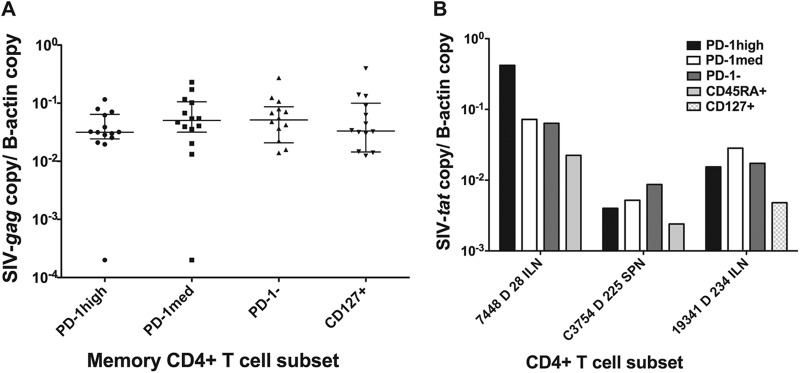
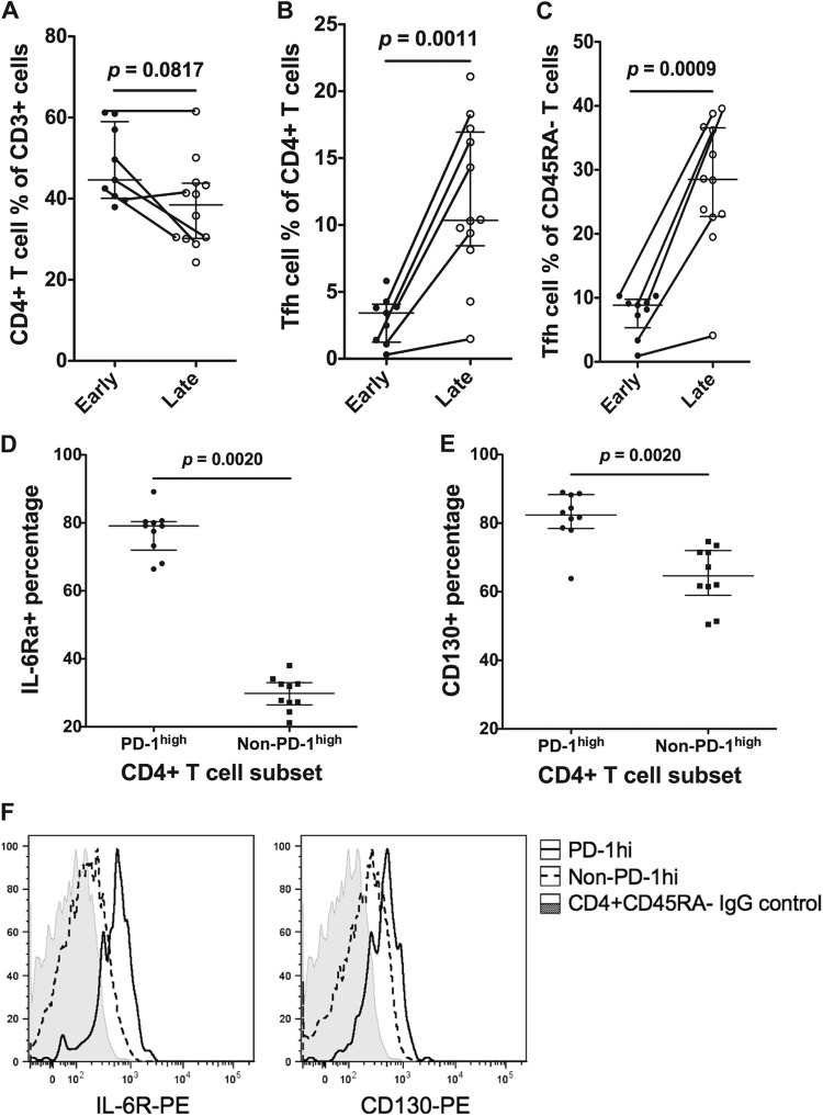
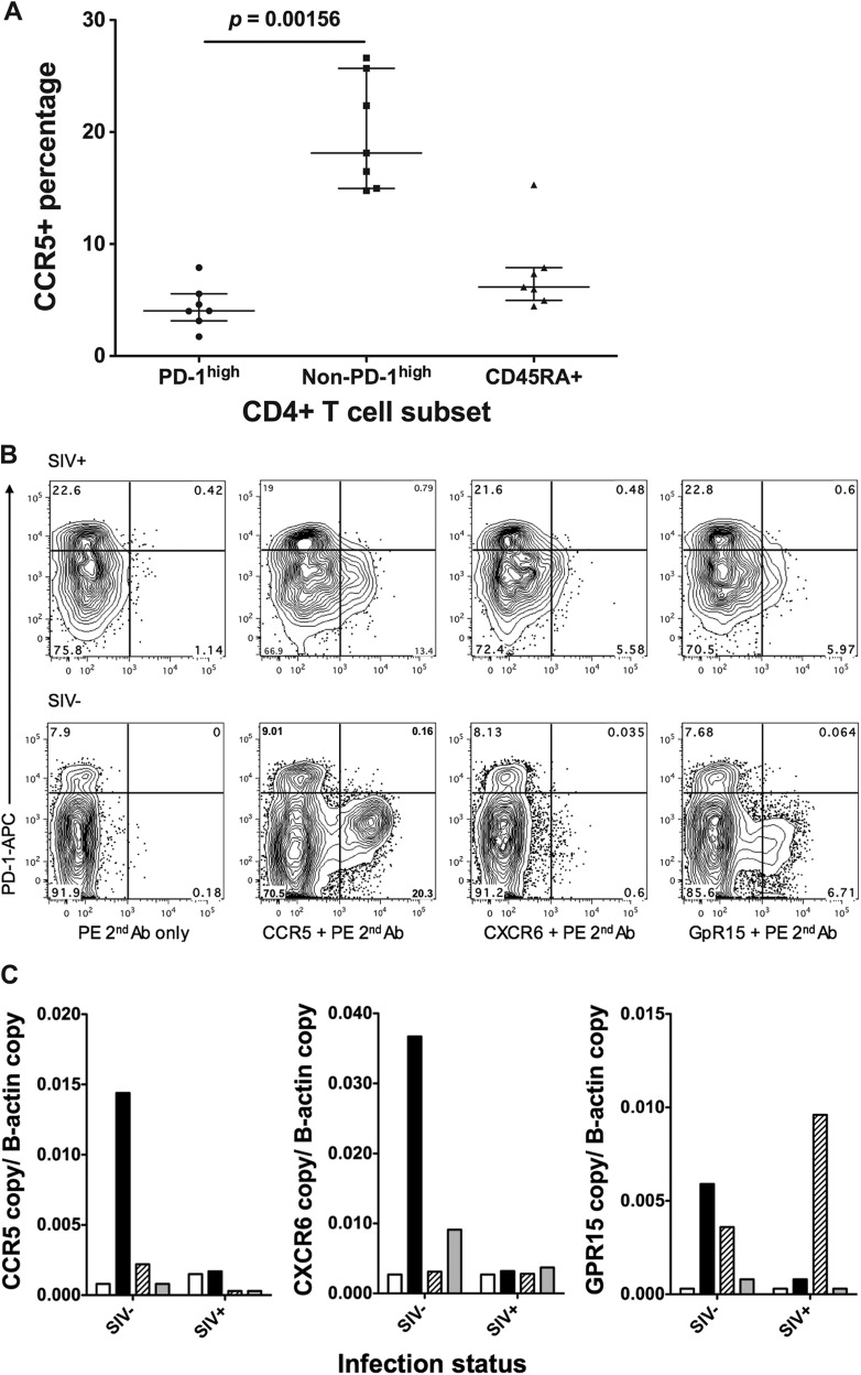
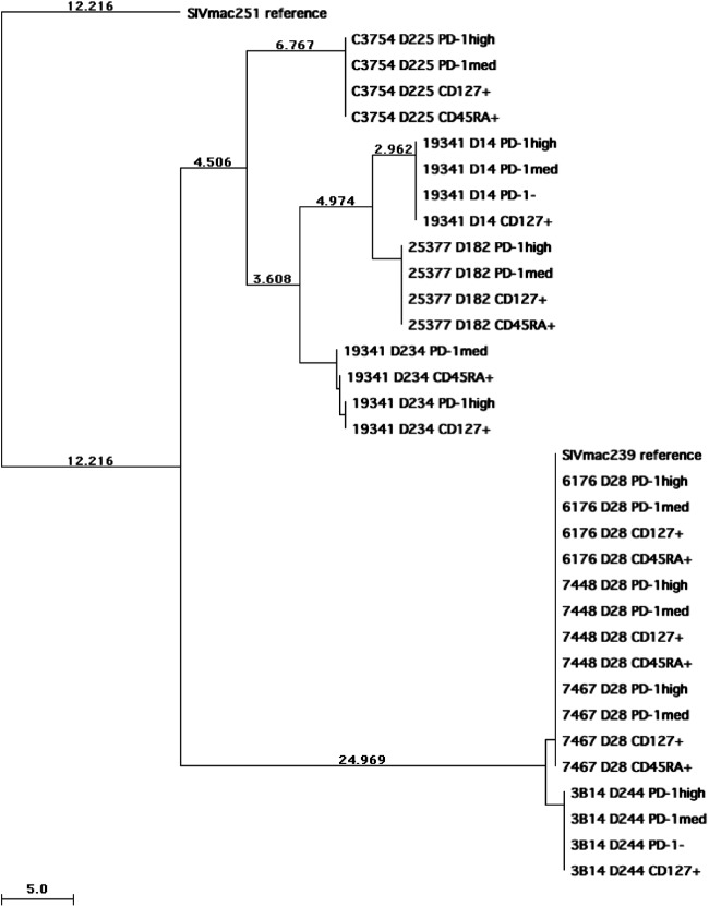
T follicular helper (Tfh) cells are a specialized subset of memory CD4(+) T cells that are found exclusively within the germinal centers of secondary lymphoid tissues and are important for adaptive antibody responses and B cell memory. Tfh cells do not express CCR5, the primary entry coreceptor for both human immunodeficiency virus type 1 (HIV-1) and simian immunodeficiency virus (SIV), and therefore, we hypothesized that these cells would avoid infection. We studied lymph nodes and spleens from pigtail macaques infected with pathogenic strain SIVmac239 or SIVmac251, to investigate the susceptibility of Tfh cells to SIV infection. Pigtail macaque PD-1(high) CD127(low) memory CD4(+) T cells have a phenotype comparable to that of human Tfh cells, expressing high levels of CXCR5, interleukin-21 (IL-21), Bcl-6, and inducible T cell costimulator (ICOS). As judged by either proviral DNA or cell-associated viral RNA measurements, macaque Tfh cells were infected with SIV at levels comparable to those in other CD4(+) memory T cells. Infection of macaque Tfh cells was evident within weeks of inoculation, yet we confirmed that Tfh cells do not express CCR5 or either of the well-known alternative SIV coreceptors, CXCR6 and GPR15. Mutations in the SIV envelope gp120 region occurred in chronically infected macaques but were uniform across each T cell subset investigated, indicating that the viruses used the same coreceptors to enter different cell subsets. Early infection of Tfh cells represents an unexpected focus of viral infection. Infection of Tfh cells does not interrupt antibody production but may be a factor that limits the quality of antibody responses and has implications for assessing the size of the viral reservoir.


Figures

Fig 1
Fig 1
Identification of Tfh cells in pigtail macaques. (A, top) Representative plots acquired from a BD FACSAria cell sorter showing the gating strategy for sorting of putative Tfh cells. Lymphocytes were first gated based on forward and side scatter (not shown) and then displayed on the first plot (SSC, side scatter). CD3+ T cells were subsequently gated as CD45RA memory cells and then displayed as PD-1–PE versus CD127-eFluor 450 (top right). (Bottom) Representative PD-1-versus-CD127 plots, acquired by using a BD LSRII flow cytometer, for memory CD4+ T cells from macaque spleen, mesenteric lymph node, inguinal lymph node, and peripheral blood, from left to right. PD-1high CD127low cells were present at high levels in lymphoid tissues, but they were rare in peripheral blood. (B) Significantly higher copy numbers of CXCR5 mRNA, normalized to β-actin copy numbers, in purified PD-1high CD127low putative Tfh cells than in other subsets of lymphoid tissue memory CD4+ T cells, sorted using gating as depicted in panel A, top (n = 21). (C) Significantly higher copy numbers of IL-21 mRNA, normalized to β-actin copy numbers, in purified PD-1high CD127low putative Tfh cells than in other subsets of lymphoid tissue memory CD4+ T cells, sorted using gating as depicted in panel A, top (n = 21). (D) PD-1high CD127low putative Tfh cells are CXCR5+ by flow cytometry, with intermediate expression compared to B cells and a higher mean fluorescence intensity than other memory CD4+ T cells. The no-antibody control (no ab) shows negative staining of B cells. Results are representative of two independent experiments. (E) PD-1high CD127low putative Tfh cells are both Bcl-6+ (left) and ICOS+ (right) by flow cytometry. Data are representative of two experiments.
Fig 2
Fig 2
SIV-gag proviral DNA copy numbers normalized to gDNA input. (A) SIV-gag proviral DNA levels in the four memory CD4+ T cell subsets, sorted as described in the legend of Fig. 1A. Twenty-two samples from nine infected macaques were sorted. (B) SIV-gag proviral DNA levels in three memory CD4+ T cell subsets, PD-1high CD127low, PD-1med CD127low, and PD-1low CD127low, and in naïve CD4+ T cells were obtained from 12 samples from 6 infected macaques. Naïve CD4+ T cells had significantly lower SIV-gag proviral DNA levels than the other three memory CD4+ T cell subsets. (C) Longitudinal data from ILN samples from 4 macaques taken at an earlier time point (14 to 28 days) and then again at a later time point (77 to 469 days).
Fig 3
Fig 3
(A) SIV-gag cell-associated RNA copy numbers normalized to β-actin copy numbers. There was no significant difference (n = 14) among the 4 memory CD4+ T cell subsets. (B) Spliced-tat RNA copy numbers normalized to β-actin copy numbers from three macaque samples. Spliced-tat levels in cells from another three macaque samples were above the limit of detection but below the limit of quantification and are not shown.
Fig 4
Fig 4
CD4+ T cell and Tfh cell frequencies during the early stage (≤28 days postinfection) and late stage (≥77 days postinfection) in ILNs. Lines connecting two data points indicate sequential ILN biopsy specimens from the same macaque. (A) The CD4+ T cell proportion as a percentage of CD3+ T cells decreased in ILNs from the early stage (n = 9) to the late stage (n = 12) but was not statistically significant. (B) The Tfh cell proportion as a percentage of CD4+ T cells significantly increased in ILNs from the early stage (n = 9) to the late stage (n = 12). (C) The Tfh cell proportion as a percentage of memory (CD45RA) T cells significantly increased in ILNs from the early stage (n = 9) to the late stage (n = 12). (D) In memory CD4+ T cells, the PD-1high subset expressed significantly higher levels of IL-6Rα than the non-PD-1high subset (n = 10). (E) In memory CD4+ T cells, the PD-1high subset expressed significantly higher levels of CD130 than the non-PD-1high subset (n = 10). (F) Representative staining of CD126 (left) and CD130 (right) on macaque memory CD4+ T cells. The IgG staining control is shown as shaded histograms, PD-1high CD127low cells are shown as solid lines, and the remaining memory CD4+ T cells are shown as dashed lines. Results are representative of 10 experiments.
Fig 5
Fig 5
Expression levels of CCR5, CXCR6, and GPR15 determined by flow cytometry and RT-qPCR. (A) CCR5 protein levels on CD4+ T cell subsets on macaque tissues (n = 7), measured by flow cytometry. (B) Representative plots for CCR5 (second panel), CXCR6 (third panel), and GPR15 (far right) staining on memory CD4+ T cells from SIV+ (top) and SIV-negative (bottom) macaque tissues. Plots on the left show negative controls, in which the first antibody was not added. (C) mRNA levels of CCR5 (left), CXCR6 (middle), and GPR15 (right) on CD4+ T cell subsets measured by RT-qPCR (white bars, PD-1high; black bars, PD-1med; patterned bars, CD127+; gray bars, CD45RA+).
Fig 6
Fig 6
Neighbor-joining tree of gp120 sequences from proviral DNA isolated from CD4+ T cell subsets (PD-1high CD127low, PD-1med CD127low, PD-1 CD127low, and CD127+ memory CD4+ T cells as well as CD45RA+ naïve CD4+ T cells) of macaques infected with SIVmac239 or SIVmac251. SIVmac239 and SIVmac251 reference sequences were downloaded from GenBank, and SIVmac251 was assigned to be the root of the tree. The sample name shows the macaque identification, days postinoculation, and subset name in order. Branches are scaled in absolute numbers of differences according to the scales at the bottom of the trees.
Fig 7
Fig 7
Anti-SIV antibody production in 13 SIV-infected macaques. (A) Five macaques were euthanized within 6 months postinfection. Antibody levels reached maximum at the time of euthanasia. (B) Four macaques, monitored for longer than 6 months postinfection, had antibody levels that were still increasing at the time of euthanasia. (C) Four macaques, monitored for longer than 6 months postinfection, had antibody levels that reached a maximum between days 35 and 97 postinfection and then decreased by the time of euthanasia. OD, optical density.

Similar articles

Cited by

References

    1. King C. 2009. New insights into the differentiation and function of T follicular helper cells. Nat. Rev. Immunol. 9:757–766 - PubMed
    1. Crotty S. 2011. Follicular helper CD4 T cells (TFH). Annu. Rev. Immunol. 29:621–663 - PubMed
    1. Cooper DA, Imrie AA, Penny R. 1987. Antibody response to human immunodeficiency virus after primary infection. J. Infect. Dis. 155:1113–1118 - PubMed
    1. Tindall B, Cooper DA. 1991. Primary HIV infection: host responses and intervention strategies. AIDS 5:1–14 - PubMed
    1. Fiebig EW, Wright DJ, Rawal BD, Garrett PE, Schumacher RT, Peddada L, Heldebrant C, Smith R, Conrad A, Kleinman SH, Busch MP. 2003. Dynamics of HIV viremia and antibody seroconversion in plasma donors: implications for diagnosis and staging of primary HIV infection. AIDS 17:1871–1879 - PubMed

Publication types

MeSH terms

LinkOut - more resources