. 2013 Feb;140(4):906-15.
doi: 10.1242/dev.087148. Epub 2013 Jan 16.

Axon tracts guide zebrafish facial branchiomotor neuron migration through the hindbrain

Affiliations

Axon tracts guide zebrafish facial branchiomotor neuron migration through the hindbrain

Sarah J Wanner et al. Development. 2013 Feb.

Abstract

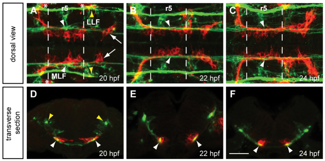
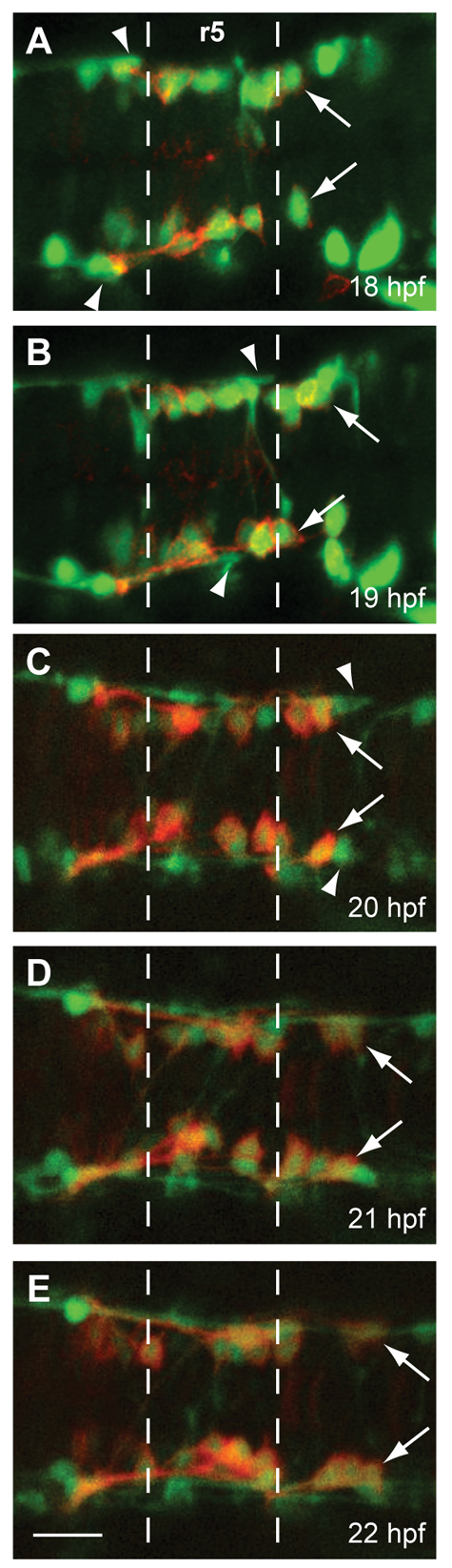
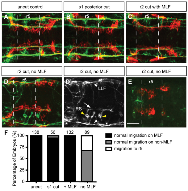
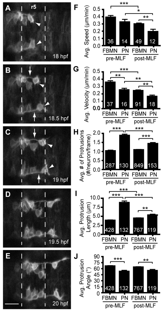
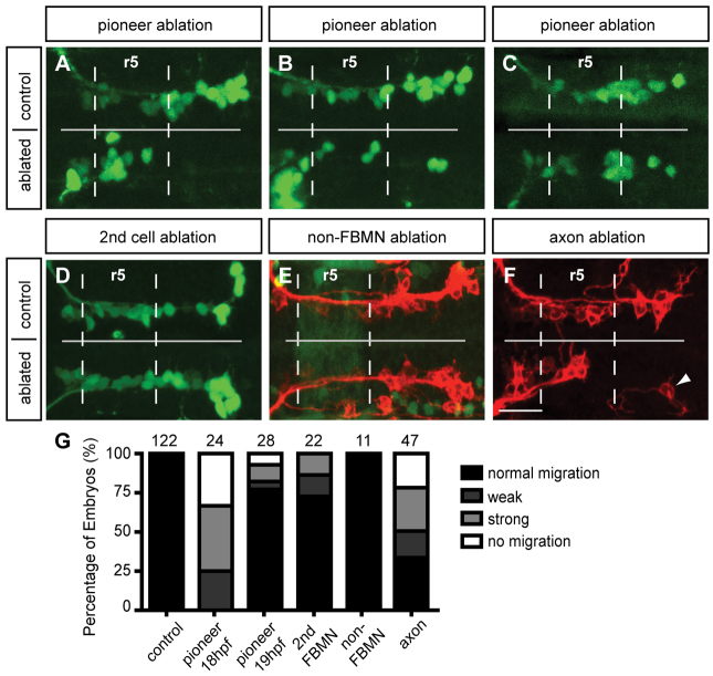
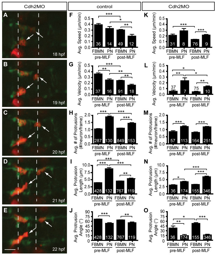
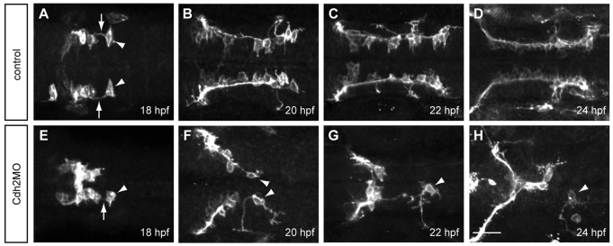
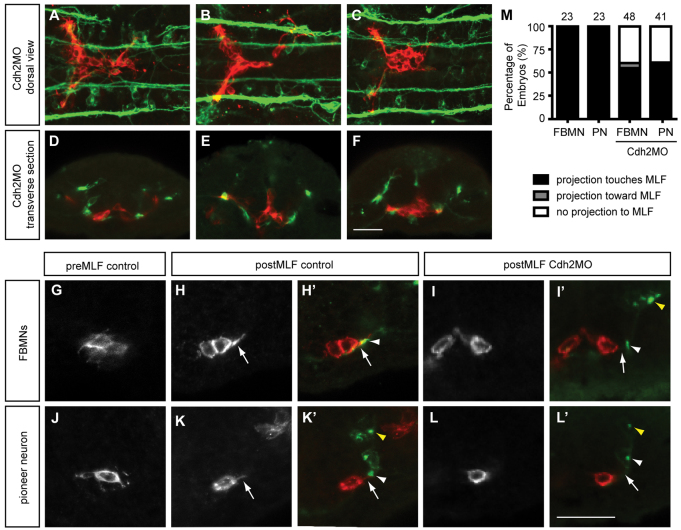
Appropriate localization of neurons within the brain is a crucial component of the establishment of neural circuitry. In the zebrafish hindbrain, the facial branchiomotor neurons (FBMNs) undergo a chain-like tangential migration from their birthplace in rhombomere (r) 4 to their final destination in r6/r7. Here, we report that ablation of either the cell body or the trailing axon of the leading FBMN, or 'pioneer' neuron, blocks the migration of follower FBMNs into r5. This demonstrates that the pioneer neuron and its axon are crucial to the early migration of FBMNs. Later migration from r5 to r6 is not dependent on pioneer neurons but on the medial longitudinal fasciculus (MLF), a bundle of axons lying ventral to the FBMNs. We find that MLF axons enter r5 only after the pioneer neuron has led several followers into this region; the MLF is then contacted by projections from the FBMNs. The interactions between FBMNs and the MLF are important for migration from r5 to r6, as blocking MLF axons from entering the hindbrain can stall FBMN migration in r5. Finally, we have found that the adhesion molecule Cdh2 (N-cadherin) is important for interactions between the MLF and FBMNs, as well as for interactions between the trailing axon of the pioneer neuron and follower FBMNs. Interestingly, migration of pioneer neurons is independent of both the MLF and Cdh2, suggesting pioneer migration relies on independent cues.


Figures

Fig. 1.
Fig. 1.
FBMNs migrate in close apposition to the MLF. (A-C) Maximum projection dorsal views of Tg(zCREST1:membRFP) embryos immunostained using zn-12 antibody show that FBMNs lie in close proximity to the MLF throughout their migration. zn-12 (green) labels the MLF (white arrowheads) and lateral longitudinal fasciculus (LLF, yellow arrowheads). (A) At 20 hpf, FBMNs (red) have migrated into r5 and the first FBMNs to migrate have reached r6 (arrows). (B) By 22 hpf, migration has continued with more FBMNs reaching r6. (C) At 24 hpf, FBMN migration into r6 continues. (D-F) Transverse sections of zCREST1:membRFP embryos at r5 stained using zn-12 show direct interaction between the MLF and FBMNs (white arrowheads) throughout FBMN migration at (D) 20 hpf, (E) 22 hpf and (F) 24 hpf. Asterisks highlight the otic vesicle. Broken lines indicate r5 boundaries for all figures. Scale bar: 20 μm.
Fig. 2.
Fig. 2.
FBMN migration into r5 and r6 precedes the MLF entering the r4-r6 hindbrain region. Time-lapse imaging of Tg(zCREST1:membRFP/HuC-GFP) embryos from 18-22 hpf. (A-E) Still images taken at 1-hour intervals from two representative time-lapse movies: (A,B) a 18-20 hpf movie; (C-E) a 20-22 hpf movie. Arrows indicate the first FBMN to migrate. Arrowheads indicate the growth cone of the first MLF axon to enter the hindbrain. Scale bar: 20 μm.
Fig. 3.
Fig. 3.
Loss of MLF axons in the hindbrain blocks FBMN migration. Maximum projection dorsal views of FBMNs (red) and the MLF (green) in 22 hpf Tg(zCREST1:membRFP) embryos stained using zn-12 antibody. (A) Uncut control embryos at 22 hpf show that FBMNs migrate in close proximity to the MLF. (B) Cuts made posterior to the hindbrain at the first somite (s1) do not affect FBMN migration. (C) Incomplete severing at r2 allows normal MLF axon and FBMN migration. (D) Severing of the embryo at r2 often results in FBMN migration into r6 on the LLF or projections of the reticulospinal (RS) neurons. (D′) zn-12 staining labels the LLF (white arrowheads), and the RS neurons (yellow arrowheads) and their projections (arrow). (E) Severing the MLF causes FBMN migration to stall in r5. (F) Percentage of embryos affected by manipulations. The number of embryos scored is indicated for each manipulation. Scale bar: 20 μm.
Fig. 4.
Fig. 4.
The pioneer neuron behaves differently than follower FBMNs. (A-E) Still images from time-lapse imaging of Tg(zCREST1:membRFP) embryos from 18-22 hpf shown at 30-minute intervals. Arrowheads indicate the pioneer neuron and arrows indicate the trailing axon of the pioneer neuron. (F-J) Analysis of pioneer and follower FBMN behavior before (pre-MLF) and after (post-MLF) the MLF has entered the hindbrain region. (F) Average speed of migration. (G) Average posterior velocity. (H) Average number of protrusions per neuron per 5-minute time point. (I) Average length of protrusions per 5-minute time point. (J) Average angle of protrusions, where 0° is towards the posterior in the direction of migration and 90° is towards the midline. Error bars indicate s.e.m. Significance values: *P<0.05, **P<0.01, ***P<0.001. Scale bar: 20 μm.
Fig. 5.
Fig. 5.
Pioneer neuron ablation blocks migration of follower FBMNs. (A-C) Maximum projection images of Tg(islet1:GFP) (green) embryos at 24 hpf, after ablation of the pioneer neuron on one side of the embryo at 18-19 hpf. (A) Pioneer neuron ablation blocks follower FBMN migration. (B) Strong partial migration phenotype results in two to four FBMNs reaching r6. (C) Weak partial migration phenotype results in migration of five to eight FBMNs into r6. (D) Ablation of the second FBMN to migrate does not affect FBMN migration in most embryos. (E) Tg(zCREST1:membRFP/pGFP5.3) embryos label FBMNs (red) and neuroepithelial cells in r5 (green), respectively. Ablation of a single r5 neuroepithelial cell in the path of FBMN migration does not affect migration. (F) Ablation of the trailing axon of the pioneer neuron, using Tg(zCREST1:membRFP) embryos, blocks FBMN migration; arrowhead indicates presumed pioneer neuron. (G) Percentage of embryos affected by ablation. Scale bar: 20 μm.
Fig. 6.
Fig. 6.
Pioneer neuron migration is independent of Cdh2-mediated cell adhesion. (A-E) Still images from a representative time-lapse movie of Tg(zCREST1:membRFP/HuC-GFP) embryos injected with Cdh2MO from 18-22 hpf, shown at 1-hour intervals. Arrows indicate the pioneer neurons. (F-J) Analysis of pioneer and follower FBMN behavior before and after the MLF has entered the hindbrain region in uninjected controls. (K-O) Analysis of Cdh2MO time-lapse data comparing pioneer neuron behavior with follower FBMNs before and after the MLF is present. (F,K) Average speed. (G,L) Average posterior velocity. (H,M) Average number of protrusions per neuron per 5-minute time point. (I,N) Average protrusion length per 5-minute time point. (J,O) Average protrusion angle. Error bars indicate s.e.m. *P<0.05, **P<0.01, ***P<0.001. Scale bar: 20 μm.
Fig. 7.
Fig. 7.
Cdh2 is required for maintaining the interaction between the pioneer trailing axon and follower FMBNs. Maximum projection images of Tg(zCREST1:membRFP) embryos. (A-D) In uninjected control embryos at (A) 18 hpf, (B) 20 hpf, (C) 22 hpf and (D) 24 hpf, the trailing axon (arrows) of the pioneer neuron (arrowheads) maintains contact with follower FBMNs. (E-H) In Cdh2MO-injected embryos, the interaction between the trailing axon of the pioneer neuron and follower FBMNs is reduced or absent; examples at (E) 18 hpf, (F) 20 hpf, (G) 22 hpf and (H) 24 hpf. Arrowheads indicate the pioneer neuron. Arrow indicates the trailing axon. Scale bar: 20 μm.
Fig. 8.
Fig. 8.
Depletion of Cdh2 reduces interactions between the FBMNs and MLF. Maximum projections of Tg(zCREST1:membRFP) embryos stained using zn-12. (A-C) Dorsal views of Cdh2MO-injected embryos at (A) 20 hpf, (B) 22 hpf and (C) 24 hpf show reduced interaction between FBMNs (red) and the MLF (green). (D-F) Transverse sections of Cdh2-depleted embryos at r5 show decreased interactions between the MLF and FBMNs at (D) 20 hpf, (E) 22 hpf and (F) 24 hpf. (G-L′) High-magnification images of transverse sections at 19.5 hpf. Yellow arrowheads indicate the LLF. (G) Before the MLF is present, FBMNs send projections in random directions. (H,H′) Once the MLF (white arrowhead) is present, FBMNs send a projection to the MLF (arrow). (I,I′) In Cdh2-depleted embryos, FBMNs in the midline do not send projections to the MLF. (J) The pioneer neuron, before the MLF has entered r5, projects randomly. (K,K′) Once the MLF is present, the pioneer neuron sends a projection towards it. (L,L′) In Cdh2-depleted embryos, the pioneer has reduced interactions with the MLF. (M) Percentage of embryos with neural projections contacting the MLF. Scale bar: 20 μm.

Similar articles

Cited by

References

    1. Bate C. M. (1976). Pioneer neurones in an insect embryo. Nature 260, 54-56 - PubMed
    1. Bingham S., Higashijima S., Okamoto H., Chandrasekhar A. (2002). The Zebrafish trilobite gene is essential for tangential migration of branchiomotor neurons. Dev. Biol. 242, 149-160 - PMC - PubMed
    1. Bingham S. M., Toussaint G., Chandrasekhar A. (2005). Neuronal development and migration in zebrafish hindbrain explants. J. Neurosci. Methods 149, 42-49 - PMC - PubMed
    1. Carreira-Barbosa F., Concha M. L., Takeuchi M., Ueno N., Wilson S. W., Tada M. (2003). Prickle 1 regulates cell movements during gastrulation and neuronal migration in zebrafish. Development 130, 4037-4046 - PubMed
    1. Chandrasekhar A. (2004). Turning heads: development of vertebrate branchiomotor neurons. Dev. Dyn. 229, 143-161 - PMC - PubMed

Publication types