. 2013 Feb;140(4):751-64.
doi: 10.1242/dev.090159. Epub 2013 Jan 16.

Spatiotemporal patterns of multipotentiality in Ptf1a-expressing cells during pancreas organogenesis and injury-induced facultative restoration

Affiliations

Spatiotemporal patterns of multipotentiality in Ptf1a-expressing cells during pancreas organogenesis and injury-induced facultative restoration

Fong Cheng Pan et al. Development. 2013 Feb.

Abstract

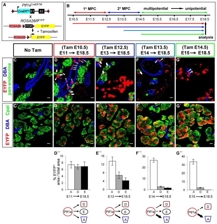
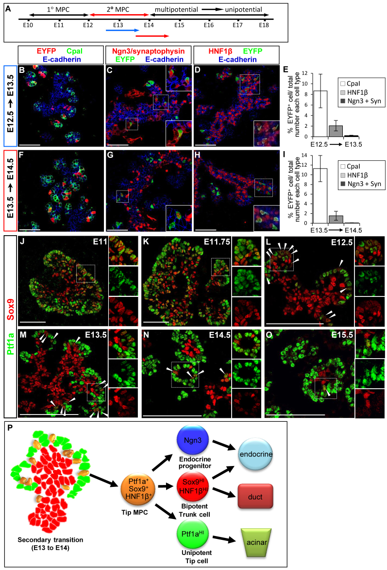
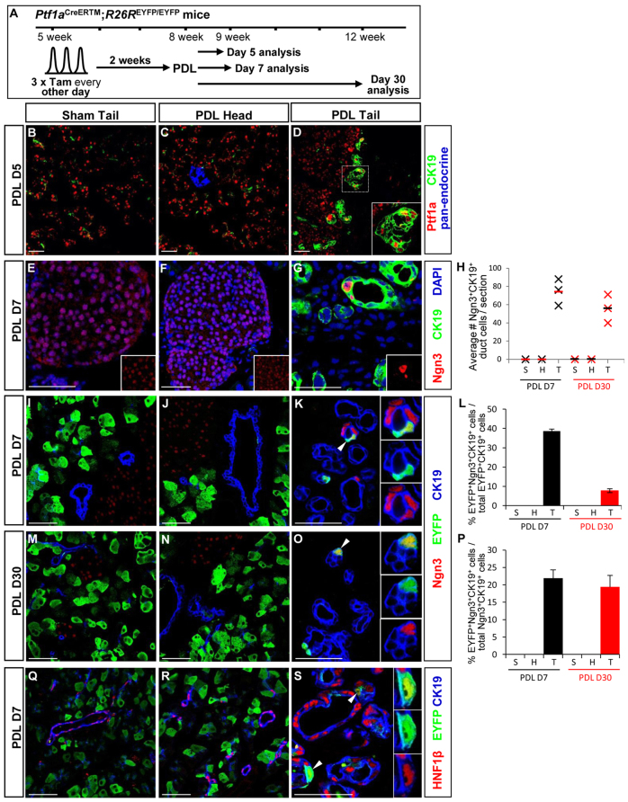
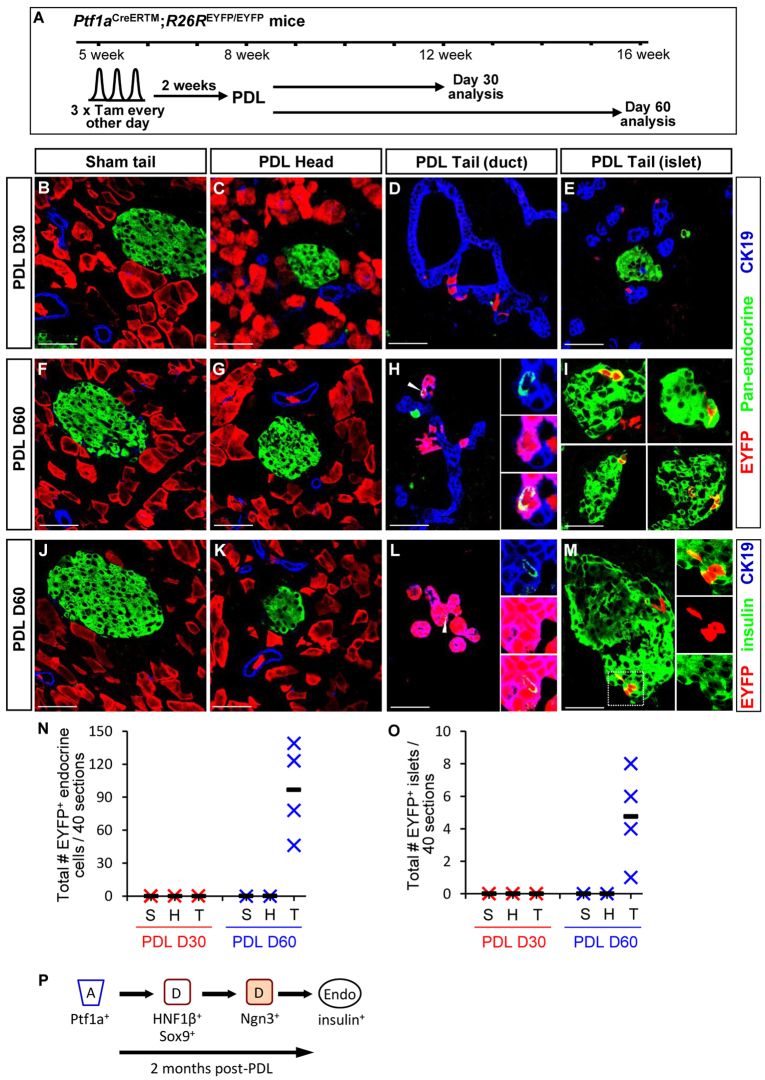
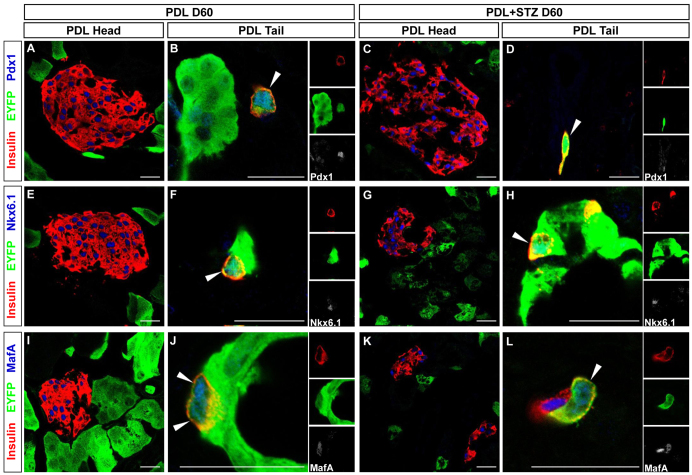
Pancreatic multipotent progenitor cells (MPCs) produce acinar, endocrine and duct cells during organogenesis, but their existence and location in the mature organ remain contentious. We used inducible lineage-tracing from the MPC-instructive gene Ptf1a to define systematically in mice the switch of Ptf1a(+) MPCs to unipotent proacinar competence during the secondary transition, their rapid decline during organogenesis, and absence from the mature organ. Between E11.5 and E15.5, we describe tip epithelium heterogeneity, suggesting that putative Ptf1a(+)Sox9(+)Hnf1β(+) MPCs are intermingled with Ptf1a(HI)Sox9(LO) proacinar progenitors. In the adult, pancreatic duct ligation (PDL) caused facultative reactivation of multipotency factors (Sox9 and Hnf1β) in Ptf1a(+) acini, which undergo rapid reprogramming to duct cells and longer-term reprogramming to endocrine cells, including insulin(+) β-cells that are mature by the criteria of producing Pdx1(HI), Nkx6.1(+) and MafA(+). These Ptf1a lineage-derived endocrine/β-cells are likely formed via Ck19(+)/Hnf1β(+)/Sox9(+) ductal and Ngn3(+) endocrine progenitor intermediates. Acinar to endocrine/β-cell transdifferentiation was enhanced by combining PDL with pharmacological elimination of pre-existing β-cells. Thus, we show that acinar cells, without exogenously introduced factors, can regain aspects of embryonic multipotentiality under injury, and convert into mature β-cells.


Figures

Fig. 1.
Fig. 1.
Lineage tracing Ptf1a-expressing cells. (A) Tam-dependent Ptf1aCreERTM recombination of R26REYFP irreversibly labels Ptf1a-expressing cells and progeny. (B) Color-coded E10.5-15.5 lineage-tracing scheme. (C,C′) Ptf1aCreERTM recombination is strictly Tam-dependent. (D-F′) Ptf1a-expressing cells pulse-labeled at (D,D′) E11, (E,E′) E13 or (F,F′) E14 produced endocrine, duct and acinar cells in various proportions (arrow: EYFP+ endocrine+ cells; open arrowhead: EYFP+DBA+ duct cells). (G,G′) E15 labeling produced acini and small numbers of duct cells. (D″-G″) Histogram representation of EYFP+ cells in (A, acinar; D, duct; E, endocrine) compartments after Tam labeling at E11 (D″; n=4), E13 (E″; n=3), E14 (F″; n=5), and E15 (G″; n=3). Quantitation is total EYFP+ area of each cell type over total area of each cell type. Scale bars: 20 μm.
Fig. 2.
Fig. 2.
Ptf1a+ tip cell contribution to trunk and endocrine progenitor/precursor compartment, and heterogeneity in 2°-transition tip epithelium (with putative Ptf1aLO MPC and Ptf1aHI proacinar cells). (A) Short-term lineage-tracing schematic. (B-D,F-H) EYFP-labeled CpaI+ tip cells, Hnf1β+ trunk cells, and Ngn3+/Synaptophysin+ (Syn) endocrine precursors were detected 24 hours post-labeling of Ptf1a-expressing cells at (B-D) E12.5 or (F-H) E13.5. (E,I) Percentage EYFP+ cells in each lineage with labeling at E12.5 (E; n=4) or E13.5 (I; n=4). (J-O) Ptf1a/Sox9 immunodetection between E11-15.5 showing tip epithelium with two Ptf1a+ populations: Ptf1a+Sox9+ putative MPC (arrowhead), and Ptf1aHISox9LO proacinar progenitors. Numbers of Ptf1a+Sox9+ cells decreased rapidly as organogenesis progressed. (P) Schematic, lineage potency of Ptf1a+Sox9+Hnf1β+ tip MPC during the 2° transition. Scale bars: 50 μm.
Fig. 3.
Fig. 3.
Ptf1a-expressing cells are acinar-restricted after birth. (A) Postnatal/adult lineage-tracing schematic. (B,C) Ptf1aCreERTM;R26REYFP embryos Tam-pulsed at E18.5, analyzed 3 weeks after birth showing EYFP exclusively in acini. (D) P21; percentage of each pancreatic cell type expressing EYFP. (E,F,H,I) 1-month-old Ptf1aCreERTM;R26REYFP mice treated with Tam were chased for (E,F) 1 week or (H,I) 6 months; EYFP was only in CpaI+ acinar cells. (G,J) Comparing EYFP+ traced cells at P36 (G) or 7 months (J) showed similar acinar labeling, suggesting homeostatic acinar self-replication. (K-L) EYFP was absent from (K,K′) Ck19+ or (L) Hnf1β+ CAC cells after 6-month chase; diagrammed as Ptf1a-expressing cells not producing CACs. (M-M‴) Ptf1a protein was undetectable in Sox9+Ck19+ CACs. Scale bars: 50 μm.
Fig. 4.
Fig. 4.
PDL-induced acinar-to-ductal transdifferentiation and de novo Ngn3 activation. (A) PDL lineage-tracing schematic. (B-F) At D5, Ptf1a protein was found only in acini in (B) sham tail and (C) PDL head, but was in (D) PDL tail duct cells. Ngn3 was found only in islets of (E) sham tail or (F) PDL head. (G) De novo activation of Ngn3 in Ck19+ duct cells, PDL tail. (H) Quantification, average number Ngn3+Ck19+ duct cells per section, PDL D7 and D30 (∼10-15 sections entirely counted per PDL tail; n=3); solid line indicates mean. (I,J,M,N) EYFP+Ngn3+ cells were absent from sham tail and PDL head at (I,J) PDL D7 or (M,N) D30. (K,O) A small fraction of the Ngn3+Ck19+ duct cells were derived from Ptf1a-lineage-labeled cells (EYFP+, arrowhead) in PDL tail at (K) PDL D7 and (O) D30. (L,P) Quantification of (L) percentage of EYFP+Ck19+ cells that were also Ngn3+, and (P) percentage of EYFP+Ngn3+Ck19+ cells over total Ngn3+Ck19+ cells at PDL D7 and D30. (Q,R) EYFP+Ck19+Hnf1β+ duct cells were absent from (Q) sham tail or (R) PDL head. (S) Ptf1a-lineage-derived ducts (EYFP+Ck19+) were Hnf1β+ (arrowhead) in PDL tail at PDL D7, indicating acinar-to-ductal transdifferentiation. Scale bars: 50 μm.
Fig. 5.
Fig. 5.
Ptf1a+ acini transdifferentiate into endocrine cells, likely via Ck19+ ductal intermediates, 2 months post-PDL. (A) Lineage-tracing schematic. (B-M) EYFP+ cells were absent from duct and endocrine compartment in (B) sham tail, and (C) PDL head, at post-PDL D30. Ptf1a-lineage-labeled cells (EYFP+) were detected in (D) Ck19+ ducts, but not (E) endocrine cells in PDL tail at PDL D30. At post-PDL D60, while EYFP+ cells were found only in acini in (F,J) sham tail or (G,K) PDL head, EYFP+hormone+ cells were detected (H) in or closely apposed to Ck19+ ducts, and (I) in islets in PDL tail. Some of these cells are insulin+ in (L) ducts and (M) islets. (N,O) Total number of (N) EYFP+ endocrine cells and (O) EYFP+ islets (40 sections, ∼10% of PDL tail or head; n=4) at PDL D30 and D60; solid line indicates mean. (P) Summary of PDL lineage-tracing results. S, sham tail; H,T: PDL head, tail. Scale bars: 50 μm.
Fig. 6.
Fig. 6.
[PDL+STZ] enhances acinar-to-endocrine transdifferentiation. (A) [PDL+STZ] lineage-tracing schematic. (B-M) At [PDL+STZ] D30, no EYFP+ cells were present in duct or endocrine compartment in either (B) sham tail or (C) PDL head. EYFP+hormone+ cells were detected 30 days post-[PDL+STZ] in (D) the vicinity of ducts but (E) not in islets. At post-[PDL+STZ] D60, EYFP+ cells were only in acini in (F,J) sham tail and (G,K) PDL head, but numerous EYFP+endocrine-hormone+ cells in PDL tail were (H) apposed to ducts or (I) in islets. Of these, EYFP+insulin+ cells were found (L) near/in ducts or (M) in small islet clusters. (N,O) Quantitation of (N) total number EYFP+ endocrine cells and (O) total number EYFP+ islets (40 sections, ∼10% of PDL tail or head; n=3) at post-[PDL+STZ] D30 and D60; solid line indicates mean. S, sham tail; H, T: PDL head, tail. Scale bars: 50 μm.
Fig. 7.
Fig. 7.
Ptf1a-lineage-labeled insulin+ cells produced mature β-cell transcription factors. Mature islet β-cells in PDL head tissue produced (A,C) Pdx1HI, (E,G) Nkx6.1, (I,K) MafA. EYFP+insulin+ cells from tail tissues of PDL D60 and [PDL+STZ] D60 were similarly (B,D) Pdx1HI, (F,H) Nkx6.1+ and (J,L) Mafa+. Scale bars: 20 μm.

References

    1. Ahn S., Joyner A. L. (2004). Dynamic changes in the response of cells to positive hedgehog signaling during mouse limb patterning. Cell 118, 505-516 - PubMed
    1. Baeyens L., De Breuck S., Lardon J., Mfopou J. K., Rooman I., Bouwens L. (2005). In vitro generation of insulin-producing beta cells from adult exocrine pancreatic cells. Diabetologia 48, 49-57 - PubMed
    1. Bertelli E., Bendayan M. (1997). Intermediate endocrine-acinar pancreatic cells in duct ligation conditions. Am. J. Physiol. 273, C1641-C1649 - PubMed
    1. Bouwens L., Rooman I. (2005). Regulation of pancreatic beta-cell mass. Physiol. Rev. 85, 1255-1270 - PubMed
    1. Burlison J. S., Long Q., Fujitani Y., Wright C. V., Magnuson M. A. (2008). Pdx-1 and Ptf1a concurrently determine fate specification of pancreatic multipotent progenitor cells. Dev. Biol. 316, 74-86 - PMC - PubMed

Publication types