. 2013 Mar;24(6):734-47.
doi: 10.1091/mbc.E12-10-0754. Epub 2013 Jan 16.

Collagen remodeling by phagocytosis is determined by collagen substrate topology and calcium-dependent interactions of gelsolin with nonmuscle myosin IIA in cell adhesions

Affiliations

Collagen remodeling by phagocytosis is determined by collagen substrate topology and calcium-dependent interactions of gelsolin with nonmuscle myosin IIA in cell adhesions

P D Arora et al. Mol Biol Cell. 2013 Mar.

Abstract

We examine how collagen substrate topography, free intracellular calcium ion concentration ([Ca(2+)]i, and the association of gelsolin with nonmuscle myosin IIA (NMMIIA) at collagen adhesions are regulated to enable collagen phagocytosis. Fibroblasts plated on planar, collagen-coated substrates show minimal increase of [Ca(2+)]i, minimal colocalization of gelsolin and NMMIIA in focal adhesions, and minimal intracellular collagen degradation. In fibroblasts plated on collagen-coated latex beads there are large increases of [Ca(2+)]i, time- and Ca(2+)-dependent enrichment of NMMIIA and gelsolin at collagen adhesions, and abundant intracellular collagen degradation. NMMIIA knockdown retards gelsolin recruitment to adhesions and blocks collagen phagocytosis. Gelsolin exhibits tight, Ca(2+)-dependent binding to full-length NMMIIA. Gelsolin domains G4-G6 selectively require Ca(2+) to interact with NMMIIA, which is restricted to residues 1339-1899 of NMMIIA. We conclude that cell adhesion to collagen presented on beads activates Ca(2+) entry and promotes the formation of phagosomes enriched with NMMIIA and gelsolin. The Ca(2+) -dependent interaction of gelsolin and NMMIIA in turn enables actin remodeling and enhances collagen degradation by phagocytosis.


Figures

FIGURE 1:
FIGURE 1:
(A) Representative images of gelsolin and NMMIIA show minimal colocalization with vinculin-stained focal adhesions in wild-type cells plated on collagen-coated planar substrates. There was 30 and 40% colocalization, respectively, between gelsolin and vinculin and between NMMIIA and vinculin. (B) Wild-type cells plated on 2-μm collagen-coated beads show colocalization of gelsolin and NMMIIA with vinculin at collagen bead adhesion sites. Insets show targeting of gelsolin and NMMIIA to developing phagosomes. (C) Quantitative immunoblot analysis of collagen bead–associated proteins. Data are mean ± SEM (in picomoles per cell) of β-actin, gelsolin, and NMMIIA. (D) Gelsolin strongly colocalizes with vinculin at collagen bead–binding sites in wild-type embryonic stem cells but not in NMMIIA-null embryonic stem cells.
FIGURE 2:
FIGURE 2:
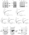
(A) Pelleting assays show association of full-length NMMIIA with full-length gelsolin in presence or absence of EGTA. (B) Coomassie-stained SDS-polyacrylamide gel shows that high-salt (600 mM KCl) buffer prevents NMMIIA binding to gelsolin bound to GST-Sepharose beads. (C) Association between full-length NMMIIA and gelsolin domains G1–G3 and G4–G6 in pull-down assays. Pellets were visualized on Coomassie-stained SDS–PAGE gel and show that the interaction between GST-gelsolin domains G4–G6 and full-length NMMIIA requires Ca2+. (D) Quantification of the binding signal of gelsolin to NMMIIA shows that gelsolin (G1–G6) binds to NMMIIA efficiently with Kd = 0.158 ± 0.082 μM. (E) In the presence of EGTA the binding was reduced (Kd = 0.423 ± 0.152 μM). (F, G) The N- and C-terminal halves G1–G3 and G4–G6 showed binding with Kd = 0.220 ± 0.049 and 0.212 ± 0.063 μM, respectively. (H, I) In the presence of EGTA, N- and C-terminal halves G1–G3 and G4–G6 showed reduced binding with Kd = 0.495 ± 0.178 and 1.312 ± 0.256 μM, respectively, suggesting that binding of G4–G6 to NMMIIA requires calcium. (J) Pull-down assays show pellets in Coomassie-stained gels. GST-Sepharose bead–bound gelsolin did not bind to HMM IIA (1–1337) in the presence or absence of EGTA. (K) NMMIIA-His-thioredoxin residues 1339–1891 associated with full-length gelsolin G1-G6–Sepharose beads in absence of EGTA. (L) NMMIIA residues 1899–1960 bound to GST-Sepharose beads did not bind to gelsolin in the presence or absence of EGTA.
FIGURE 3:
FIGURE 3:
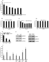
(A) Histogram shows loss of fluorescence of pyrene-labeled actin filaments (0.3 μM) as a result of severing by gelsolin (0.1 μM) in the presence of 1 mM CaCl2. Addition of purified NMMIIA (0.01, 0.02, 0.05, 0.1, 0.2, and 0.4 μM) and gelsolin (0.1 μM) complex show decreased reduction of fluorescence compared with gelsolin alone. (B–D) In experiments of similar design, NMMIIA domains consisting of residues 1–1338, 1339–1891, or 1899–1960 were incubated with gelsolin, and actin severing was measured. (E) Lysates prepared from cells spreading on collagen-coated beads show increase in severing of pyrene-labeled actin filaments compared with cells grown overnight on collagen-coated tissue culture plates. Inset, G-purified gelsolin standard; S, Q, gelsolin expression levels in cell lysates prepared from spreading (S) and quiescent (Q) cells. (F) Total cell lysates from gelsolin-null and wild-type cells were immunoblotted for the indicated proteins. (G) Cells transfected with NMMIIA siRNA show 75% reduction in NMMIIA protein levels compared with cells treated with nontargeted siRNA. (H) Wild-type or gelsolin-null cells transfected with control siRNA or NMMIIA siRNA were maintained in quiescence or were spread on collagen. Cell lysates were analyzed for actin severing by loss of pyrene fluorescence. Wild-type cells transfected with NMMIIA siRNA show enhanced severing of actin filaments in actively spreading cells compared with cells transfected with irrelevant, control siRNA.
FIGURE 4:
FIGURE 4:
(A) Comparison of collagen degradation on collagen-coated planar surface and collagen-coated 2-μm beads. Histogram shows FITC-collagen associated with discrete cellular compartments. The data are percentage of FITC-collagen released into media, cell-surface bound, internalized, or remaining (nondegraded) adherent to substrate (planar or bead surfaces). (B, C) Collagen internalization was retarded at low temperatures and in the presence of MMP inhibitor. (D, E) Histograms show collagen on planar or irregular surface metabolized by gelsolin-null, WT cells transfected with myosin IIA siRNA.
FIGURE 5:
FIGURE 5:
(A, D) Fura-2/AM–loaded cells plated on collagen-coated beads or collagen-coated planar surface show increase in intracellular calcium levels ([Ca2+]i) over time. Images show estimates of intracellular calcium levels in pseudocolor. (B, E) Treatment of cells with the stretch-activated channel (SAC) inhibitor GsMTx-4 (10 μM) reduced [Ca2+]i increases in response to plating on collagen. (C, F) In the presence of EGTA-containing buffer, the increase in [Ca2+]i was minimal in cells plated on collagen beads or collagen-coated planar surface. Chelation of intracellular Ca2+ with BAPTA/AM also reduced Ca2+ transients. (G, H) Lack of effect of GsMTx-4 on store-operated channels. Cells were subjected to store depletion of intracellular calcium stores with low Ca2+ buffer, followed by thapsigargin (1 μM), and treated with either vehicle (G) or GsMTx-4 (10 μM; H).
FIGURE 6:
FIGURE 6:
(A, B) Quantitative analysis of localized fluorescent regions of interest around bead attachment sites. The data show the percentage of positively stained regions of interest around bound collagen beads for cells immunostained with gelsolin and NMMIIA. Data were collected over time in the presence or absence of EGTA. (C) Pretreatment of cells with GsMTx-4 (10 μM) blocked colocalization of NMMIIA and gelsolin at bead sites. (D) Loss of fluorescence of pyrene-labeled actin filaments (0.3 μM) as a result of severing by wild-type gelsolin (0.1 μM) or the severing mutant (Gsn 100/210). (E) Pelleting assays show association of full-length NMMIIA with gelsolin severing mutant (Gsn 100/210) in the presence or absence of EGTA in a Coomassie-stained, SDS-polyacrylamide gel. (F) Gelsolin null cells transfected with gelsolin cDNA and severing mutant showing localization of gelsolin and NMMIIA to collagen beads. (G, H) Histogram showing requirement of severing activity of gelsolin for collagen internalization during compartmentalization of FITC-collagen coated on beads. Severing mutant (Gsn 100/210) shows retarded collagen internalization. (I) Stretch-activated channel inhibitor (GsMTx-4) prevents internalization and collagen degradation.
FIGURE 7:
FIGURE 7:
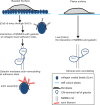
Schematic model of calcium regulation of gelsolin interaction with NMMIIA. Cell adhesion to topologically complex collagen substrate induces [Ca2+]i flux, which promotes interaction of NMMIIA rods with gelsolin domains G1–G3 and G4–G6. This interaction enhances actin remodeling by gelsolin at collagen adhesion sites to enable collagen phagocytosis. Low [Ca2+]i inhibits binding of NMMIIA with gelsolin domains G4–G6, which results in reduced actin remodeling and inhibition of collagen phagocytosis.

Similar articles

Cited by

References

    1. Alexandrova AY, Arnold K, Schaub S, Vasiliev JM, Meister JJ, Bershadsky AD, Verkhovsky AB. Comparative dynamics of retrograde actin flow and focal adhesions: formation of nascent adhesions triggers transition from fast to slow flow. PLoS One. 2008;3:e3234. - PMC - PubMed
    1. Arora PD, Bibby KJ, McCulloch CA. Slow oscillations of free intracellular calcium ion concentration in human fibroblasts responding to mechanical stretch. J Cell Physiol. 1994;161:187–200. - PubMed
    1. Arora PD, Chan MW, Anderson RA, Janmey PA, McCulloch CA. Separate functions of gelsolin mediate sequential steps of collagen phagocytosis. Mol Biol Cell. 2005;16:5175–5190. - PMC - PubMed
    1. Arora PD, Conti MA, Ravid S, Sacks DB, Kapus A, Adelstein RS, Bresnick AR, McCulloch CA. Rap1 activation in collagen phagocytosis is dependent on nonmuscle myosin II-A. Mol Biol Cell. 2008a;19:5032–5046. - PMC - PubMed
    1. Arora PD, Fan L, Sodek J, Kapus A, McCulloch CA. Differential binding to dorsal and ventral cell surfaces of fibroblasts: effect on collagen phagocytosis. Exp Cell Res. 2003;286:366–380. - PubMed

Publication types