Prenatal cerebral ischemia disrupts MRI-defined cortical microstructure through disturbances in neuronal arborization
- PMID: 23325800
- PMCID: PMC3857141
- DOI: 10.1126/scitranslmed.3004669
Prenatal cerebral ischemia disrupts MRI-defined cortical microstructure through disturbances in neuronal arborization
Abstract
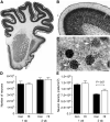
Children who survive preterm birth exhibit persistent unexplained disturbances in cerebral cortical growth with associated cognitive and learning disabilities. The mechanisms underlying these deficits remain elusive. We used ex vivo diffusion magnetic resonance imaging to demonstrate in a preterm large-animal model that cerebral ischemia impairs cortical growth and the normal maturational decline in cortical fractional anisotropy (FA). Analysis of pyramidal neurons revealed that cortical deficits were associated with impaired expansion of the dendritic arbor and reduced synaptic density. Together, these findings suggest a link between abnormal cortical FA and disturbances of neuronal morphological development. To experimentally investigate this possibility, we measured the orientation distribution of dendritic branches and observed that it corresponds with the theoretically predicted pattern of increased anisotropy within cases that exhibited elevated cortical FA after ischemia. We conclude that cortical growth impairments are associated with diffuse disturbances in the dendritic arbor and synapse formation of cortical neurons, which may underlie the cognitive and learning disabilities in survivors of preterm birth. Further, measurement of cortical FA may be useful for noninvasively detecting neurological disorders affecting cortical development.
Figures


Comment in
-
Brain maturation after preterm birth.Sci Transl Med. 2013 Jan 16;5(168):168ps2. doi: 10.1126/scitranslmed.3005379. Sci Transl Med. 2013. PMID: 23325799
Similar articles
-
Brain maturation after preterm birth.Sci Transl Med. 2013 Jan 16;5(168):168ps2. doi: 10.1126/scitranslmed.3005379. Sci Transl Med. 2013. PMID: 23325799
-
Transient Hypoxemia Disrupts Anatomical and Functional Maturation of Preterm Fetal Ovine CA1 Pyramidal Neurons.J Neurosci. 2019 Oct 2;39(40):7853-7871. doi: 10.1523/JNEUROSCI.1364-19.2019. Epub 2019 Aug 27. J Neurosci. 2019. PMID: 31455661 Free PMC article.
-
Prenatal cerebral ischemia triggers dysmaturation of caudate projection neurons.Ann Neurol. 2014 Apr;75(4):508-24. doi: 10.1002/ana.24100. Epub 2014 Mar 13. Ann Neurol. 2014. PMID: 24395459 Free PMC article.
-
What brakes the preterm brain? An arresting story.Pediatr Res. 2014 Jan;75(1-2):227-33. doi: 10.1038/pr.2013.189. Epub 2013 Oct 31. Pediatr Res. 2014. PMID: 24336432 Review.
-
The role of diffusion tensor imaging in the evaluation of ischemic brain injury - a review.NMR Biomed. 2002 Nov-Dec;15(7-8):561-9. doi: 10.1002/nbm.786. NMR Biomed. 2002. PMID: 12489102 Review.
Cited by
-
Morphometric Analysis of Brain in Newborn with Congenital Diaphragmatic Hernia.Brain Sci. 2021 Apr 2;11(4):455. doi: 10.3390/brainsci11040455. Brain Sci. 2021. PMID: 33918479 Free PMC article.
-
Emerging Roles of miRNAs in Brain Development and Perinatal Brain Injury.Front Physiol. 2019 Mar 28;10:227. doi: 10.3389/fphys.2019.00227. eCollection 2019. Front Physiol. 2019. PMID: 30984006 Free PMC article. Review.
-
Cerebral white and gray matter injury in newborns: new insights into pathophysiology and management.Clin Perinatol. 2014 Mar;41(1):1-24. doi: 10.1016/j.clp.2013.11.001. Clin Perinatol. 2014. PMID: 24524444 Free PMC article. Review.
-
Diffusion Tensor Imaging Colour Mapping Threshold for Identification of Ventilation-Induced Brain Injury after Intrauterine Inflammation in Preterm Lambs.Front Pediatr. 2017 Apr 5;5:70. doi: 10.3389/fped.2017.00070. eCollection 2017. Front Pediatr. 2017. PMID: 28424764 Free PMC article.
-
Primary neuronal dysmaturation in preterm brain: Important and likely modifiable.J Neonatal Perinatal Med. 2021;14(1):1-6. doi: 10.3233/NPM-200606. J Neonatal Perinatal Med. 2021. PMID: 33136070 Free PMC article. No abstract available.
References
-
- Ment LR, Hirtz D, Hüppi PS. Imaging biomarkers of outcome in the developing preterm brain. Lancet Neurol. 2009;8:1042–1055. - PubMed
-
- Ferriero DM. Neonatal brain injury. N. Engl. J. Med. 2004;351:1985–1995. - PubMed
-
- Hack M, Taylor HG, Drotar D, Schluchter M, Cartar L, Andreias L, Wilson-Costello D, Klein N. Chronic conditions, functional limitations, and special health care needs of school-aged children born with extremely low-birth-weight in the 1990s. JAMA. 2005;294:318–325. - PubMed
-
- Zubiaurre-Elorza L, Soria-Pastor S, Junque C, Segarra D, Bargalló N, Mayolas N, Romano-Berindoague C, Macaya A. Gray matter volume decrements in preterm children with periventricular leukomalacia. Pediatr. Res. 2011;69:554–560. - PubMed
Publication types
MeSH terms
Grants and funding
- F30 NS066704/NS/NINDS NIH HHS/United States
- P51 RR000163/RR/NCRR NIH HHS/United States
- 1F30NS066704/NS/NINDS NIH HHS/United States
- 1R01NS054044/NS/NINDS NIH HHS/United States
- R01 NS070022/NS/NINDS NIH HHS/United States
- R01NS070022/NS/NINDS NIH HHS/United States
- P30 NS061800/NS/NINDS NIH HHS/United States
- R01 NS045737/NS/NINDS NIH HHS/United States
- R37 NS045737/NS/NINDS NIH HHS/United States
- P51RR000163/RR/NCRR NIH HHS/United States
- R01 NS054044/NS/NINDS NIH HHS/United States
- T32 AG023477/AG/NIA NIH HHS/United States
- R37NS045737-06S1/06S2/NS/NINDS NIH HHS/United States
LinkOut - more resources
Full Text Sources
Other Literature Sources
Miscellaneous

