Differentiating tumor recurrence from treatment necrosis: a review of neuro-oncologic imaging strategies
- PMID: 23325863
- PMCID: PMC3635510
- DOI: 10.1093/neuonc/nos307
Differentiating tumor recurrence from treatment necrosis: a review of neuro-oncologic imaging strategies
Abstract
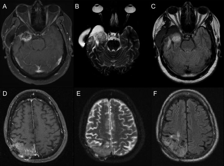
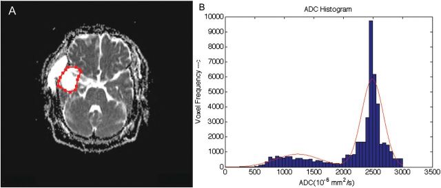
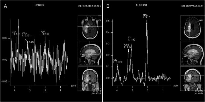
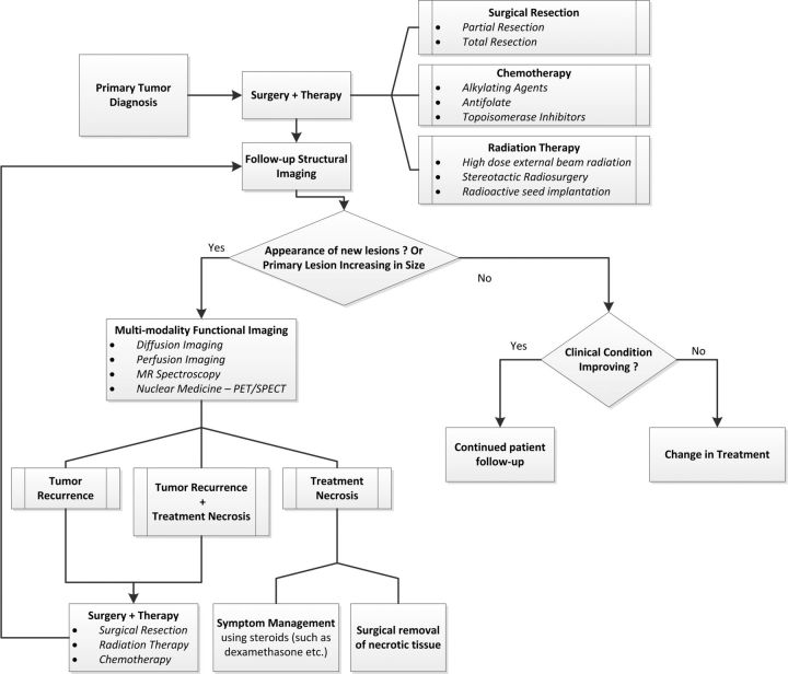
Differentiating treatment-induced necrosis from tumor recurrence is a central challenge in neuro-oncology. These 2 very different outcomes after brain tumor treatment often appear similarly on routine follow-up imaging studies. They may even manifest with similar clinical symptoms, further confounding an already difficult process for physicians attempting to characterize a new contrast-enhancing lesion appearing on a patient's follow-up imaging. Distinguishing treatment necrosis from tumor recurrence is crucial for diagnosis and treatment planning, and therefore, much effort has been put forth to develop noninvasive methods to differentiate between these disparate outcomes. In this article, we review the latest developments and key findings from research studies exploring the efficacy of structural and functional imaging modalities for differentiating treatment necrosis from tumor recurrence. We discuss the possibility of computational approaches to investigate the usefulness of fine-grained imaging characteristics that are difficult to observe through visual inspection of images. We also propose a flexible treatment-planning algorithm that incorporates advanced functional imaging techniques when indicated by the patient's routine follow-up images and clinical condition.
Figures


Similar articles
-
Molecular imaging based on differential protein content in differentiating glioma from radiation necrosis.Neurosurgery. 2011 Jun;68(6):N16-7. doi: 10.1227/01.neu.0000398208.56326.19. Neurosurgery. 2011. PMID: 21778943 Review. No abstract available.
-
Differentiating brain radionecrosis from tumour recurrence: a role for contrast-enhanced ultrasound?Acta Neurochir (Wien). 2017 Dec;159(12):2405-2408. doi: 10.1007/s00701-017-3306-x. Epub 2017 Sep 5. Acta Neurochir (Wien). 2017. PMID: 28875373
-
Distinguishing glioma recurrence from treatment effect after radiochemotherapy and immunotherapy.Neurosurg Clin N Am. 2010 Jan;21(1):181-6. doi: 10.1016/j.nec.2009.08.003. Neurosurg Clin N Am. 2010. PMID: 19944976 Review.
-
The diagnosis and treatment of pseudoprogression, radiation necrosis and brain tumor recurrence.Int J Mol Sci. 2014 Jul 3;15(7):11832-46. doi: 10.3390/ijms150711832. Int J Mol Sci. 2014. PMID: 24995696 Free PMC article. Review.
-
Treatment induced necrosis versus recurrent/progressing brain tumor: going beyond the boundaries of conventional morphologic imaging.J Neurooncol. 2010 Oct;100(1):17-29. doi: 10.1007/s11060-010-0139-3. Epub 2010 Feb 24. J Neurooncol. 2010. PMID: 20179990 Review.
Cited by
-
Co-Occurrence Conundrum: Brain Metastases from Lung Adenocarcinoma, Radiation Necrosis, and Gliosarcoma.Case Rep Oncol. 2021 Mar 18;14(1):487-492. doi: 10.1159/000514297. eCollection 2021 Jan-Apr. Case Rep Oncol. 2021. PMID: 33976625 Free PMC article.
-
Combined iron oxide nanoparticle ferumoxytol and gadolinium contrast enhanced MRI define glioblastoma pseudoprogression.Neuro Oncol. 2019 Mar 18;21(4):517-526. doi: 10.1093/neuonc/noy160. Neuro Oncol. 2019. PMID: 30277536 Free PMC article.
-
Phytosomal curcumin causes natural killer cell-dependent repolarization of glioblastoma (GBM) tumor-associated microglia/macrophages and elimination of GBM and GBM stem cells.J Exp Clin Cancer Res. 2018 Jul 25;37(1):168. doi: 10.1186/s13046-018-0792-5. J Exp Clin Cancer Res. 2018. PMID: 30041669 Free PMC article.
-
MRI phenotypes of glioblastomas early after treatment are suggestive of overall patient survival.Neurooncol Adv. 2023 Oct 12;5(1):vdad133. doi: 10.1093/noajnl/vdad133. eCollection 2023 Jan-Dec. Neurooncol Adv. 2023. PMID: 37908765 Free PMC article.
-
Diagnostic accuracy of magnetic resonance imaging techniques for treatment response evaluation in patients with high-grade glioma, a systematic review and meta-analysis.Eur Radiol. 2017 Oct;27(10):4129-4144. doi: 10.1007/s00330-017-4789-9. Epub 2017 Mar 22. Eur Radiol. 2017. PMID: 28332014 Free PMC article.
References
-
- Kumar AJ, Leeds NE, Fuller GN, et al. Malignant gliomas: MR imaging spectrum of radiation therapy- and chemotherapy-induced necrosis of the brain after treatment. Radiology. 2000;217(2):377–384. - PubMed
-
- Curnes J, Laster D, Ball M, et al. MRI of radiation injury to the brain. Am J Roentgenol. 1986;147(1):119–124. - PubMed
-
- Brismar J, Roberson GH, Davis KR. Radiation necrosis of the brain. Neuroradiological considerations with computed tomography. Neuroradiology. 1976;12(2):109–113. - PubMed
-
- Mikhael M. Radiation necrosis of the brain: correlation between computed tomography, pathology, and dose distribution. J comput Assist Tomogr. 1978;2(1):71–80. - PubMed
Publication types
MeSH terms
LinkOut - more resources
Full Text Sources
Other Literature Sources
Medical