. 2013 Feb 15;190(4):1501-9.
doi: 10.4049/jimmunol.1200750. Epub 2013 Jan 16.

Id2 influences differentiation of killer cell lectin-like receptor G1(hi) short-lived CD8+ effector T cells

Affiliations

Id2 influences differentiation of killer cell lectin-like receptor G1(hi) short-lived CD8+ effector T cells

Jamie Knell et al. J Immunol. .

Abstract

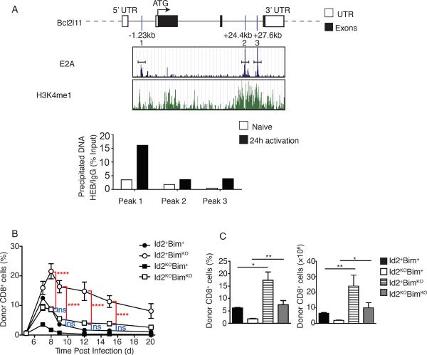
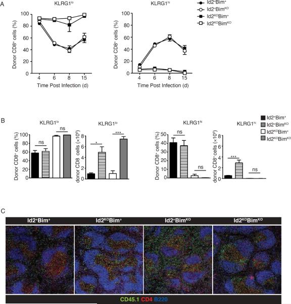
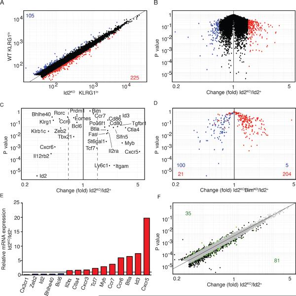
CD8(+) T cells play a crucial role in the clearance of intracellular pathogens through the generation of cytotoxic effector cells that eliminate infected cells and long-lived memory cells that provide enhanced protection against reinfection. We have previously shown that the inhibitor of E protein transcription factors, Id2, is necessary for accumulation of effector and memory CD8(+) T cells during infection. In this study, we show that CD8(+) T cells lacking Id2 did not generate a robust terminally differentiated killer cell lectin-like receptor G1 (KLRG1)(hi) effector population, but displayed a cell-surface phenotype and cytokine profile consistent with memory precursors, raising the question as to whether loss of Id2 impairs the differentiation and/or survival of effector memory cells. We found that deletion of Bim rescued Id2-deficient CD8(+) cell survival during infection. However, the dramatic reduction in KLRG1(hi) cells caused by loss of Id2 remained in the absence of Bim, such that Id2/Bim double-deficient cells form an exclusively KLRG1(lo)CD127(hi) memory precursor population. Thus, we describe a role for Id2 in both the survival and differentiation of normal CD8(+) effector and memory populations.


Figures

Figure 1
Figure 1. Id2-deficient CD8+ T cells display an altered phenotype during infection
A 1:1 mixture of OT-I Id2-knockout cells (CD45.2) and OT-I Id2-wild type cells (CD45.1) were transferred into congenically distinct hosts 1 day before infection with Lm-OVA. (A) KLRG1 and CD127 expression by Id2+ and Id2KO OT-I cells recovered from spleen on days 7 and 12 post infection. Flow cytometry plots display surface phenotype of CD8+ and CD45.2+ or CD45.1+ gated cells. Numbers indicate percentage of cells in each quadrant (left). Average (±SEM) percentage donor CD8+ and total numbers of KLRG1hi and KLRG1lo cells summarizing flow cytometry data (bar graphs, right). Data are from two independent experiments, four mice per timepoint. (B) Histograms of CXCR6 and CD43 expression by donor cells, average (±SEM) of mean fluorescence intensity (right). Statistical significance was determined using unpaired two-tailed t test where ns, not significant, *, P < 0.05, **, P < 0.005, ***, P < 0.0005. Knell et al.
Figure 2
Figure 2. Bim-deficiency rescues survival of Id2-deficient CD8+ T cells during infection
(A) E2A occupancy (blue peaks) and H3K4me1 occupancy (green peaks) for the Bcl2l11 gene as reported by Lin et al. (17). E box binding sites are indicated relative to the transcriptional start site (ATG = 0kb). Immunoprecipitation of chromatin (with anti-HEB or immunologlobulin G, IgG) isolated from purified OT-I Id2+ cells directly ex vivo or 24 h after in vitro activation with OVAp followed by quantitative PCR analysis of input or precipitated DNA for indicated E-box sites in the Bcl2l11 gene. Data are representative of two independent experiments, n = 3. (B) Percentage of CD8+ OT-I Id2+Bim+, BimKO, Id2KO or Id2KOBimKO CD45.2 over the course of infection in PBL. CD45.1+ C57BL/6 mice received OT-I Id2+Bim+, BimKO, Id2KO or Id2KOBimKO (CD45.2+) cells (2 × 10⁴) 1 day before infection with Lm-OVA, average (±SEM). Two-way ANOVA analysis indicated no significant difference between expansion and contraction of Id2+Bim+ and Id2KOBimKO cells throughout the course of infection. These analyses also revealed significant difference between Id2+BimKO and Id2KOBimKO cells throughout the course of infection. . (C) Bar graphs summarizing frequency of donor cells among CD8+ cells and total number of indicated donor cells recovered from spleen on day 8 of infection. Statistical significance was determined using unpaired two-tailed t test where ns, not significant, *, P < 0.05, **, P < 0.005, ***, P < 0.0005. Data are representative of three independent experiments with three mice per time point. Knell et al.
Figure 3
Figure 3. Bim-deficiency does not rescue phenotypic defects in Id2KO CD8+ T cells during infection
Flow cytometric analysis of cell-surface phenotype of donor CD8+CD45.2+ splenocytes on day 15 of infection KLRG1, CD127, CD62L, CD44, CD27, CXCR3, CD43, CD122 (gated on Ly6C+ population). CD45.1+ C57BL/6 mice received OT-I Id2+Bim+, BimKO, Id2KO or Id2KOBimKO (CD45.2+) cells (2 × 10⁴) 1 day before infection with Lm-OVA. CXCR6 expression was examined on day 9 and CD25 expression was examined on day 4 of infection. Knell et al.
Figure 4
Figure 4. Bim-deficiency does not restore KLRG1hi phenotype in Id2KO CD8+ T cells during infection
(A) Percentage of KLRG1lo (left graph) or KLRG1hi (right graph) CD8+ OT-I Id2+Bim+, BimKO, Id2KO or Id2KOBimKO CD45.2 over the course of infection in spleen. CD45.1+ C57BL/6 mice received OT-I Id2+Bim+, BimKO, Id2KO or Id2KOBimKO (CD45.2+) cells (2 × 10⁴) 1 day before infection with Lm-OVA, average (±SEM). Data are representative of one (day 4 and 6) to three (day 8 and 15) independent experiments, with three mice per timepoint. (B) Bar graphs summarizing average (±SEM) frequency of KLRG1hi or KLRG1lo donor cells among total donor CD8+ cells and total number of KLRG1hi and KLRG1lo donor OT-I donor cells recovered from spleen on day 15 of infection. (C) Confocal images of serial spleen sections of Id2+Bim+, Id2KOBim+, Id2+BimKO and Id2KOBimKO donor OT-I recipients on day 8 of Lm-OVA infection. Sections were stained for CD45.2, CD4 and B220. Images collected at 10× magnification. Data are representative of 2 independent experiments. Statistical significance was determined using unpaired two-tailed t test where ns, not significant, *, P < 0.05, **, P < 0.005, ***, P < 0.0005 relative to CD8+ OT-I Id2+Bim+ cells. Data are representative of three independent experiments, with three mice per timepoint. Knell et al.
Figure 5
Figure 5. Id2-deficiency alters cytokine production by CD8+ T cells during infection
Flow cytometric analysis of (A) IL-2 (B) IFNγ and (C) TNFα production by OT-I Id2+Bim+, BimKO, Id2KO or Id2KOBimKO (CD45.2+) splenocytes. CD45.1+ C57BL/6 mice received OT-I Id2+Bim+, BimKO, Id2KO or Id2KOBimKO (CD45.2+) cells (2 × 10⁴) 1 day before infection with Lm-OVA. Splenocytes isolated on day 15 post infection were restimulated for 6 h in vitro with OVA peptide (solid line) or incubated in media alone (dotted line) followed by surface and intracellular staining. Bar graphs indicating average (±SEM) percentage of cytokine producing cells among donor cells. (D) E2A occupancy (blue peaks) and H3K4me1 occupancy (green peaks) in A12 cells overexpressing E47 as reported by Lin et al. (17). Immunoprecipitation of chromatin (with anti-HEB or IgG) isolated from purified OT-I Id2+ cells directly ex vivo or 24 h after in vitro activation with OVAp followed by quantitative PCR analysis of input or precipitated DNA for indicated E-box sites. Data are representative of three independent experiments. Knell et al.
Figure 6
Figure 6. Gene-expression profile of Id2+ versus Id2KO KLRG1lo cells during infection
Gene expression analysis of WT versus Id2KO effector cells. CD45.1.2+ mice received CD45.1 OT-I WT (4 × 10⁴) cells mixed with CD45.2 OT-I BimKO, Id2KO or Id2KOBimKO (4 × 10⁴) cells 1 day before infection with Lm-OVA. KLRG1hi and KLRG1lo cells were sorted on day 6. (A) Fold change plot of WT KLRG1lo versus Id2KO KLRG1lo CD8+ T cells. Numbers in corners indicate genes with a difference in expression of 1.7-fold or more. Genes upregulated in Id2KO KLRG1lo cells are indicated in red, genes downregulated are indicated in blue. Data are pooled from 3 independent experiments. (B) `Volcano' plot of expression data in (A). (C) Gene expression of key genes in WT KLRG1lo versus Id2KO KLRG1lo CD8+ T cells. Dotted lines indicate 1.5-fold difference. (D) Gene expression of genes 1.7 fold up- or downregulated in WT KLRG1lo versus Id2KOBimKO KLRG1lo CD8+ T cells plotted for wild-type versus Id2-knockout cells. Genes upregulated in Id2KOBimKO KLRG1lo CD8+ T cells are indicated in red, genes downregulated are indicated in blue. (E) mRNA expression of relevant genes in Id2KO compared to Id2+ KLRG1lo cells on day 6 of Lm-OVA infection. (F) Gene expression in WT KLRG1lo versus Id2KO KLRG1lo CD8+ T cells; green indicates E2A occupancy. Numbers in corners indicate number of genes with E2A occupancy. Knell et al.

Similar articles

Cited by

References

    1. Harty JT, Badovinac VP. Shaping and reshaping CD8+ T-cell memory. Nat Rev Immunol. 2008;8:107–119. - PubMed
    1. Jameson SC, Masopust D. Diversity in T cell memory: an embarrassment of riches. Immunity. 2009;31:859–871. - PMC - PubMed
    1. Joshi NS, Kaech SM. Effector CD8 T cell development: a balancing act between memory cell potential and terminal differentiation. J Immunol. 2008;180:1309–1315. - PubMed
    1. Joshi NS, Cui W, Chandele A, Lee HK, Urso DR, Hagman J, Gapin L, Kaech SM. Inflammation directs memory precursor and short-lived effector CD8(+) T cell fates via the graded expression of T-bet transcription factor. Immunity. 2007;27:281–295. - PMC - PubMed
    1. Kaech SM, Hemby S, Kersh E, Ahmed R. Molecular and functional profiling of memory CD8 T cell differentiation. Cell. 2002;111:837–851. - PubMed

Publication types

MeSH terms

Associated data