. 2013 Feb 15;190(4):1725-36.
doi: 10.4049/jimmunol.1200867. Epub 2013 Jan 16.

A chemokine self-presentation mechanism involving formation of endothelial surface microstructures

Affiliations

A chemokine self-presentation mechanism involving formation of endothelial surface microstructures

Catherine Whittall et al. J Immunol. .

Abstract

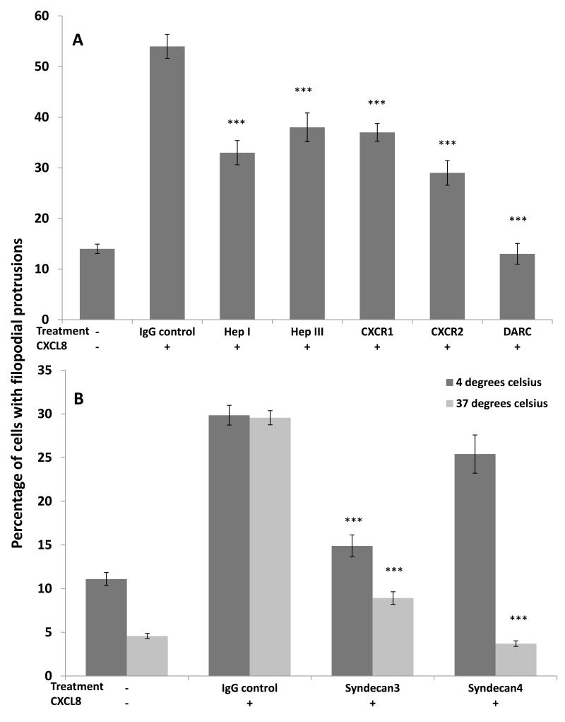
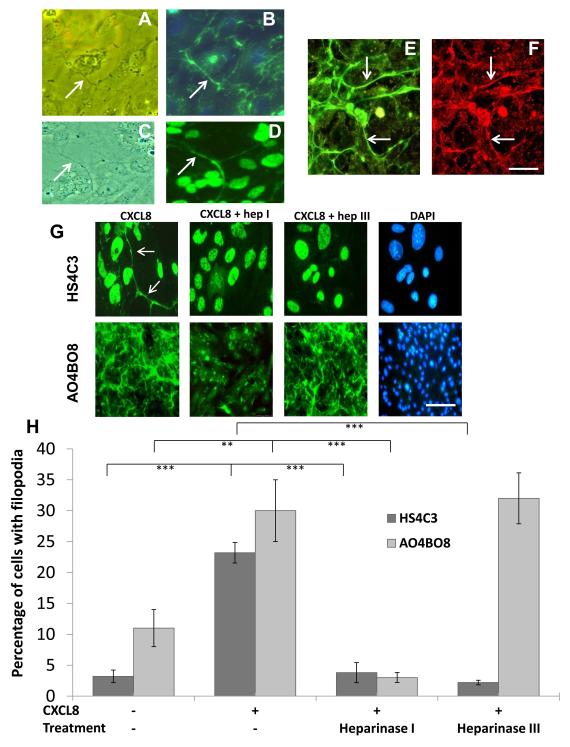
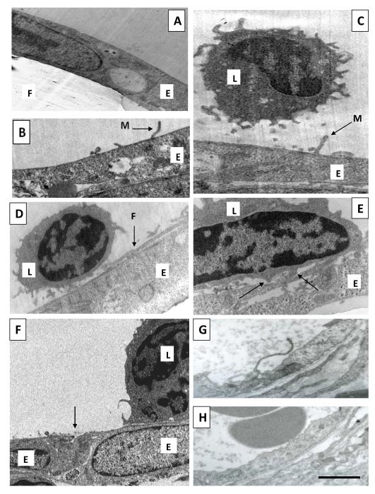
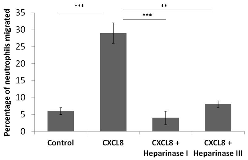
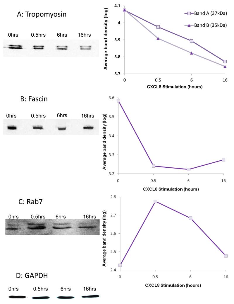
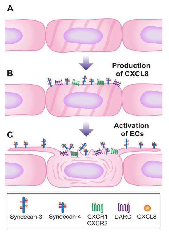
Endothelial surface microstructures have been described previously under inflammatory conditions; however, they remain ill-characterized. In this study, CXCL8, an inflammatory chemokine, was shown to induce the formation of filopodia-like protrusions on endothelial cells; the same effects were observed with CXCL10 and CCL5. Chemokines stimulated filopodia formation by both microvascular (from bone marrow and skin) and macrovascular (from human umbilical vein) endothelial cells. Use of blocking Abs and degradative enzymes demonstrated that CXCL8-stimulated filopodia formation was mediated by CXCR1 and CXCR2, Duffy Ag/receptor for chemokines, heparan sulfate (HS), and syndecans. HS was present on filopodial protrusions appearing as a meshwork on the cell surface, which colocalized with CXCL8, and this glycosaminoglycan was 2,6-O- and 3-O-sulfated. Transmission electron microscopy revealed that CXCL8-stimulated filopodial and microvilli-like protrusions that interacted with leukocytes before transendothelial migration and removal of HS reduced this migration. iTRAQ mass spectrometry showed that changes in the levels of cytoskeletal, signaling, and extracellular matrix proteins were associated with CXCL8-stimulated filopodia/microvilli formation; these included tropomyosin, fascin, and Rab7. This study suggests that chemokines stimulate endothelial filopodia and microvilli formation, leading to their presentation to leukocytes and leukocyte transendothelial migration.


Figures

Figure 1
Figure 1. Chemokine-stimulated formation of filopodial protrusions
Human bone marrow (HBMEC), human dermal microvascular (HMVEC) and human umbilical vein (HUVEC) endothelial cells were stimulated with varying concentrations of chemokine (or no chemokine for the negative controls). After 30 minutes at 37°C, the percentage of cells with filopodial protrusions was determined using phase contrast microscopy at a magnification of ×200. (A) is an example of a control HBMEC in the absence of CXCL8 showing lack of filopodia. (B) is in the presence of CXCL8 (100ng/ml) showing cells with filopodial protrusions at the cell periphery (examples are arrowed). Bar = 25μm in A and B. (C) Endothelial cells were treated with varying concentrations of CXCL8, CXCL10, CCL5, or no chemokine (control) and the percentage of cells with filopodial protrusions was calculated. Data show the percentage mean ± standard error (SE) from n=10 fields of view and are representative of three independent experiments, *p=<0.05 ***p=<0.0001 compared to untreated controls.
Figure 2
Figure 2. HBMEC filopodial protrusion formation after blocking or degrading CXCL8 binding sites
(A) CXCL8-stimulated cells (100ng/ml) were pre-treated with anti-CXCR1 (2μg/ml), anti-CXCR2 (2.5 μg/ml), anti-DARC (50ng/ml), heparinase I (10U/ml) or heparinase III (2U/ml). As a positive control endothelial cells were treated with CXCL8 in the presence of mouse IgG, as a negative control endothelial cells were left unstimulated. ***p=<0.0001 compared to CXCL8-treated IgG control. Data show the percentage mean ± standard error (SE) of n=10 fields of view and are representative of three independent experiments. (B) CXCL8-stimulated endothelial cells were treated with anti-syndecan-3 or anti-syndecan-4 (both 5μg/ml) and mouse IgG as a control; unstimulated cells were also used to determine background filopodia formation. The assay was performed at both +4°C and +37°C prior to endothelial cells being fixed and the percentage of cells with filopodia determined using phase contrast microscopy. ***p=<0.0001 compared to CXCL8-treated IgG control. Data show the means ± standard errors (SE) of the percentage of cells with filopodia in 10 fields of view (at ×200 magnification) from one experiment representative of two independent experiments.
Figure 3
Figure 3. Localisation of heparan sulphate 2,6-O and 3-O sulphated motifs on endothelial filopodia
(A) CXCL8-stimulated HBMECs analysed by phase contrast showing a filopodial protrusion. (B) same image as (A) using immunofluorescence with an antibody against the 2,6-O-sulphated heparan sulphate epitope (AO4BO8). Arrows show the localisation of heparan sulphate to the filopodium. (C) CXCL8-stimulated HBMECs analysed by phase contrast showing a filopodial extension. (D) same image as (B) using immunofluorescence with an antibody against the 3-O sulphated heparan sulphate epitope (HS4C3). Arrows show the localisation of heparan sulphate to the filopodium. Double labelling using antibodies to heparan sulphate (E) and CXCL8 (F) showing colocalisation (arrows). Bar =5μm in (A-F). (G) CXCL8-stimulated HBMECs pre-treated with and without heparinase I (10U/ml) and heparinase III (2U/ml) and analysed by immunofluorescence with antibodies against 3-O-sulphated epitopes (HS4C3) and 2,6-O sulphated epitopes (AO4BO8)(both green) with DAPI showing nuclear staining (blue). With HS4C3 antibody arrows show the filopodia. Bar = 5μm for HS4C3 and 20μm for AO4BO8. (H) Analysis of immunofluorescence shown in (G) with the antibodies described, the percentage of cells with positive staining on the filopodia was quantified. Data show the means ± standard errors (SE) of the percentage of cells with filopodia in 10 fields of view (at ×200 magnification) from one experiment representative of two independent experiments. **p=<0.001 and ***p=<0.0001.
Figure 4
Figure 4. Endothelial filopodial and microvillous protrusions and leukocyte interaction
For A-F endothelial cells (HBMECs) were cultured on 3μm pore transwell filters and treated with 100ng/ml CXCL8 for 60 minutes in the basal compartment of transwells. Leukocytes were added to the apical compartment for the final 30 minutes before fixation and analysis using transmission electron microscopy. (A) An unstimulated HBMEC monolayer (E) grown on a transwell filter (F). (B) Microvillous protrusion (M) formed by the endothelial cell (E) in response to CXCL8 stimulation. (C) A CXCL8-stimulated endothelial cell layer (E) with a leukocyte (L) in close proximity to endothelial microvilli (M). (D) A leukocyte (L) interacting with a filopodial protrusion (F) of HBMECs (E) stimulated with CXCL8. This microstructure is extending laterally over the endothelial cell surface whereas in (B) and (C) microvilli extend more vertically. (E) A leukocyte (L) interacting with CXCL8-stimulated HBMECs (E). Arrows indicate leukocyte podosomes. (F) A CXCL8 treated layer of HBMECs (E) in the presence of leukocytes (L), there is an absence of large intercellular gaps between endothelial cells (arrow). (G) Rabbit skin was injected with CXCL8 in vivo and after 30 minutes biopsies were taken and processed for electron microscopy. Note the presence of a protrusion similar to those seen in vitro (B-D). (H) is the same as (G) except that skin was vehicle-injected instead of CXCL8. Bar = 2μm in (A-H).
Figure 5
Figure 5. Neutrophil transendothelial migration
In the same in vitro experiments as in Figure 4 endothelial cells (HBMECs) were cultured on 3μm pore transwell filters and treated with 100ng/ml CXCL8 for 60 minutes in the basal compartment of transwells. Leukocytes were added to the apical compartment for the final 30 minutes to allow for transendothelial migration. The percentage of neutrophils that migrated was determined by flow cytometry. HBMECs were also pre-treated with heparinase I (10U/ml) or heparinase III (2U/ml) prior to leukocyte migration. The percentage of neutrophils that migrated through an unstimulated endothelial monolayer (control), or a CXCL8-stimulated endothelial monolayer is shown. The graph indicates significant reductions when heparan sulphate was cleaved with heparinase I or III. The data represent percentage means ± SE (n=3 endothelial monolayers) and are representative of two individual experiments, ***p=<0.0001 and **p=<0.01 comparing treatments indicated.
Figure 6
Figure 6. Western blot analysis of unstimulated and CXCL8-stimulated HBMECs
Western blot analysis of HBMEC cell extracts in the absence or after addition of CXCL8 for the indicated time; total cell extracts were analysed using the indicated antibodies. Graphical data represent the log average band intensities from the western blots (Y-axis) for each of the time-points (X-axis) normalised for equal loading: (A) Tropomyosin (band A 37kDa, band B 35kDa isoforms), representative blot of three individual experiments, (B) Fascin, representative of two individual experiments, (C) Rab7, the lower of the two bands was quantitated which corresponded to the expected molecular weight of the protein. Data are representative of two individual experiments. (D) Representative blot probed for GAPDH which was used a loading control.
Figure 7
Figure 7. Schematic model of chemokine-stimulated endothelial filopodial and microvillous protrusion formation
(A) Smooth endothelium in non-inflamed tissue. (B) In inflammation CXCL8 production leads to binding of the chemokine to heparan sulphate chains on syndecan-3/syndecan-4, CXCR1, CXCR2 and DARC. (C) Upon CXCL8 binding to the receptors, there are changes in signalling pathways, reorganisation of the cytoskeleton and the formation of filopodial and microvillous protrusions. Filopodial protrusions are long laterally extending structures that can mere to form a mesh. On these structures the chemokine is presented to leukocytes in the blood by heparan sulphate on syndecan-3 and -4. Microvillous protrusions are shorter and more vertically orientated. They also present chemokines probably in association with heparan sulphate (9).

Similar articles

Cited by

References

    1. Middleton J, Americh L, Gayon R, Julien D, Aguilar L, Amalric F, Girard JP. Endothelial cell phenotypes in the rheumatoid synovium: activated, angiogenic, apoptotic and leaky. Arthritis Res Ther. 2004;6:60–72. - PMC - PubMed
    1. De Smet F, Segura I, De Bock K, Hohensinner PJ, Carmeliet P, P Mechanisms of vessel branching: filopodia on endothelial tip cells lead the way. Arterioscler Thromb Vasc Biol. 2009;29:639–649. - PubMed
    1. Petrovic N, Schacke W, Gahagan JR, O’Conor CA, Winnicka B, Conway RE, Mina-Osorio P, Shapiro LH. CD13/APN regulates endothelial invasion and filopodia formation. Blood. 2007;110:142–150. - PMC - PubMed
    1. Sainson RC, Johnston DA, Chu HC, Holderfield MT, Nakatsu MN, Crampton SP, Davis J, Conn E, Hughes CC. TNF primes endothelial cells for angiogenic sprouting by inducing a tip cell phenotype. Blood. 2008;111:4997–5007. - PMC - PubMed
    1. Sattar A, umar KP, Kumar S. Rheumatoid- and osteo-arthritis: quantitation of ultrastructural features of capillary endothelial cells. J. Pathol. 1986;148:45–53. - PubMed

Publication types

MeSH terms

LinkOut - more resources