

Whole-brain magnetic resonance spectroscopic imaging measures are related to disability in ALS
- PMID: 23325907
- PMCID: PMC3590062
- DOI: 10.1212/WNL.0b013e318281ccec
Whole-brain magnetic resonance spectroscopic imaging measures are related to disability in ALS
Abstract
Objective: To demonstrate the sensitivity of a recently developed whole-brain magnetic resonance spectroscopic imaging (MRSI) sequence to cerebral pathology and disability in amyotrophic lateral sclerosis (ALS), and compare with measures derived from diffusion tensor imaging.
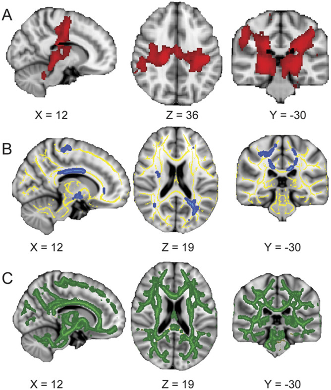
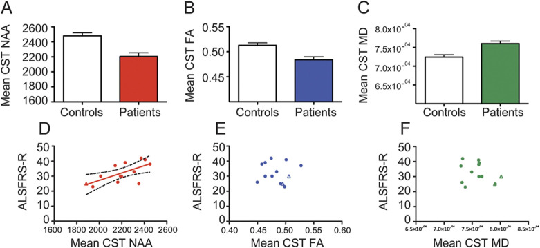
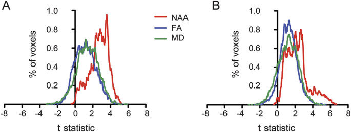
Methods: Whole-brain MRSI and diffusion tensor imaging were undertaken in 13 patients and 14 age-similar healthy controls. Mean N-acetylaspartate (NAA), fractional anisotropy, and mean diffusivity were extracted from the corticospinal tract, compared between groups, and then in relation to disability in the patient group.
Results: Significant reductions in NAA were found along the course of the corticospinal tracts on whole-brain MRSI. There were also significant changes in fractional anisotropy (decreased) and mean diffusivity (increased) in the patient group, but only NAA showed a significant relationship with disability (r = 0.65, p = 0.01).
Conclusion: Whole-brain MRSI has potential as a quantifiable neuroimaging marker of disability in ALS. It offers renewed hope for a neuroimaging outcome measure with the potential for harmonization across multiple sites in the context of a therapeutic trial.
Figures
Comment in
-
Diagnostic shifts in ALS? From clinical specter to imaging spectra.Neurology. 2013 Feb 12;80(7):606-7. doi: 10.1212/WNL.0b013e318281cd27. Epub 2013 Jan 16. Neurology. 2013. PMID: 23325912 No abstract available.
-
Motor neuron disease: whole-brain magnetic resonance spectroscopy measures hold promise as new biomarkers for disability in ALS.Nat Rev Neurol. 2013 Mar;9(3):120. doi: 10.1038/nrneurol.2013.14. Epub 2013 Feb 5. Nat Rev Neurol. 2013. PMID: 23381477 No abstract available.
References
-
- Turner MR, Kiernan MC, Leigh PN, Talbot K. Biomarkers in amyotrophic lateral sclerosis. Lancet Neurol 2009;8:94–109. - PubMed
-
- Kiernan MC, Vucic S, Cheah BC, et al. . Amyotrophic lateral sclerosis. Lancet 2011;377:942–955. - PubMed
-
- Verstraete E, Veldink JH, Hendrikse J, Schelhaas HJ, van den Heuvel MP, van den Berg LH. Structural MRI reveals cortical thinning in amyotrophic lateral sclerosis. J Neurol Neurosurg Psychiatry 2012;83:383–388. - PubMed
-
- Turner MR, Agosta F, Bede P, Govind V, Lule D, Verstraete E. Neuroimaging in amyotrophic lateral sclerosis. Biomark Med 2012;6:319–337. - PubMed
Publication types
MeSH terms
Substances
Grants and funding
LinkOut - more resources
Full Text Sources
Other Literature Sources
Medical
Miscellaneous