Review
. 2013:8:203-20.
doi: 10.2147/IJN.S37348. Epub 2013 Jan 9.

Nanodiamonds as novel nanomaterials for biomedical applications: drug delivery and imaging systems

Affiliations
Review

Nanodiamonds as novel nanomaterials for biomedical applications: drug delivery and imaging systems

Randeep Kaur et al. Int J Nanomedicine. 2013.

Abstract

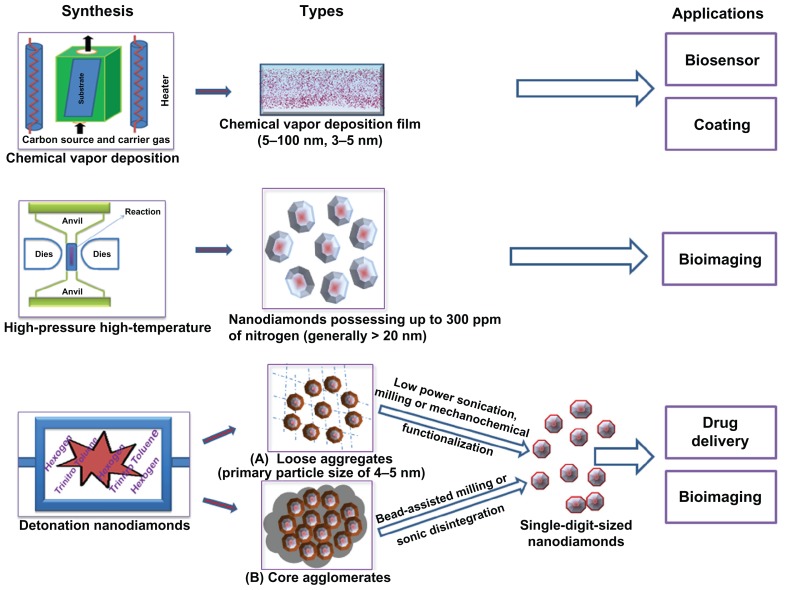
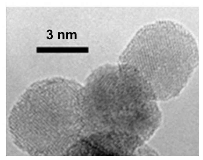
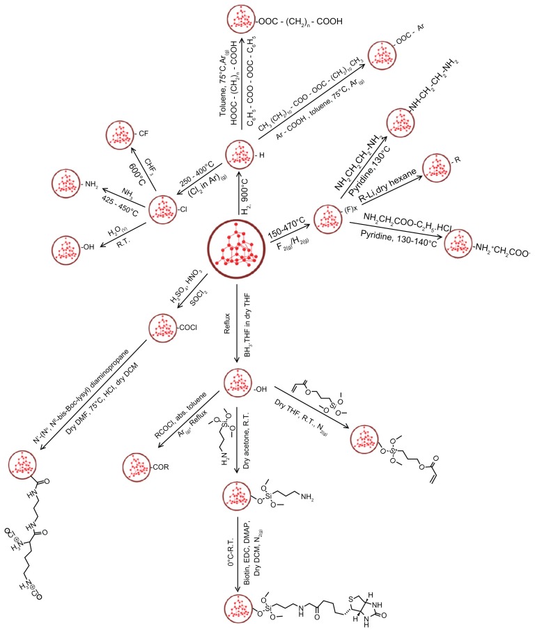
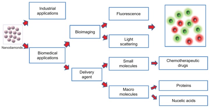
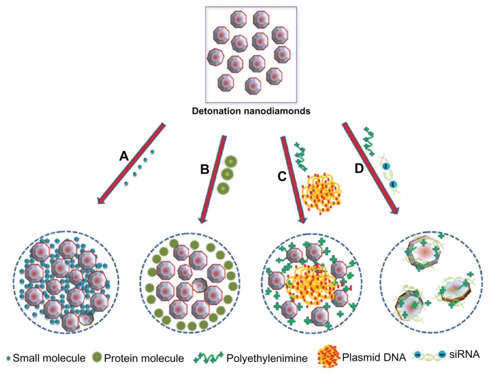
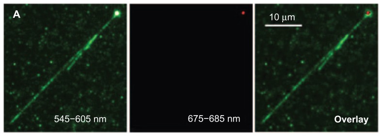
Detonation nanodiamonds (NDs) are emerging as delivery vehicles for small chemical drugs and macromolecular biotechnology products due to their primary particle size of 4 to 5 nm, stable inert core, reactive surface, and ability to form hydrogels. Nanoprobe technology capitalizes on the intrinsic fluorescence, high refractive index, and unique Raman signal of the NDs, rendering them attractive for in vitro and in vivo imaging applications. This review provides a brief introduction of the various types of NDs and describes the development of procedures that have led to stable single-digit-sized ND dispersions, a crucial feature for drug delivery systems and nanoprobes. Various approaches used for functionalizing the surface of NDs are highlighted, along with a discussion of their biocompatibility status. The utilization of NDs to provide sustained release and improve the dispersion of hydrophobic molecules, of which chemotherapeutic drugs are the most investigated, is described. The prospects of improving the intracellular delivery of nucleic acids by using NDs as a platform are exemplified. The photoluminescent and optical scattering properties of NDs, together with their applications in cellular labeling, are also reviewed. Considering the progress that has been made in understanding the properties of NDs, they can be envisioned as highly efficient drug delivery and imaging biomaterials for use in animals and humans.

Keywords: carriers; dispersion; fluorescence; light scattering; surface functionalization; toxicity.


Figures

Figure 1
Figure 1
A schematic representation of nanodiamond types based on their methods of synthesis and applications.
Figure 2
Figure 2
Scanning electron microscopic image of (A) nanocrystalline and (B) ultrananocrystalline diamond films grown on silicon substrate. A is reprinted with permission from: Philip J, Hess P, Feygelson T, et al. Elastic, mechanical, and thermal properties of nanocrystalline diamond films. J Appl Phys. 2003;93(4):2164–2171, Copyright (2003), American Institute of Physics. B is reprinted with permission from: Sumant AV, Grierson DS, Gerbi JE, Carlisle JA, Auciello O, Carpick RW. Surface chemistry and bonding configuration of ultrananocrystalline diamond surfaces and their effects on nanotribological properties. Phys Rev B Condens Matter Mater Phys. 2007;76(23):235429-1–235429-11, Copyright (2007) by the American Physical Society.
Figure 3
Figure 3
Phase diagram for carbon depicting the pressure and temperature requirements for synthesis of detonation nanodiamond (1); cooling profile of products produced by wet detonation synthesis (2) and dry detonation synthesis (3). Notes: “TD” represents Debye temperature. Reproduced courtesy of IOP Publishing Ltd, from: Baidakova M, Vul A. New prospects and frontiers of nanodiamond clusters. J Phys D Appl Phys. 2007;40(20):6300–6311.
Figure 4
Figure 4
High-resolution transmission electron microscopic image of detonation nanodiamonds. Note: Image is courtesy of Bogdan Palosz, IHPP, Warsaw, Poland.
Figure 5
Figure 5
A schematic representation of approaches used for modification of nanodiamond surfaces. Abbreviations: R.T., room temperature; R, alkyl group; Ar, aromatic group; THF, tetrahydrofuran; DMAP, (dimethylamino)pyridine; EDC, (1-ethyl-3-[(3-dimethylamino)propyl]carbodiimidehydrochloride).
Figure 6
Figure 6
A schematic representation of the applications of the nanodiamonds.
Figure 7
Figure 7
A schematic diagram representing the binding of detonation nanodiamonds with (A) small molecules,, (B) proteins, (C) plasmid DNA,, and (D) siRNA. ©2012 Dove Medical Press. Adapted with permission from Kaur R, Chitanda JM, Michel D, et al. Lysine-functionalized nanodiamonds: synthesis, physiochemical characterization, and nucleic acid binding studies. Int J Nanomedicine. 2012;7:3851–3866.
Figure 8
Figure 8
Overlay image (far right) demonstrating the wrapping of TOTO-1 dye-labeled T4DNA (green color, V-shape) around the surface of a single polyL-lysine-coated fluorescent nanodiamond (red color). Reprinted with permission from: Fu CC, Lee HY, Chen K, et al. Characterization and application of single fluorescent nanodiamonds as cellular biomarkers. Proc Natl Acad Sci U S A. 2007;104(3):727–732. Copyright (2007) National Academy of Sciences, USA.
Figure 9
Figure 9
Images of a single HeLa cell treated with ND-transferrin complex: (A) bright-field image, (B) confocal scanning image obtained by collecting all the fluorescence emissions above a wavelength of 550 nm, and (C) confocal scanning image obtained by collecting only the fluorescence emissions at wavelengths of 663–738 nm. Note: A 514.5 nm laser was used as excitation source. Reprinted from: Weng MF, Chiang SY, Wang NS, Niu H. Fluorescent nanodiamonds for specifically targeted bioimaging: application to the interaction of transferrin with transferrin receptor. Diam Relat Mater. 2009;18(2–3):587–591. Copyright (2008), with permission from Elsevier. Abbreviation: ND, nanodiamonds.
Figure 10
Figure 10
(I) Images of Caenorhabditis elegans: (A) an untreated worm with labeled organ morphology; worms after oral administration of bare nanodiamonds (NDs) for (B) 2 hours and (C) 12 hours; worms fed with bare NDs for 2 hours with subsequent administration of Escherichia coli for (D) 20 minutes (E) and 40 minutes. (BE) upper show epifluorescence images; lower show epifluorescence and differential interference contrast merged images. (II) Epifluorescence and differential interference contrast merged images of Caenorhabditis elegans: worms after oral administration of (A) dextran-conjugated NDs, (B) bovine serum albumin-conjugated NDs, and worms exposed to Escherichia Coli after administration of (C) dextran-conjugated NDs and (D) bovine serum albumin-conjugated NDs. Notes: Blue arrows indicate internalization of NDs in intestinal cells; yellow arrow indicate localization of NDs in lumen of worm. Reprinted with permission from: Mohan N, Chen CS, Hsieh HH, Wu YC, Chang HC. In vivo imaging and toxicity assessments of fluorescent nanodiamonds in Caenorhabditis elegans. Nano Lett. 2010;10(9):3692–3699. Copyright (2010) American Chemical Society.
Figure 11
Figure 11
Lysozymal interaction sites revealed with Escherichia coli: (I) carboxylated nanodiamonds (NDs) and Escherichia coli, (II) carboxylated ND-lysozyme complex and Escherichia coli. (A) Optical images identifying Escherichia coli; (B) confocal Raman mapping identifying NDs present in the square area outlined in optical image; (C) merging of the optical image with Raman mapping, demonstrating the interaction of the lysozymes with Escherichia coli. Reproduced from: Perevedentseva E, Cheng CY, Chung PH, Tu JS, Hsieh YH, Cheng CL. The interaction of the protein lysozyme with bacteria E. coli observed using nanodiamond labelling. Nanotechnology. 2007;18(31):315102, courtesy of IOP Publishing Ltd.

Similar articles

Cited by

References

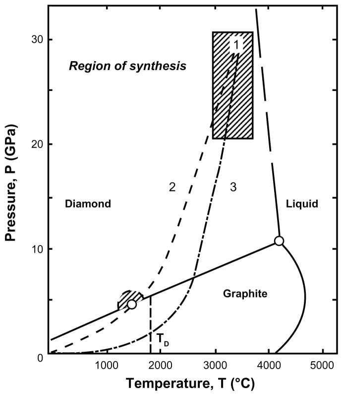
    1. Shenderova OA, McGuire G. Types of nanocrystalline diamond. In: Shenderova OA, Gruen DM, editors. Ultrananocrystalline Diamond: Synthesis Properties and Applications. New York, NY: William Andrew; 2006. pp. 79–114.
    1. Mochalin VN, Shenderova O, Ho D, Gogotsi Y. The properties and applications of nanodiamonds. Nat Nanotechnol. 2012;7(1):11–23. - PubMed
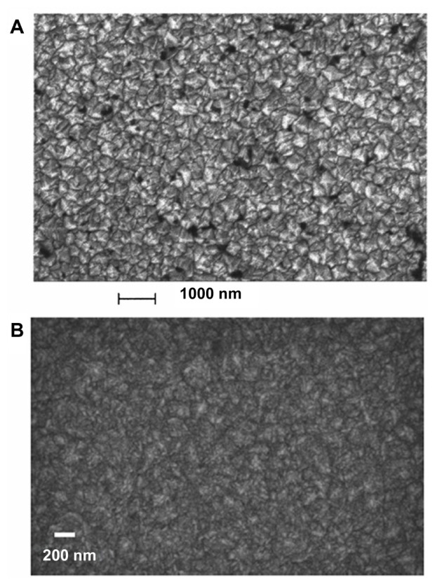
    1. Gracio JJ, Fan QH, Madaleno JC. Diamond growth by chemical vapour deposition. J Phys D Appl Phys. 2010;43:374017.
    1. Philip J, Hess P, Feygelson T, et al. Elastic, mechanical, and thermal properties of nanocrystalline diamond films. J Appl Phys. 2003;93(4):2164–2171. Available at: http://dx.doi.org/10.1063/1.1537465. - DOI
    1. Zhou D, Gruen DM, Qin LC, McCauley TG, Krauss AR. Control of diamond film microstructure by Ar additions to CH4/H2 microwave plasmas. J Appl Phys. 1998;84(4):1981–1989.