Review
. 2013 Jan;9(1):e1003057.
doi: 10.1371/journal.ppat.1003057. Epub 2013 Jan 10.

Viral and bacterial interactions in the upper respiratory tract

Affiliations
Review

Viral and bacterial interactions in the upper respiratory tract

Astrid A T M Bosch et al. PLoS Pathog. 2013 Jan.

Abstract

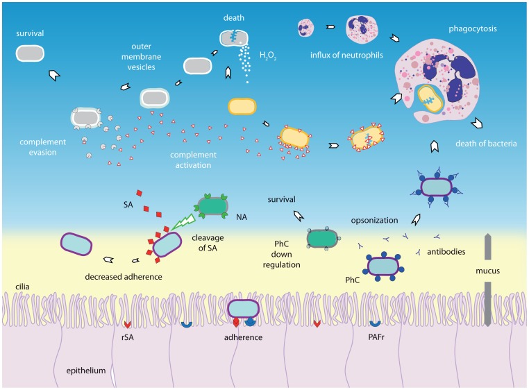
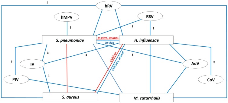
Respiratory infectious diseases are mainly caused by viruses or bacteria that often interact with one another. Although their presence is a prerequisite for subsequent infections, viruses and bacteria may be present in the nasopharynx without causing any respiratory symptoms. The upper respiratory tract hosts a vast range of commensals and potential pathogenic bacteria, which form a complex microbial community. This community is assumed to be constantly subject to synergistic and competitive interspecies interactions. Disturbances in the equilibrium, for instance due to the acquisition of new bacteria or viruses, may lead to overgrowth and invasion. A better understanding of the dynamics between commensals and pathogens in the upper respiratory tract may provide better insight into the pathogenesis of respiratory diseases. Here we review the current knowledge regarding specific bacterial-bacterial and viral-bacterial interactions that occur in the upper respiratory niche, and discuss mechanisms by which these interactions might be mediated. Finally, we propose a theoretical model to summarize and illustrate these mechanisms.


Conflict of interest statement

DB and KT declare to have received consulting fees from Pfizer. EAMS declares to have received unrestricted research support from Pfizer and Baxter, consulting fees from Pfizer and GlaxoSmithKline, lecturing fees from Pfizer, and grant support for vaccine studies from Pfizer and GlaxoSmithKline. None of the fees or grants listed here were received for the research described in this paper. For all other authors no conflicts of interest were declared. This does not alter our adherence to all PLOS Pathogens policies on sharing data and materials.

Figures

Figure 1
Figure 1. Bacterial–bacterial interactions.
The composition of nasopharyngeal microbiota is constantly subject to interactions between species. Bacterial species can interact with other bacterial species by competition and synergism. Synergism can be characterized by, for instance, the production of components that favors another species, as shown for the production of outer membrane vesicles. These may contain factors that are able to inactivate complement factor C3, thereby allowing another species to escape the immune system. Production of substances by one species, for example hydrogen peroxide (H2O2), may eliminate its competitor. The immune system may also be involved in competition, as one bacterium has fewer escape mechanisms to evade the immune system than another and therefore may use co-inhabitants to survive, whereas the reverse phenomenon (i.e., one species may trigger the immune system to combat the other species) may also occur. In addition, since PhC (phosphorylcholine) is shown to be immunogenic and some species may be able to switch off PhC expression whereas others cannot, there might be a selective advantage. Another form of competition involves competition for the same host receptor, as demonstrated for PhC and its receptor platelet activating factor receptor (PAFr). Moreover, one species may use neuraminidase to cut off the sialic acids (SA) that other bacteria may require for attachment to host receptors, therefore inhibiting adherence of the other bacterial species. H2O2, hydrogen peroxide; PAFr, platelet activating factor receptor; PhC, phosphorylcholine; NA, neuraminidase; SA, sialic acid (SA); rSA, receptor for sialic acids; Ab, antibodies.
Figure 2
Figure 2. Viral–bacterial interactions.
(A) Viral–bacterial interaction on the respiratory epithelial surface. Viral presence is thought to predispose the respiratory niche to bacterial colonization by different mechanisms. First, viruses may render the epithelium more susceptible to bacterial colonization by altering the mucosal surfaces. Ciliae may be damaged, leading to decreased mucociliar function of the respiratory epithelium. Additionally, due to viral-induced damage and loss of integrity of the epithelium layer, bacterial colonization may be enhanced and translocation may be increased. Virus-infected cells may decrease the expression of antimicrobial peptides, as shown for β-defensins, thereby affecting the natural defense of the host epithelium. Viral neuraminidase (NA) activity is able to cleave sialic acids residues, thereby giving access to bacterial receptors that were covered by these residues. Finally, viruses may induce bacterial colonization and replication both directly and indirectly, the latter by inducing upregulation of various receptors required for bacterial adherence, including PAFr, CAECAM-1, P5F, ICAM-1, and G-protein. PAFr, platelet activating factor receptor; ICAM-1, intracellular adhesion molecule 1; P5 fimbriae, outer membrane protein P5-homologous fimbriae; CAECAM-1, carcinoembryonic adhesion molecule-1; PhC, phosphorylcholine; SA, sialic acids; rSA, receptor for sialic acids; NA, neuraminidase; mRNA, messenger RNA, AMPs, antimicrobial peptides. (B) Viral–bacterial interaction in relation to the host immune system. Viruses may also induce changes in immune function favorable to bacterial invasion: fewer NK cells may be recruited into the tissue and their functionality may be suboptimal as a consequence of viral infection. Virus-induced IFN-α and IFN-β may impair recruitment and functionality of neutrophils, and subsequently induce apoptosis of neutrophils recruited to combat the viral invader. Furthermore, IFN-γ seems to negatively affect the activity of macrophages. Viral-infected monocytes appear less effective in ingesting and killing bacteria, predisposing them to bacterial overgrowth and invasion. Viral infection seems to impair TLR pathways, induce production of the anti-inflammatory cytokine IL-10, and decrease the concentration of the pro-inflammatory cytokine TNF-α, generally affecting adequate immune responses to bacterial infections. Black arrows indicate increased (↑) or decreased (↓) activity or functionality of a cytokine. IFN, interferon; TNF, tumor necrosis factor; TLR, toll like receptor; IL, interleukin; NK cell, natural killer cell.
Figure 3
Figure 3. Proposed model of bacterial and viral interactions.
This model represents the cumulative dynamics of bacterial and viral interactions occurring within the nasopharyngeal niche during asymptomatic episodes as observed in all cumulative literature references. All available information on the four main potential pathogenic bacteria (Streptococcus pneumoniae, Haemophilus influenzae, Staphylococcus aureus, and Moraxella catarrhalis) and seven common respiratory viruses (rhinoviruses (hRV), respiratory syncytial virus (RSV), adenoviruses (AdV), coronavirus (CoV), influenza viruses (IV), para-influenza viruses (PIV), and human metapneumovirus (hMPV)) are depicted. Red lines represent a negative association of co-colonization (competition), blue lines represent a positive association of co-colonization (synergism). For all depicted associations, evidence is available from human (surveillance) studies, except for those indicated with ‡, where evidence is only available from in vitro and/or animal studies.

References

    1. World Health Organization Geneva, Health Statistics and Informatics Department (2011) Causes of death 2008. In: World health statistics 2011. 170 pages, ISBN 978 92 4 156419 9 (NLM classification: WA 900.1). pp. 57–76. Available: http://www.who.int/gho/publications/world_health_statistics/EN_WHS2011_F.... Accessed 7 December 2012.
    1. Black RE, Cousens S, Johnson HL, Lawn JE, Rudan I, et al. (2010) Global, regional, and national causes of child mortality in 2008: A systematic analysis. child health epidemiology reference group of WHO and UNICEF. Lancet 375(9730) 1969–1987. - PubMed
    1. Vergison A, Dagan R, Arguedas A, Bonhoeffer J, Cohen R, et al. (2010) Otitis media and its consequences: Beyond the earache. Lancet Infect Dis 10(3) 195–203 10.1016/S1473-3099(10)70012-8. - PubMed
    1. Watson KBH, Carville KMA, Bowman JBA, Jacoby P, Riley TV, et al. (2006) Upper respiratory tract bacterial carriage in aboriginal and non-aboriginal children in a semi-arid area of western Australia. Pediatr Infect Dis J 25(9) 782–790 10.1097/01.inf.0000232705.49634.68. - PubMed
    1. Blaser MJ, Falkow S (2009) What are the consequences of the disappearing human microbiota? Nat Rev Microbiol 7(12):887–894 10.1038/nrmicro2245. - PMC - PubMed

Publication types

MeSH terms