. 2013 Jan;9(1):e1003109.
doi: 10.1371/journal.ppat.1003109. Epub 2013 Jan 10.

Lipid exchange between Borrelia burgdorferi and host cells

Affiliations

Lipid exchange between Borrelia burgdorferi and host cells

Jameson T Crowley et al. PLoS Pathog. 2013 Jan.

Abstract

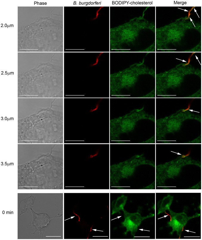
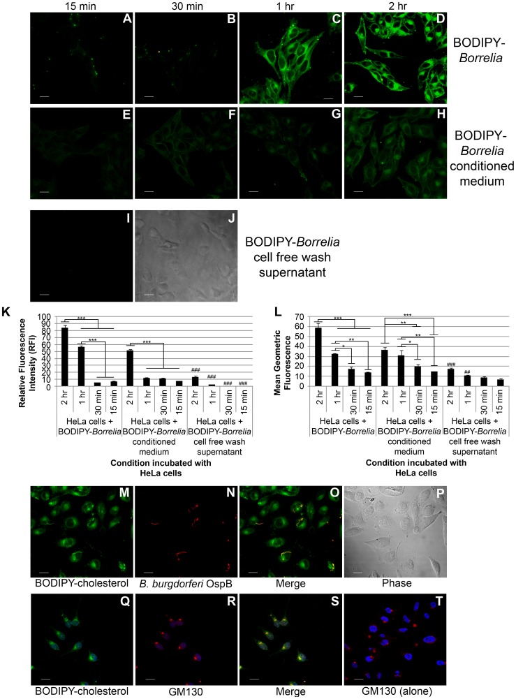
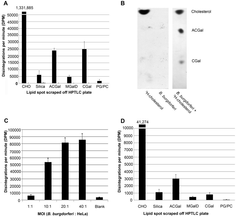
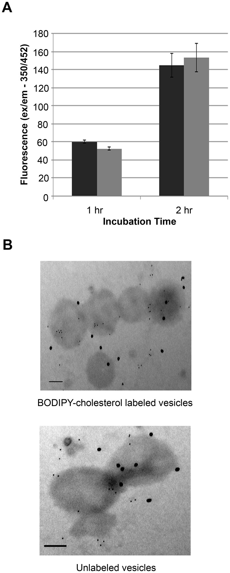
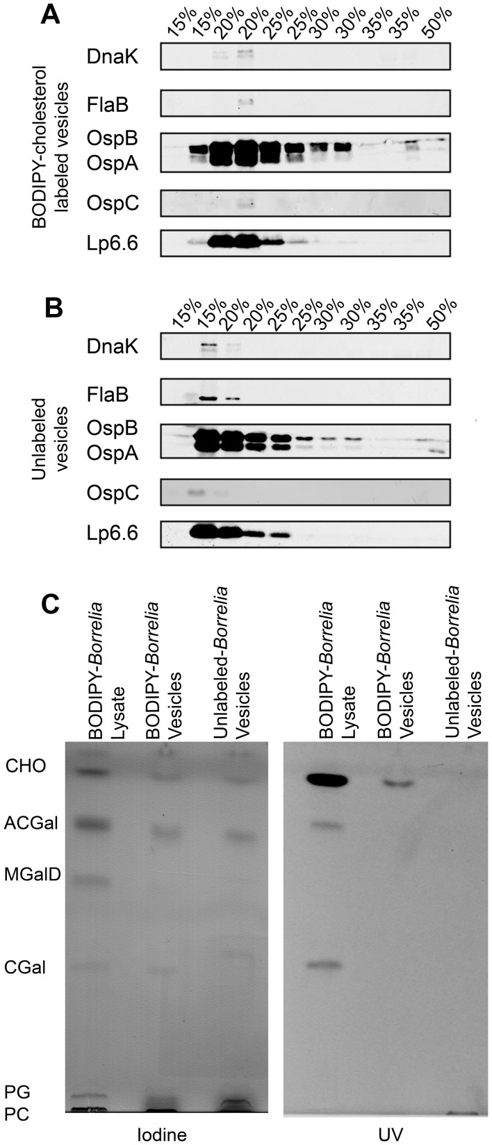
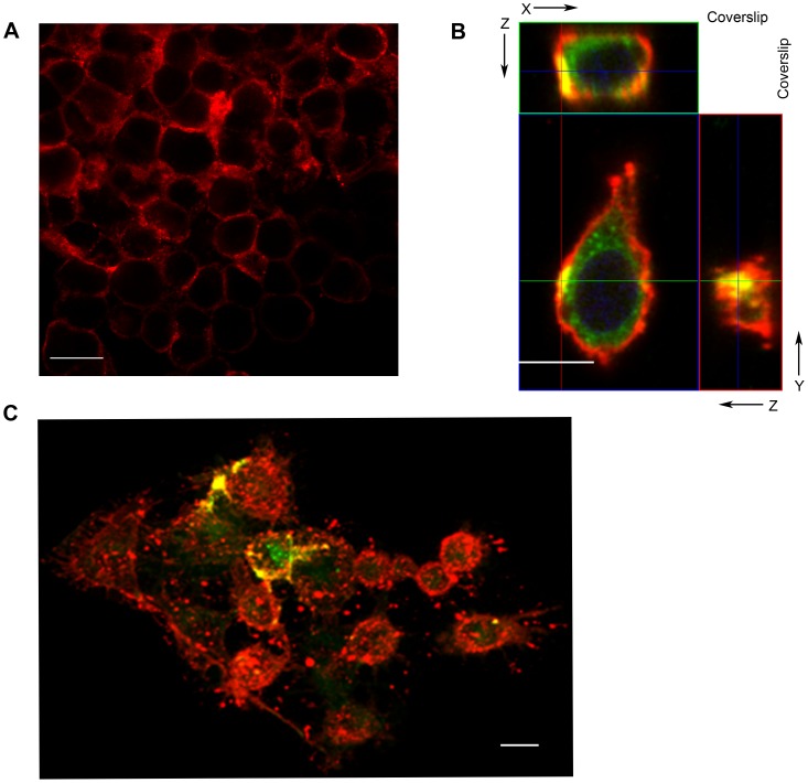
Borrelia burgdorferi, the agent of Lyme disease, has cholesterol and cholesterol-glycolipids that are essential for bacterial fitness, are antigenic, and could be important in mediating interactions with cells of the eukaryotic host. We show that the spirochetes can acquire cholesterol from plasma membranes of epithelial cells. In addition, through fluorescent and confocal microscopy combined with biochemical approaches, we demonstrated that B. burgdorferi labeled with the fluorescent cholesterol analog BODIPY-cholesterol or (3)H-labeled cholesterol transfer both cholesterol and cholesterol-glycolipids to HeLa cells. The transfer occurs through two different mechanisms, by direct contact between the bacteria and eukaryotic cell and/or through release of outer membrane vesicles. Thus, two-way lipid exchange between spirochetes and host cells can occur. This lipid exchange could be an important process that contributes to the pathogenesis of Lyme disease.


Conflict of interest statement

The authors have declared that no competing interests exist.

Figures

Figure 1
Figure 1. B. burgdorferi attach to HeLa cells and acquire cholesterol from the epithelial cell membranes.
HeLa cells incubated with BODIPY-cholesterol (green) and washed with MβCD were incubated with B. burgdorferi (red) for 1 hr at an MOI of 40∶1. Cells were fixed, stained with CB2 (red) and examined by confocal fluorescence microscopy. Four individual optical sections are shown at 2.0 µm, 2.5 µm, 3.0 µm and 3.5 µm. Confocal micrographs show colocalization (yellow, arrows) of BODIPY-cholesterol (green) and OspB (red) on the spirochete at the point of attachment. Scale bars = 10 µm. Confocal micrographs taken at the start of the experiment, 0 min panels (bottom), show that acquisition of BODIPY-cholesterol (green) has not occurred on the spirochetes (red) because there is no colocalization (yellow). The lack of colocalization is highlighted in the individual panels (arrows). Scale bars = 20 µm.
Figure 2
Figure 2. B. burgdorferi incorporate BODIPY-cholesterol into its outer membrane as a component of the cholesterol-glycolipids.
A. Spirochetes were grown for 6 hrs in BSK-II media lacking cholesterol and their fluorescence was measured by spectrophotometry. 4.0 mg/L BODIPY-cholesterol in BSK-II without free cholesterol (green); 0.25% DMSO in BSK-II without free cholesterol (purple); 2.0 mg/L BODIPY-cholesterol BSK-II without free cholesterol (red); 0.175% DMSO in BSK-II without free cholesterol (dark blue); 0.2 mg/L BODIPY-cholesterol in BSK-II without free cholesterol (light blue); 0.0125% DMSO in BSK-II without free cholesterol (black). B. B. burgdorferi incubated with and without BODIPY-cholesterol were analyzed by flow cytometry. ANOVA ***p<0.001. C. Unfixed, live spirochetes grown in the presence of 4.0 mg/L BODIPY-cholesterol for 4 hrs were observed by fluorescent microscopy. The spirochetes did not exhibit morphological or motility defects. Scale bar = 20 µm. D. Following incubation of B. burgdorferi with 4.0 mg/L BODIPY-cholesterol for 4 hrs, the spirochetes were grown in BSK-II for up to 96 hrs, and there were no significant differences in growth compared to controls. Untreated control (black diamond); labeled with BODIPY-cholesterol (blue square); Incubated with 0.25% DMSO (red triangle). E. B. burgdorferi were treated and incubated with 4.0 mg/L. B. burgdorferi pellets were extracted by Bligh and Dyer lipid extraction and analyzed using a chloroform-methanol (85/15) HPTLC. The HPTLC plate was developed using iodine and exposure to UV light. F. B. burgdorferi were grown in BSK-II media lacking free cholesterol with BODIPY-cholesterol or cholesterol as the primary source of sterol in the media to assess long term growth and cytotoxicity in the presence of exogenously added sterol. BSK-II (green); 4.0 mg/L BODIPY-cholesterol in cholesterol free BSK-II without serum (blue); 4.0 mg/L cholesterol in cholesterol free BSK-II without serum (black). Experiments A, B, D, and F represent the mean ± standard error of the mean from three separate experiments.
Figure 3
Figure 3. B. burgdorferi exchange BODIPY-cholesterol to HeLa cells.
B. burgdorferi labeled with BODIPY-cholesterol were incubated with HeLa cells for 15 min, 30 min, 1 hr, and 2 hr. A–D: HeLa cells exposed to B. burgdorferi labeled with BODIPY-cholesterol. E–H: HeLa cells exposed to conditioned medium from B. burgdorferi labeled with BODIPY-cholesterol. I–J: HeLa cells exposed to cell free wash supernatant from B. burgdorferi labeled with BODIPY-cholesterol for 2 hrs. I. Photograph of negative control representative of all temperatures; J. Phase contrast. Scale Bar = 20 µm. K. The mean relative fluorescence intensity (RFI) +/− standard error of the mean of HeLa cells from 10 microscope fields were calculated from the different experimental conditions. ANOVA ***p<0.001, ###p<0.001(negative control is significantly less than associated condition). L. Mean geometric fluorescence +/− standard error of the mean from three separate flow cytometry analysis of HeLa cells incubated following experimental conditions. ANOVA *p<0.05, *p<0.01 ***p<0.001, ##p<0.01, ###p<0.001(negative control is significantly less than associated experimental condition). M–P. Single confocal microscopy optical section illustrates that during the 2 hr incubation the B. burgdorferi (yellow) adhered to HeLa cells (green). Scale bar = 20 µm. Q–T. B. burgdorferi derived cholesterol are processed by the HeLa cell and localize to the Golgi complex. Confocal microscopy demonstrated that the cis-Golgi marker, GM130 (red), co-localizes (yellow) with the BODIPY-cholesterol fluorescence (green). In the control image (T) where no Borrelia were added, the perinuclear localization (DAPI nuclear stain, blue) of GM130 is consistent with the other images. Scale bar = 20 µm.
Figure 4
Figure 4. B. burgdorferi transfer 3H-cholesterol to HeLa cells.
A. B. burgdorferi utilized 3H-cholesterol as a substrate for cholesterol glycolipid synthesis; DPM's derived from scraped silica from HPTLC plate. B. burgdorferi incubated with 3H-cholesterol (black bars) utilized 3H-cholesterol as a substrate for cholesterol-glycolipid synthesis and incorporated the radiolabeled sterol into their lipid fraction when compared to unlabeled B. burgdorferi (gray bars). B. Exposure of the HPTLC plate to Kodak BioMax MR film without scraping demonstrates that the 3H-cholesterol was incorporated into the cholesterol-glycolipids. Radioactive cholesterol was not observed in B. burgdorferi incubated with unlabeled cholesterol (middle lane). This data supports the results from HPTLC exposed to UV light from Figure 1E. C. Spirochetes labeled with 10.0 µCi 3H-cholesterol transfer membrane lipids to HeLa cells. Spirochetes were incubated at an MOI of 1∶1, 10∶1, 20∶1, and 40∶1 with HeLa cells for 2 hours at 37°C in BSK-II. HeLa cells were analyzed for transfer by liquid scintillation counting. D. B. burgdorferi transfer 3H-cholesterol labeled glycolipids to HeLa cells. Radiolabeled B. burgdorferi were incubated with HeLa cells. The bacteria were removed and the HeLa cell lipid extracts were analyzed by liquid scintillation. Experiments A, C, and D represent the mean ± standard error of the mean from three separate experiments.
Figure 5
Figure 5. B. burgdorferi labeled with BODIPY-cholesterol release the fluorescent cholesterol probe in OMV.
A. BODIPY-cholesterol labeled and unlabeled B. burgdorferi release similar amounts of membrane material. DPH was added to the supernatants collected from B. burgdorferi labeled with BODIPY-cholesterol and unlabeled spirochetes to measure released OMV. Using the SpectraMaxM2, the DPH fluorescence was calculated for each sample at the specific time points. Supernatant from labeled B. burgdorferi (black); supernatant from unlabeled B. burgdorferi (gray). B. Transmission electron micrograph showing isolated vesicles from both labeled and unlabeled B. burgdorferi OMV. Immunogold labeling of OspB (18 nm) and B. burgdorferi glycolipids (6 nm) is seen throughout the vesicles. Scale Bar = 100 nM. Experiment A represents the mean ± standard error of the mean from three separate experiments.
Figure 6
Figure 6. Purified OMV from B. burgdorferi contain outer membrane proteins and labeled cholesterol-glycolipids.
OMV from labeled and unlabeled B. burgdorferi were purified using an Optiprep density gradient. Each percentage (15%–35%) of the discontinuous gradient was collected in two fractions. A. SDS-PAGE western blot of the 11 fractions isolated from the supernatants of BODIPY-cholesterol labeled B. burgdorferi. Vesicles isolated from fractions 2–5 contain OspB, OspA, and lp6.6. B. SDS-PAGE western blot of the 11 fractions isolated from the supernatants of unlabeled B. burgdorferi. Vesicles in fractions 2–4 contain OspB, OspA, and lp6.6. C. Left: Chloroform-methanol (85/15) HPTLC of isolated B. burgdorferi OMV. Fractions 15–25% of both labeled and unlabeled OMV were pooled and analyzed for glycolipid content with iodine staining. Labeled and unlabeled OMV have both phospholipids, free cholesterol, ACGal, and CGal. Right: UV exposed chloroform-methanol (85/15) HPTLC of isolated B. burgdorferi OMV. Vesicles contain the cholesterol-glycolipids and phospholipids, but no MGalD. Vesicles derived from labeled B. burgdorferi have the fluorescent cholesterol analog BODIPY-cholesterol.
Figure 7
Figure 7. OMV from B. burgdorferi labeled with BODIPY-cholesterol contribute to the transfer of bacterial lipids to HeLa cells.
Vesicles purified from B. burgdorferi labeled with BODIPY-cholesterol were incubated with adherent HeLa cells for 2 hrs. A. Vybrant Cell Labeling Solution DiI labels the plasma membrane of HeLa cells. B. Single image of OMV labeled with BODIPY-cholesterol and Vybrant Cell Label Solution DiI. Incubation of labeled OMV (green) colocalize (yellow) at the plasma membrane (red). C. Merged three dimensional confocal micrograph demonstrate Vybrant Cell Labeling Solution DiI (red) is located at the plasma membrane of the HeLa cell. There is colocalization (yellow) of labeled OMV (green) on the surface of the HeLa cells. Scale Bar = 20 µm.

Similar articles

Cited by

References

    1. Burgdorfer W, Barbour AG, Hayes SF, Benach JL, Grunwaldt E, et al. (1982) Lyme disease-a tick-borne spirochetosis? Science 216: 1317–1319. - PubMed
    1. Benach JL, Bosler EM, Hanrahan JP, Coleman JL, Habicht GS, et al. (1983) Spirochetes isolated from the blood of two patients with Lyme disease. N Engl J Med 308: 740–742. - PubMed
    1. Hossain H, Wellensiek HJ, Geyer R, Lochnit G (2001) Structural analysis of glycolipids from Borrelia burgdorferi. Biochimie 83: 683–692. - PubMed
    1. Belisle JT, Brandt ME, Radolf JD, Norgard MV (1994) Fatty acids of Treponema pallidum and Borrelia burgdorferi lipoproteins. J Bacteriol 176: 2151–2157. - PMC - PubMed
    1. Radolf JD, Goldberg MS, Bourell K, Baker SI, Jones JD, et al. (1995) Characterization of outer membranes isolated from Borrelia burgdorferi, the Lyme disease spirochete. Infect Immun 63: 2154–2163. - PMC - PubMed

Publication types

MeSH terms