. 2013 Jan;9(1):e1003117.
doi: 10.1371/journal.ppat.1003117. Epub 2013 Jan 10.

Assembly of the type II secretion system such as found in Vibrio cholerae depends on the novel Pilotin AspS

Affiliations

Assembly of the type II secretion system such as found in Vibrio cholerae depends on the novel Pilotin AspS

Rhys A Dunstan et al. PLoS Pathog. 2013 Jan.

Abstract

The Type II Secretion System (T2SS) is a molecular machine that drives the secretion of fully-folded protein substrates across the bacterial outer membrane. A key element in the machinery is the secretin: an integral, multimeric outer membrane protein that forms the secretion pore. We show that three distinct forms of T2SSs can be distinguished based on the sequence characteristics of their secretin pores. Detailed comparative analysis of two of these, the Klebsiella-type and Vibrio-type, showed them to be further distinguished by the pilotin that mediates their transport and assembly into the outer membrane. We have determined the crystal structure of the novel pilotin AspS from Vibrio cholerae, demonstrating convergent evolution wherein AspS is functionally equivalent and yet structurally unrelated to the pilotins found in Klebsiella and other bacteria. AspS binds to a specific targeting sequence in the Vibrio-type secretins, enhances the kinetics of secretin assembly, and homologs of AspS are found in all species of Vibrio as well those few strains of Escherichia and Shigella that have acquired a Vibrio-type T2SS.


Conflict of interest statement

The authors have declared that no competing interests exist.

Figures
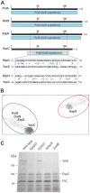
Figure 1
Figure 1. YacC is a novel member of the PulS-OutS family of proteins.
(A) The conserved domain architecture tool (CDART) was used to map the regions of PulS from Klebsiella oxytoca 10-5250 (EHT07154.1), OutS from Dickeya dadantii 3937 (YP_003883937.1), EtpO from Shiga toxin-producing E. coli (STEC) O157:H7 (CAA70966.1) and YacC (CAS07673.1) from EPEC. Numbers refer to the amino acids of each protein sequence, and the broken blue bar denotes that the N-terminal and C-terminal residues of YacC diverge from the consensus features of the PulS-OutS family. Pairwise sequence alignment over 80 residues showing similarity of the previously characterized EtpO (CAA70966.1) and YacC (CAS07673.1). Identical residues are highlighted between the two sequences, and conserved substitutions are shown (+). (B) CLANS analysis graphically depicts homology in large datasets of proteins, utilizing all-against-all pairwise BLAST to cluster representations (colored dots) of individual protein sequences in three-dimensional space. Lines are shown between the most similar sequences, with an E-value cut-off of 1e−5. The analysis shows that proteins from diverse species cluster into two groups: the PulS/OutS group and YacC-related proteins (blue), and the AspS-related proteins (red). There are numerous relationships between the PulS/OutS proteins and YacC proteins, but no relationship links these to the AspS group of proteins. (C) Wild-type EPEC (WT), and the indicated mutants of EPEC were grown in culture and post-cell supernatants containing secreted proteins (200 µg of protein) were analyzed by SDS-PAGE and Coomassie blue staining. Mass spectrometry was used to identify SslE, FliC and EspC, consistent with a previous study .
Figure 2
Figure 2. The kinetics of assembly, measured in vivo, for GspD in EPEC.
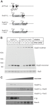
(A) The indicated strains of EPEC, complemented with the plasmid encoding GspD-C4 were cultured in medium to an OD600∼0.6 and arabinose was then added to the culture (0.1%, final concentration). At the indicated time-point cell extracts were prepared from the culture, resuspended in sample buffer containing Lumio reagent and analysed by SDS-PAGE. The polyacrylamide gels were then imaged by fluorimetry (Argon Blue 488 nm laser and 520 nm BP40 filter). Positions of molecular weight markers and the 21 kDa protein SlyD are indicated. (B) Wild-type EPEC (WT), ΔgspD mutant EPEC, and the ΔgspD mutant EPEC complemented with the plasmid encoding GspD-C4 were grown in culture and post-cell supernatants containing secreted proteins (200 µg of protein) were analyzed by SDS-PAGE and Coomassie blue staining. (C) Strains of EPEC: ΔgspD, ΔgspDΔyacC or ΔgspDΔaspS, were complemented with the plasmid encoding GspD-C4 under control of the tet promoter and were cultured to an OD600∼1.0, extracted and then fractionated by sucrose density centrifugation. Identical samples were analysed by SDS-PAGE for detection of GspD multimers with Lumio reagent and immunoblotting for the outer membrane protein BamA and the inner membrane β-subunit of the F1Fo-ATP synthase (F1β).
Figure 3
Figure 3. Subcellular targeting of GspD to the outer membrane depends on AspS.
(A) The expression cassette in pETDuet plasmids GspD-C4, GspD-C4+AspS and GspD-C4+YacC are represented diagrammatically. The pETDuet-1 vector (Novagen) has two multi-cloning sites (MCS) represented as black squares, and cloning into the NcoI and NdeI sites is optimal with respect to the ribosome-binding sites: NcoI and HindIII sites were used to clone the open-reading frame corresponding to GspD-C4; NdeI and XhoI sites were used to clone the open-reading frame corresponding to AspS and YacC. The T7 terminator sequence (T) in the plasmid is represented by a black triangle. (B) E. coli BL21(DE3)(ΔgspDaspS) complemented with pETDuet (GspD-C4) or pETDuet (GspD-C4+AspS) were cultured to an OD600 of ∼0.6 and IPTG was added to the culture (0.1 mM, final concentration). At the indicated time-point cell extracts were prepared from the cultures using Lumio analysed by SDS-PAGE and imaged by fluorimetry. (C) The strains described above were cultured to an OD600∼1, extracted and then fractionated by sucrose density centrifugation. Replicate samples were analysed by SDS-PAGE for detection of GspD multimers with Lumio reagent and immunoblotting for BamA and F1β.
Figure 4
Figure 4. Phylogenetic analysis reveals distinct types of T2SS secretins.
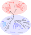
Phylogenetic tree reconstruction was performed with PhyML v3.0 using 500 bootstrap calculations and shown as percentage values (for further details see Methods). Based on the strong statistical support in the division, the Vibrio-type and Klebsiella type secretin sub-families are highlighted with colour. These have also been labelled as “GspDα” and “GspDβ” in accordance with a new nomenclature recently proposed for ETEC str. H10407 . The branch to the secretin SttD in Dickeya spp. is not shown to full scale. For a full list of the sequence accession numbers see Table S2.
Figure 5
Figure 5. The S-domains of Vibrio-type secretins have diagnostic sequence features.
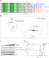
(A) Alignment of a representative subset of the secretin sequences used in this study to demonstrate sequence conservation (darker to lighter shades of green represent higher to lower levels of sequence conservation). Accession numbers for all secretins investigated in this study are given in Table S2. (B) S-domain sequences from the secretins were subject to CLANS analysis . The position corresponding to each S-domain sequence from the Vibrio-type EpsD and GspD proteins is represented by red dots, ExeD by orange dots and the group circled in red. The position corresponding to each sequence from the Klebsiella-type PulD, OutD, EtpD and GspD proteins is colour-coded in blue. The connections shown represent an E-value cut-off of 1e−10. (C) E. coli BL21(DE3)(ΔgspDaspS) complemented with either pETDuet (GspD-C4+AspS) or pETDuet (GspDΔS-C4+AspS) were cultured to an OD600 of ∼0.6 and IPTG was added to the culture (0.1 mM, final concentration). At the indicated time-point cell extracts were prepared from the cultures and incubated with a modified sample buffer containing Lumio reagent, analysed by SDS-PAGE and imaged by fluorimetry. (D) Size-exclusion chromatography profiles of the purified AspS (red), the purified MBP-S-domain fusion (green) and the complex of AspS and MBP-S-domain fusion (blue) on a Superdex200 column. An SDS-PAGE gel of the peak fractions of AspS-MBP-S-domain complex shows an approximately stoichiometric ratio of the two proteins. A280, absorbance at 280 nm; mAU, milli absorbance units. Figure S5 shows the results of the control experiment, where AspS and MBP without S-domain do not interact.
Figure 6
Figure 6. Structure of the pilotin AspS.
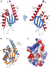
(A) Ribbon representation of the structure of V. cholerae AspS. α-helices, α1–α4, are in crimson; β-strands, β1–β5, are in light blue. Zn2+ ions are shown as grey spheres. Acetate ions are shown in stick representation. Residues coordinating Zn2+ and acetate ions are in stick representation with oxygen and nitrogen atoms color-coded red and blue, respectively. The position of the disulphide bond Cys74–Cys111 is shown. (B) A superposition of AspS (light blue) and PA3611 (orange) structures in ribbon representation. Note the outward movement of β-strands β1 and β2 in PA3611 structure. (C) Electrostatic surface potential of PA3611 structure (positive = blue; negative = red). The buffer CAPS molecules are shown in stick representation.
Figure 7
Figure 7. Pilotins distinguished for each class of T2SS secretin.
In K. oxytoca, PulD has three characterized domains: the N-domains (blue) that dock it to the inner membrane components of the T2SS, the secretin domain (pink) responsible for multimerization, and the C-terminal S-domain, which is critical for PulD to engage the pilotin PulS –. The predicted domain structure of GspD from EPEC is similarly shown, including the S-domain demonstrated to be necessary and sufficient for AspS binding. Also indicated are the T2SS secretins HxcQ and XcpQ from Pseudomonas, each of which has a C-terminal extension beyond the recognizable secretin domain which may or may not serve for binding of the protein of unknown function, PA3611.

Similar articles

Cited by

References

    1. Bos MP, Robert V, Tommassen J (2007) Biogenesis of the gram-negative bacterial outer membrane. Annu Rev Microbiol 61: 191–214. - PubMed
    1. Knowles TJ, Scott-Tucker A, Overduin M, Henderson IR (2009) Membrane protein architects: the role of the BAM complex in outer membrane protein assembly. Nat Rev Microbiol 7: 206–214. - PubMed
    1. Hagan CL, Silhavy TJ, Kahne D (2011) beta-Barrel membrane protein assembly by the Bam complex. Annu Rev Biochem 80: 189–210. - PubMed
    1. Tokuda H, Matsuyama S (2004) Sorting of lipoproteins to the outer membrane in E. coli. Biochim Biophys Acta 1693: 5–13. - PubMed
    1. Okuda S, Tokuda H (2011) Lipoprotein sorting in bacteria. Annu Rev Microbiol 65: 239–259. - PubMed

Publication types

MeSH terms