. 2013;9(1):e1003175.
doi: 10.1371/journal.pgen.1003175. Epub 2013 Jan 10.

A Drosophila model of high sugar diet-induced cardiomyopathy

Affiliations

A Drosophila model of high sugar diet-induced cardiomyopathy

Jianbo Na et al. PLoS Genet. 2013.

Abstract

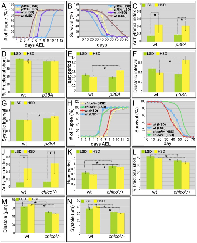
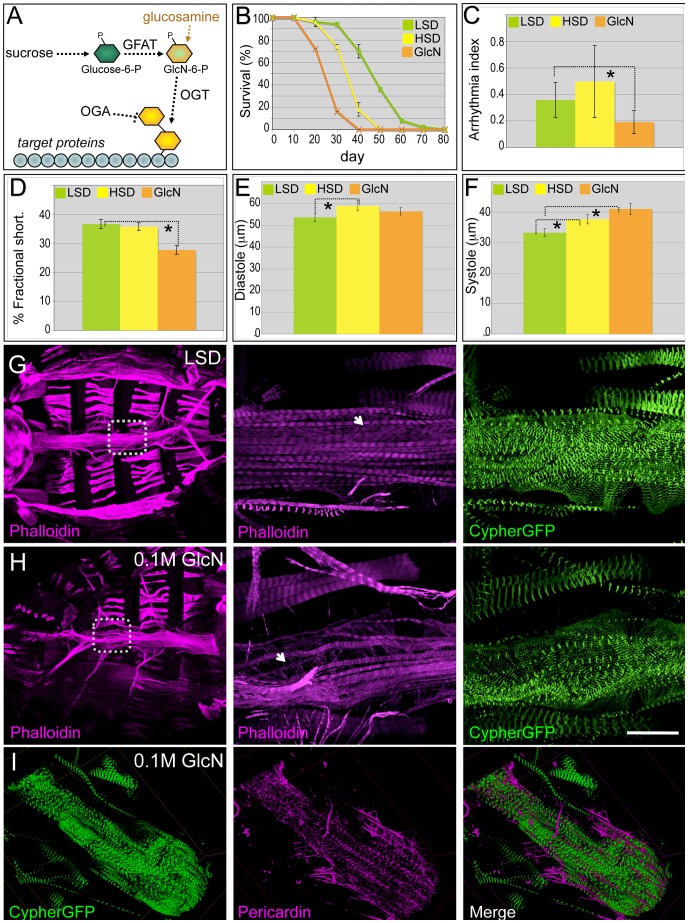
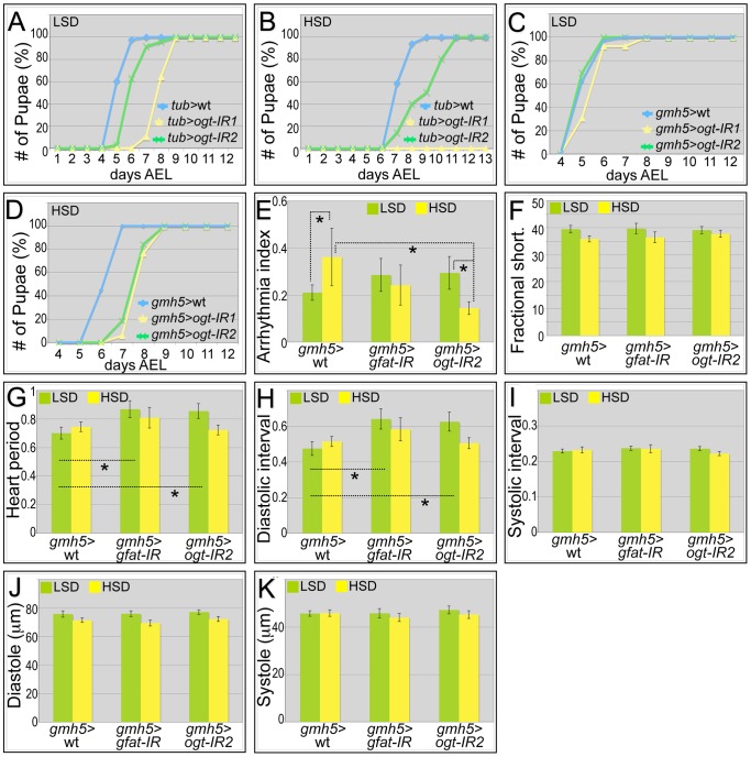
Diets high in carbohydrates have long been linked to progressive heart dysfunction, yet the mechanisms by which chronic high sugar leads to heart failure remain poorly understood. Here we combine diet, genetics, and physiology to establish an adult Drosophila melanogaster model of chronic high sugar-induced heart disease. We demonstrate deterioration of heart function accompanied by fibrosis-like collagen accumulation, insulin signaling defects, and fat accumulation. The result was a shorter life span that was more severe in the presence of reduced insulin and P38 signaling. We provide evidence of a role for hexosamine flux, a metabolic pathway accessed by glucose. Increased hexosamine flux led to heart function defects and structural damage; conversely, cardiac-specific reduction of pathway activity prevented sugar-induced heart dysfunction. Our data establish Drosophila as a useful system for exploring specific aspects of diet-induced heart dysfunction and emphasize enzymes within the hexosamine biosynthetic pathway as candidate therapeutic targets.


Conflict of interest statement

The authors have declared that no competing interests exist.

Figures

Figure 1
Figure 1. Drosophila model of diabetic cardiomyopathy.
(A) Ventral view of the heart tube. 3D structures are shown. The heart was stained for F-Actin with phalloidin (magenta) and for nuclei with DAPI; Cypher-GFP (green) labeled Z-lines of myofibers within cardiomyocytes. Arrowheads indicate the non-myocardial longitudinal muscle fibers, asterisks the alary muscles that support the heart, and arrows the abdominal muscles. (B) Dorsal view of the heart tube. Myocardial cells wrap in a circular fashion around the central cavity. Arrows show the ostia through which hemolymph from the abdomen enters into the heart tube and circulates. (C) HSD significantly reduced life span. w1118 male flies were raised in 0.15 M or 1.0 M sucrose diet, food was changed every 2–3 days, and flies were counted every 10 days. HSD-fed flies displayed decreased median life span, 10 days shorter than flies fed an LSD. Mean ± SE are shown; n = 50 total flies in two separate experiments. The estimated median life span of HSD flies was also expressed as the percentage of LSD flies (*p = 7.61E-08 by log rank test). See also Figure S1. (D) Hemolymph glucose concentrations in 3-week-old, control and HSD-fed w1118 adult flies. n≥6. (E) Hemolymph trehalose concentrations in 3-week-old, control and high sucrose-fed w1118 adult flies. n≥6. (F) Bodies from w1118 adults fed LSD or HSD for 3 weeks were treated with insulin (1 µM) or vehicle and visualized using antibodies against Drosophila PO4-Akt or Syntaxin. n = 10. Bands from Western blot experiments were quantified, and PO4-Akt was normalized to Syntaxin as a loading control. (G) Total triglycerides (TAG) were assayed enzymatically in 3-week-old control and high sugar-fed w1118 adult flies, and normalized to weight. n≥12. Mean ± SE are shown. An unpaired, two-tailed t-test was used to derive p-values.
Figure 2
Figure 2. High sucrose shortened Drosophila life span and was associated with increased cardiac arrhythmia and heart deterioration.
(A) Representative M-mode (5 seconds) from flies fed LSD and HSD. Three-week-old adults fed an HSD showed moderate cardiac arrhythmia; at six weeks arrhythmicity was increased. (B) Arrhythmia index obtained from w1118 flies fed LSD and HSD. Arrhythmias observed in M-mode can be quantified as arrhythmia index, which is the standard deviation of all heart periods in each record normalized to the median heart period for each fly. Mean ± SE are shown. At week three, a significant increase in arrhythmia index was observed in HSD fed flies (0.44) compared to low sucrose fed flies (0.16) (*P = 1.54E-17 by F-test). Arrhythmia index of six-week-old flies increased to 0.66 in HSD and 0.26 in low sucrose diet, respectively (*P = 6.01E-12 by F-test). Data are means ± SE. (C) Heart period of adult files fed low vs. high dietary sucrose. At three weeks of age, no difference was observed between HSD- and LSD-fed flies. Heart period was significantly increased at six weeks of age in both HSD- and LSD-fed flies (*P = 9.64E-06 and 1.44E-09, respectively, by t-test). Interestingly at six weeks of age, heart period of HSD-fed flies was shorter than that of low sucrose fed flies (*P = 0.015 by t-test). Data are means ± SE. (D) Fractional shortening of adult flies fed low vs. high dietary sucrose. At three weeks of age, no difference was observed between HSD- and LSD-fed flies; however, fractional shortening was significantly decreased in flies fed HSD- vs. LSD-fed (*P = 0.043 by t-test). Data are means ± SE. (E) Quantification of Pericardin level of adult heart by Western blot. Eight hearts from three-week-old LSD- and HSD-fed flies, respectively, were loaded. Pericardin level was detected by a monoclonal antibody against Pericardin, and normalized to Actin level. (F,G) Representative confocal images of three-week-old adult fly hearts expressing Cypher-GFP (posterior A2/anterior A3 segment) and stained with anti-Pericardin (magenta) antibody. Pericardin levels in hearts of flies fed an HSD were increased compared to those fed low sucrose. Note that Pericardin was detected in heart tissue but not abdominal muscles. See also Figure S4. (H) Fly hearts from w1118 adults fed LSD or HSD were treated with insulin (2.5 µM) or vehicle and visualized using antibodies against Drosophila PO4-Akt, PO4-4EBP or Actin, showing the response of the heart to exogenous insulin challenge. n = 3. Bands from Western blot experiments were quantified, and PO4-Akt and PO4-4EBP were normalized to Actin as a loading control. The ratio of HSD fed flies was then normalized to that of LSD fed flies. PO4-Akt and PO4-4EBP level were 74.3% and 13.9%, respectively, in HSD-fed flies compared to LSD-fed flies (P = 0.049 and 0.0003, respectively, by t-test). (I) Heart accumulated triglycerides (TAG) were assayed enzymatically in 15 hearts from 3-week-old LSD- and HSD-fed w1118 adult flies, and normalized to protein level. n = 2. (P = 0.028 by t-test). See also Figure S4 and Videos S1, S2, S3.
Figure 3
Figure 3. Mutations in p38A and chico attenuate high sucrose-induced heart deterioration.
(A) Mutations in p38A enhanced developmental delay of larvae fed an HSD. Eggs were collected onto LSD or HSD food for 16 hours, then permitted to mature at 25°C. Time to pupariation was scored daily. HSD feeding resulted in two days' development delay for w1118 flies. On low sucrose food, p38A mutations exhibited a slight delay compared to w1118 controls; an HSD led to a four-day delay to pupariation. (B) Mutant p38A flies fed high dietary sucrose exhibited reduced life span. Experiments for both p38A mutants and w1118 controls were performed at 22°C due to poor viability of developing p38A mutant flies grown at 25°C on an HSD. The average life span of p38A flies was ten days shorter than w1118 controls fed an HSD. (C) Loss of p38A activity did not alter high dietary sucrose-induced cardiac arrhythmia. Arrhythmia index obtained from w1118 controls and p38A mutants. The differences between LSD and HSD both in wild type controls and p38A mutants were significant (*P = 1.54E-17, and 1.20E-08, respectively, by F-test). However, there was no difference between w1118 controls and p38A mutants. Data are means ± SE. (D) No difference in fractional shortening was observed between w1118 controls and p38A mutants grown in either an LSD or an HSD. Data indicate mean ± SE. (E) Heart period was increased in p38A mutants fed an HSD. The heart period of p38A mutants fed on HSD was 0.86 second, compared to 0.60 second in w1118 controls (*P = 0.008 by t-test). Data indicate mean ± SE. (F) Diastolic interval was increased in p38A mutants fed an HSD. Diastolic interval of p38A mutants fed an HSD was 0.64 second, compared to 0.39 second in w1118 controls (*P = 0.01 by t-test). Data indicate mean ± SE. (G) Systolic interval was increased in in p38A mutants fed an HSD. Systolic interval of p38A mutants fed an HSD was 0.25 second, compared to 0.21 second in w1118 controls (*P = 0.02 by t-test). Data indicate mean ± SE. (H) No difference was observed in developmental rates between w1118 controls and chico1/+ adults fed either an LSD or HSD. Data indicate mean ± SE. (I) Increased life span was observed in chico1/+ adults compared to w1118 controls when both were fed an LSD. However, the observed average life span in HSD is 35 days in chico1/+ mutants, the same as w1118 controls. Data indicate mean ± SE. (J) chico1 mutation did not alter high dietary sucrose-induced cardiac arrhythmia. Arrhythmia index obtained from w1118 controls and chico1/+ mutants. The differences between LSD and HSD both in wild type controls and chico1/+ mutants were significant (*P = 1.54E-17, and 2.10E-10, respectively, by F-test). However, there was no difference between w1118 controls and chico1/+ mutants (P = 0.20 by F-test). Data indicate mean ± SE. (K) Heart period was increased in chico1/+ mutants fed with an LSD or HSD. Heart period of chico1/+ mutants raised on low sucrose was 0.94 second, compared to 0.68 second of w1118 controls (*P = 0.004 by t-test). Heart period of chico1/+ mutants raised on an HSD was 0.91 second, compared to 0.60 second of w1118 controls (*P = 0.0008 by t-test). Data indicate mean ± SE. (L) chico1/+ flies exhibited significantly reduced fractional shortening in both LSD and HSD. Fractional shortening of chico1/+ mutants raised on low sucrose was 37%, compared to 42% of w1118 controls (*P = 0.006 by t-test). Fractional shortening of chico1/+ mutants raised on an HSD was 34%, compared to 40% of w1118 controls (*P = 0.001 by t-test). Data are means ± SE. (M) chico1/+ flies exhibited significantly reduced diastole on both LSD and HSD. Diastole of chico1/+ mutants raised on low sucrose was 49.9 microns, compared to 62.9 microns of w1118 controls (*P = 0.0009 by t-test). Diastole of chico1/+ mutants raised on an HSD was 45.8 microns, compared to 68.1 microns of w1118 controls (*P = 2.14E-08 by t-test). Data are means ± SE. (N) Significantly reduced systole of chico1/+ flies was observed in both LSD and HSD. Systole of chico1/+ mutants raised on low sucrose was 31.6 microns, compared to 36.6 microns of w1118 controls (*P = 0.03 by t-test). Systole of chico1/+ mutants raised on an HSD was 30.2 microns, compared to 40.0 microns of w1118 controls (*P = 0.0002 by t-test). Data are means ± SE. See also Videos S4, S5.
Figure 4
Figure 4. Dietary glucosamine shortened life span and induced cardiac dysfunction.
(A) Hexosamine biosynthesis pathway (HBP). The two rate limiting enzymes GFAT and OGT convert glucose to a O-GlcNAc residue that is then targeted to protein substrates; this residue is removed by β-N-acetylglucosaminidase (OGA). Glucosamine bypasses GFAT and increases HBP flux. (B) Dietary glucosamine significantly reduced life span: the average life span of flies fed 0.1 M glucosamine was 25 days compared to 35 days on an HSD and 48 days on a (control) LSD. Data are means ± SE (n = 2 experiments, 25 flies per experiment). (C) Glucosamine-fed flies displayed decreased arrhythmia. Arrhythmia index was obtained from wild type (Canton S) in LSD, HSD, and glucosamine diet. Glucosamine diet significantly reduced arrhythmia index (*P = 0.006 by F-test). Note Canton S flies fed with low sucrose showed slightly higher arrhythmia than w1118 flies fed with low sucrose. Data are means ± SE. (D) Glucosamine reduced fractional shortening in three-week-old adult flies (*P = 0.001 by t-test). Data are means ± SE. (E) Unlike high dietary sucrose (*P = 0.042 by t-test), dietary glucosamine did not change the diastolic diameter (P = 0.32 by t-test). Data are means ± SE. (F) Diets supplemented with glucosamine (*P = 0.0009 by t-test) or HSD (*P = 0.04 by t-test) increased systolic diameter, indicating that changes in fractional shortening are due to changes in systolic diameter. Data are means ± SE. (G,H) Dietary glucosamine led to heart structure defects. F-Actin in the heart was visualized with phalloidin (red) and Cypher-GFP (green) to label the Z-lines of myofibers within cardiomyocytes (magnification = 10×). Insets magnify the boxed regions to show the myofibers at higher magnification (63×). Size bar = 250 µm. (I) Representative confocal images of a three-week old Cypher-GFP fly heart from an animal fed 0.1 M glucosamine. Visualization with anti-Pericardin antibody (magenta) indicated decreased Pericardin levels. Compare with the control shown in Figure 2E. See also Video S6.
Figure 5
Figure 5. Genetic manipulation of the hexosamine biosynthetic pathway.
(A) Global knockdown of OGT with two separate ogt-IR lines by tub-gal4 led to a delay in development to pupariation in larvae fed an LSD. (B) This delay was further enhanced in the presence of an HSD: in particular, knockdown of OGT by tub>ogt-IR1 led to lethality. (C) Heart-specific knockdown of OGT (gmh5-gal4) led to little or no effect on development rate in the presence of control food. (D) By contrast, heart-specific knockdown of OGT (gmh5>ogt-IR) led to a 1.5 day developmental delay in the presence of high dietary sucrose. (E) Heart-specific knockdown of OGT or GFAT decreased high sugar induced arrhythmia. Arrhythmia index was somewhat increased—though not statistically significantly— in flies with a heart-specific knockdown of OGT or GFAT flies raised in low sucrose diet compared to wild type controls. However, when raised on an HSD, heart-specific knockdown of OGT or GFAT led to decreased arrhythmia compared to wild type controls; especially for OGT knockdown flies, the decrease in arrhythmia index was significant (*P = 5.57E-08 by F-test). (F) Heart specific knockdown of GFAT or OGT did not alter fractional shortening. Fractional shortening of wild type controls, gmh5>gfat-IR flies and gmh5>ogt-IR2 flies are shown. Data represent means ± SE. (G) Heart period did not change in heart specific knockdown of GFAT or OGT flies raised on an HSD. However, when raised in low sucrose diet, they displayed increased heart period (*P = 0.02 for both GFAT or OGT knockdown by t-test). (H) Diastolic interval did not change in heart specific knockdown of GFAT or OGT flies raised on an HSD. However, when raised in low sucrose diet, they showed increase of diastolic interval (*P = 0.03 for GFAT knockdown and 0.04 for OGT knockdown by t-test). (I) Systolic interval did not change in heart specific knockdown of GFAT or OGT flies raised on an LSD or HSD. (J) Diastole did not change in heart specific knockdown of GFAT or OGT flies raised on an LSD or HSD. (K) Systole did not change in heart specific knockdown of GFAT or OGT flies raised on an LSD or HSD. See also Videos S7, S8, S9.

Similar articles

Cited by

References

    1. Marriott BP, Cole N, Lee E (2009) National Estimates of Dietary Fructose Intake Increased from 1977 to 2004 in the United States. The Journal of Nutrition 139: 1228S–1235S. - PubMed
    1. Mellor KM, Ritchie RH, Davidoff AJ, Delbridge LMD (2010) Elevated dietary sugar and the heart: experimental models and myocardial remodeling. Canadian Journal of Physiology and Pharmacology 88: 525–540. - PubMed
    1. Selvin E, Coresh J, Golden SH, Brancati FL, Folsom AR, et al. (2005) Glycemic Control and Coronary Heart Disease Risk in Persons With and Without Diabetes: The Atherosclerosis Risk in Communities Study. Arch Intern Med 165: 1910–1916. - PubMed
    1. Fung TT, Malik V, Rexrode KM, Manson JE, Willett WC, et al. (2009) Sweetened beverage consumption and risk of coronary heart disease in women. The American Journal of Clinical Nutrition 89: 1037–1042. - PMC - PubMed
    1. Rubler S, Dlugash J, Yuceoglu YZ, Kumral T, Branwood AW, et al. (1972) New type of cardiomyopathy associated with diabetic glomerulosclerosis. The American Journal of Cardiology 30: 595–602. - PubMed

Publication types

LinkOut - more resources