. 2013;9(1):e1003195.
doi: 10.1371/journal.pgen.1003195. Epub 2013 Jan 10.

Genome-wide screens for in vivo Tinman binding sites identify cardiac enhancers with diverse functional architectures

Affiliations

Genome-wide screens for in vivo Tinman binding sites identify cardiac enhancers with diverse functional architectures

Hong Jin et al. PLoS Genet. 2013.

Abstract

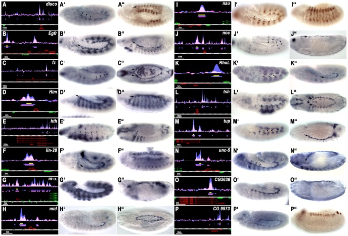
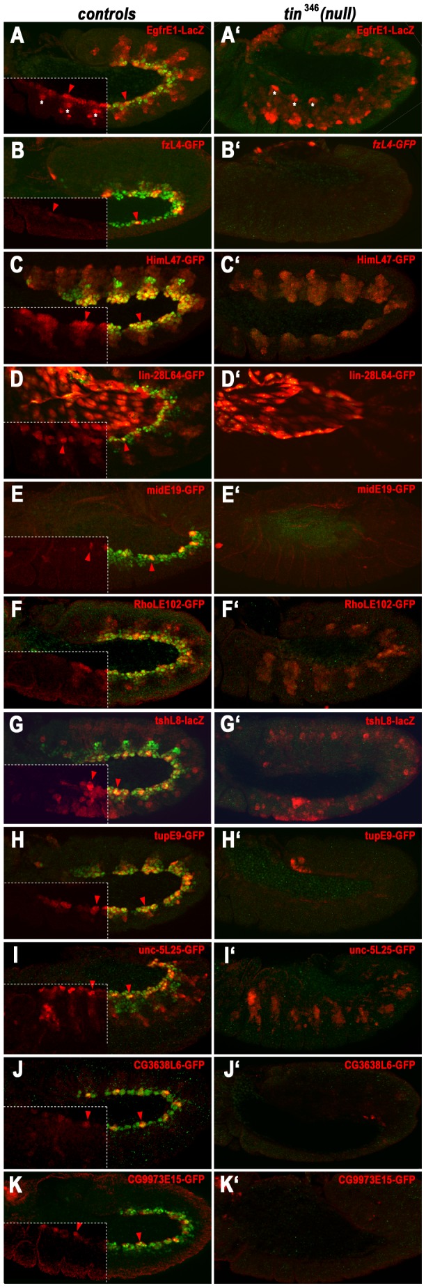
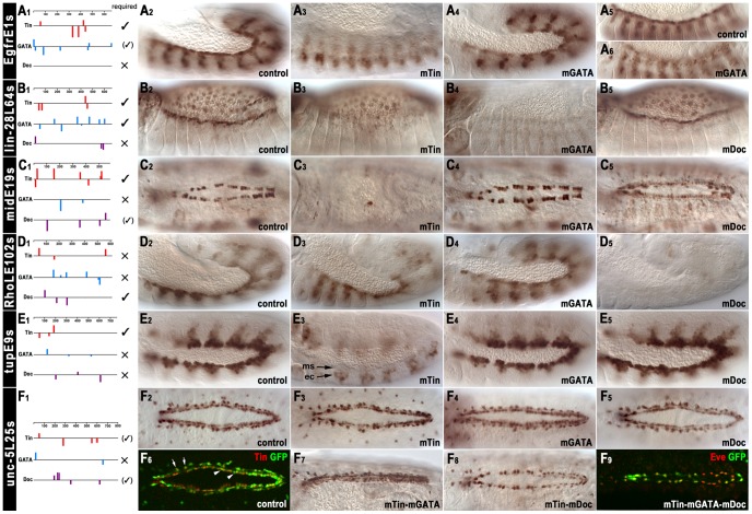
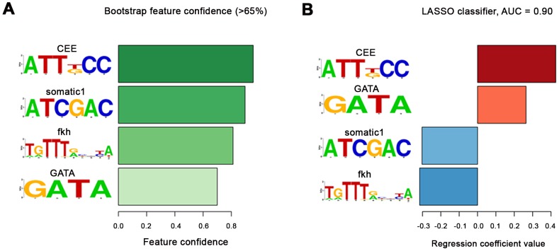
The NK homeodomain factor Tinman is a crucial regulator of early mesoderm patterning and, together with the GATA factor Pannier and the Dorsocross T-box factors, serves as one of the key cardiogenic factors during specification and differentiation of heart cells. Although the basic framework of regulatory interactions driving heart development has been worked out, only about a dozen genes involved in heart development have been designated as direct Tinman target genes to date, and detailed information about the functional architectures of their cardiac enhancers is lacking. We have used immunoprecipitation of chromatin (ChIP) from embryos at two different stages of early cardiogenesis to obtain a global overview of the sequences bound by Tinman in vivo and their linked genes. Our data from the analysis of ~50 sequences with high Tinman occupancy show that the majority of such sequences act as enhancers in various mesodermal tissues in which Tinman is active. All of the dorsal mesodermal and cardiac enhancers, but not some of the others, require tinman function. The cardiac enhancers feature diverse arrangements of binding motifs for Tinman, Pannier, and Dorsocross. By employing these cardiac and non-cardiac enhancers in machine learning approaches, we identify a novel motif, termed CEE, as a classifier for cardiac enhancers. In vivo assays for the requirement of the binding motifs of Tinman, Pannier, and Dorsocross, as well as the CEE motifs in a set of cardiac enhancers, show that the Tinman sites are essential in all but one of the tested enhancers; although on occasion they can be functionally redundant with Dorsocross sites. The enhancers differ widely with respect to their requirement for Pannier, Dorsocross, and CEE sites, which we ascribe to their different position in the regulatory circuitry, their distinct temporal and spatial activities during cardiogenesis, and functional redundancies among different factor binding sites.


Conflict of interest statement

The authors have declared that no competing interests exist.

Figures

Figure 1
Figure 1. Sequence motifs enriched in Tin binding regions.
(A) Schematic drawings of the expression domains of Tinman (red), Doc (green, hatched), and Pnr (blue, hatched) during the embryonic stages used for chromatin preparations. Grey: mesoderm not expressing any of these factors. (B) Tin motif recovered using de novo motif discovery in the top 100 peaks and full Early and Late datasets. The Tin motifs discovered in the full Early and Late dataset are almost identical, while the ones from top 100 peaks show some differences. These Tin motifs closely resemble the motifs derived from published Tin binding sites with verified in vivo functions (bottom) and previously published Tinman/Nkx2-5 motifs . The Tin motif from the full datasets is preferentially located in the center (0.5 on X-axis) of ChIP peaks (see Materials and Methods). (C) The AGATAC motif is the most enriched sixmer in both Early and Late datasets. The core of this motif is the GATA sequence to which a number of TFs, including GATA factor Pnr, bind to. The AGATAC motif is preferentially located in the centre of ChIP peaks similarly to the Tin motif, but to a slightly lesser extent. (D) The binding motifs of the T-box factors Mid and Doc2 from SELEX experiments. The Doc2 motif is also located preferentially near the ChIP peak centre, but to a lesser extent than both Tin and GATA motifs.
Figure 2
Figure 2. Tin binding and reporter activity of enhancers active in dorsal mesodermal cells, cardiac progenitors and heart progenitors.
(A–P) show the Tin binding peaks (blue: 3–5.5 hrs: pink: 5–8 hrs AEL), the location of the tested enhancers (yellow bars) and the gene models (red: genes closest to tested fragment). Y axes are at identical scales whereas x axis scales are variable (see scale bars). A′ to P′ show enhancer activities at earliest stage of appearance (arrow or arrow heads: dorsal/cardiac mesoderm) and A″ to P″ at latest stage of detection in cardiac tissues (arrows or arrow heads). Shown are GFP or lacZ in situ hybridizations except in A″, I′, I″ and P″, which show anti-GFP antibody stainings (in A″, I″ and P″ perduring GFP from earlier expression). Genes are ordered alphabetically, with CGs last. (A′) Stage 12. Expression of discoL10-GFP in heart progenitors, segmental subsets of visceral muscle precursors and (A″), stage 14, perdurance of GFP in developing visceral muscles and heart. (B′, B″) Expression of EgfrE1-lacZ in cardioblast progenitors and subsets of somatic mesodermal cells (stage 12), and in cardioblasts (stage 14). (C′, C″) Expression of fzL4-GFP in cardioblast progenitors (stage 12) and in cardioblasts and amnioserosa (stage 14). (D′, D″) Expression of HimL47-GFP in cardiogenic mesoderm and somatic mesodermal cells (stage 12), and in cardioblasts and developing somatic muscles (stage 14). (E′, E″) Expression of hthE54-GFP in heart progenitors of Md, T1–T3 segments (arrow heads) and in somatic mesoderm (with A–P graded sizes of clusters) (stage 12) and in anterior developing heart cells (arrow head) and somatic muscles (stage 14). (F′, F″) Expression of lin-28L64-GFP in cardioblasts (stage 12, stage14). (G′, G″) Expression of maL9-GFP in cardiac and somatic mesoderm (stage 12) and in heart precursors (stage 14). (H′, H″) Expression of midE19-GFP in Tin+ cardioblasts (stage 12, stage 15). (I′, I″) Expression (I′, stage 12) and perdurance (I″ stage 14) of nauL35-GFP largely in dorsal somatic mesoderm (asterisks). (J′, J″) Expression of nocL7-GFP in pericardial cell progenitors (stage 12) and pericardial cells (stage 16). (K′, K″) Expression of RhoLE102-GFP in segmented dorsal mesoderm (stage 11) and cardioblast progenitors (stage 12). (L′, L″) Expression of tshL8-lacZ in cardiogenic mesoderm and thoracic ectoderm (stage 11) and in Tin+ pericardial cells (stage 15). (M′, M″) Expression of tupE9-GFP in cardiogenic mesoderm (stage 11) and cardioblasts (stage 14). (N′, N″) Expression of unc-5L25-GFP in cardioblast and pericardial cell progenitors (stage 12) and in cardioblasts and Tin+ pericardial cell (stage 16). (O′, O″) Expression of CG3638L6-GFP in cardioblast progenitors (stage 12) and cardioblasts (stage 15). (P′, P″) Expression of CG9973E15-GFP in cardiac mesoderm (stage 11) and perdurance of GFP in developing heart (stage 14).
Figure 3
Figure 3. Dependency of early cardiac enhancer activities on tin.
Shown are stage 11–12 embryos stained for enhancer activities (anti-βGal or anti-GFP) and Tin (green). (A–K) Enhancer activities in wild type backgrounds (left corner quadrants: anti-Tin omitted for better visualization of reporter patterns; arrow heads: early cardiac expression). (A′–K′) Enhancer activities in homozygous tin 346 mutant backgrounds. (A, A′) EgfrE1-LacZ expression in cardiac mesoderm but not in somatic mesoderm (asterisks) requires tin. (B, B′) fzL4-GFP expression in cardioblast progenitors requires tin. (C, C′) High-level HimL47-GFP expression in cardiogenic mesoderm requires tin but somatic mesodermal expression does not. (D, D′) lin-28L64-GFP expression in cardiac mesoderm requires tin. Amnioserosa expression is unaffected in tin mutants. (E, E′) midE19-GFP expression in cardioblast progenitors requires tin. (F, F′) RhoLE102-GFP expression in cardiogenic mesoderm, but not in somatic mesoderm, requires tin. (G, G′) tshL8-LacZ expression in cardiogenic mesoderm, but not in somatic mesoderm, requires tin. (H, H′) tupE9-GFP expression in cardiogenic mesoderm requires tin. (I, I′) unc-5L25-GFP expression in cardiac mesoderm but not in somatic mesoderm requires tin. (J, J′) CG3638L6-GFP expression in cardioblast progenitors requires tin. (K, K′) CG9973E15-GFP expression in cardioblast progenitors requires tin.
Figure 4
Figure 4. Dependency of late cardiac enhancer activities within the dorsal vessel on tin.
Shown are reporter activities (anti-GFP, green), Tin+ cardioblasts and pericardial cells (anti-Tin, red) and Doc+ cardioblasts (anti-Doc, blue) in stage 15–16 control embryos (A–D) and in embryos specifically lacking Tin activity in cardiac cells (tinABD, tin346; A′–D′). (A) midE19-GFP is expressed specifically in the Tin+ cardioblasts. (A′) Absence of cardiac Tin expression causes a severe reduction of midE19-GFP activity. (B) tupE9-GFP is highly expressed in Tin+ cardioblasts (graded posteriorly-to-anteriorly) and, at much lower levels perduring from stage 12 expression, is present in Doc+ cardioblasts, pericardial cells, and dorsal somatic muscles. (B′) Upon loss of cardiac Tin expression almost all cardioblasts contain only low levels of perduring GFP. (C) unc-5L25-GFP expression in pericardial cells and (largely posteriorly) in cardioblasts. (C′) Absence of cardiac Tin expression causes near loss of cardioblast unc-5L25-GFP expression and a reduction of expression in pericardial cells. (D) CG3638L6-GFP is expressed in Tin+ cardioblasts (with variable intensities) and in Tin+ pericardial cells. (D′) Absence of cardiac Tin expression causes nearly complete loss of CG3638L6-GFP expression.
Figure 5
Figure 5. In vivo assays of the functions of predicted binding sites of Tin, Doc, and Pnr in identified cardiac enhancers.
(A1–F1) Positions, orientations, and scores of predicted binding sites within cardiac enhancers (identical Y-scales). In vivo results for functionality of binding sites are summarized to the right (large ✓: essential; small (✓): required for full activity or redundantly with motifs for other factors; × not required). (A2, A3) EgfrE1s-GFP activity in cardiac mesoderm (A2, stage 12) is lost upon mutation of Tin binding motifs shown in (A1). (A4–A6) Mutation of GATA motifs shown in (A1) leads to a mild reduction of EgfrE1s-GFP activity at stage 12 (A4) and a more significant reduction at stage 14 (A6). (B2, B3) lin-28L64s-GFP activity in cardioblasts (B2, stage 14) is lost upon mutation of Tin binding motifs. (B4) Mutation of GATA motifs leads to loss of lin-28L64s-GFP activity in both cardioblasts and amnioserosa cells. (B5) Mutation of Doc binding motifs affects neither cardioblast nor amnioserosa activity of lin-28L64s-GFP. (C1, C2) midE19s-GFP activity in cardioblasts (C2, stage 15) is lost upon mutation of Tin binding motifs (C3). (C4) Mutation of GATA motifs does not affect midE19s-GFP activity. (C5) Mutation of Doc binding motifs reduces cardioblast midE19s-GFP activity. (D2–D4) Mutation of the binding motifs for Tin (D3) or Pnr (D4) does not affect RhoLE102s-GFP activity in the cardiogenic and dorsal somatic mesoderm. (D5) Mutation of Doc binding motifs causes loss of RhoLE102s-GFP activity in cardiogenic and dorsal somatic mesoderm. (E2, E3) Mutation of Tin binding motifs causes loss of tupE9s-GFP activity in dorsal and cardiogenic mesoderm (ms). Ectopic tupE9s-GFP occurs in dorsal ectoderm (ec). (E4, E5) Mutation of GATA motifs (E4) or Doc binding motifs (E5) does not affect tupE9s-GFP activity. (F2–F5) Mutation of either the Tin binding motifs (F3), the GATA motifs (F4) or the Doc binding motifs (F5) does not affect unc-5L25s-GFP activity in cardioblasts and pericardial cells at stage 15. Three sequences, CCAAGGG, TCAATTG, TCGAGTG, poorly matching the Tin binding motif (Figure 1, Table S5), are still present but it is unknown whether they can bind Tin. (F6) Staining of stage 15 unc-5L25s-GFP control embryo for GFP and Tin identifies GFP-positive cells as cardioblasts (arrow heads) and Tin+ pericardial cells (arrows). (F7) Simultaneous mutation of Tin binding motifs and GATA motifs does not affect cardiac unc-5L25s-GFP activity. (F8) Simultaneous mutation of Tin and Doc binding motifs severely reduces unc-5L25s-GFP activity in cardioblasts but not in pericardial cells. (F9) Simultaneous mutation of binding motifs for Tin, Pnr, and Doc abrogates unc-5L25s-GFP activity (anti-GFP, green) in cardioblasts but not in pericardial cells (anti-Eve, red).
Figure 6
Figure 6. Cardiac enhancers classifier.
(A) The motifs with bootstrap confidence over 65%. The confidence levels are shown as bars and are colour-coded with darker color for larger confidence. The motif with the greatest confidence was the de novo CEE motif followed by another de novo motif somatic1. The GATA motif and the fkh motif from JASPAR had smaller confidence. As expected, Tin motifs were not retrieved as all fragments had been selected based upon high Tin occupancy, which in most cases involves Tin motifs, whereas the classifier was trained to distinguish between the two groups of enhancers. (In addition, the exact borders of both the cardiac and non-cardiac enhancers had been adjusted for their inclusion of one or more Tin motifs, if present within the binding peak region). (B) The regression coefficients in the final classifier that contains only the 4 motifs with >65% confidence. During classifier training the cardiac enhancers were given a target value of 1 and the rest of enhancers −1. Enhancers are scored by multiplying the regression coefficients with the standardized motif hit density (standardized to zero mean and unit variance over the whole dataset) in the sequence. Thus, a positive regression coefficient indicates that the above-average motif presence predicts cardiac enhancers, and negative that it predicts non-cardiac enhancers. The motif with the largest positive regression coefficient is the CEE motif followed by GATA. GATA (Pnr) is a known co-factor of Tin, which we further verified in our mutation analysis, while CEE was the novel predicted motif (see main text). Presence of two motifs: somatic1 and fkh predicted the non-cardiac enhancers. Somatic1 is another de novo motif we discovered (see Materials and Methods) but which we did not test functionally, while fkh indicates that a protein from the forkhead family likely binds to those Tin-bound enhancers that are not active in the cardiac cells but in other parts of the mesoderm. Note that the AUC score shown is an overly optimistic estimate of real generalization error due to selection bias .
Figure 7
Figure 7. In vivo assays of the function of the CEE motifs in selected cardiac enhancers.
(A1, B1, C1) Schematic drawings of the predicted Tin, Pnr, and Doc binding sites within cardiac enhancers as in Figure 5 and the positions of the CEE motifs relative to these (green bars). (A2–A5) Mutation of the CEE motifs causes a strong reduction of EgfrE1s-GFP activity in cardioblast progenitors at stage 12 (A2, A3) and in cardioblasts at stage 14 (A4, A5). (B2–B5) Mutation of the CEE motifs leads to the absence of lin-28L64s-GFP activity in heart precursors (and amnioserosa cells) at stage 14 (B2, B3) and a reduced activity in cardioblasts and pericardial cells at stage 16 (B4, B5). (C2, C3) Mutation of the CEE motifs causes almost complete loss of midE19s-GFP activity in Tin+ cardioblasts (stage 16).

Similar articles

Cited by

References

    1. Bodmer R, Frasch M (2010) Development and aging of the Drosophila heart. In: Harvey R, Rosenthal N, editors. Heart Development and Regeneration. Oxford: Academic Press. pp. 47–86.
    1. Bryantsev AL, Cripps RM (2009) Cardiac gene regulatory networks in Drosophila . Biochim Biophys Acta 1789: 343–353. - PMC - PubMed
    1. McCulley DJ, Black BL (2012) Transcription factor pathways and congenital heart disease. Curr Top Dev Biol 100: 253–277. - PMC - PubMed
    1. Yin Z, Xu X-L, Frasch M (1997) Regulation of the Twist target gene tinman by modular cis-regulatory elements during early mesoderm development. Development 124: 4871–4982. - PubMed
    1. Bodmer R, Jan LY, Jan YN (1990) A new homeobox-containing gene, msh-2, is transiently expressed early during mesoderm formation of Drosophila . Development 110: 661–669. - PubMed

Publication types

MeSH terms