. 2012 Dec;28(4):255-63.
doi: 10.5625/lar.2012.28.4.255. Epub 2012 Dec 26.

Human leukocytes regulate ganglioside expression in cultured micro-pig aortic endothelial cells

Affiliations

Human leukocytes regulate ganglioside expression in cultured micro-pig aortic endothelial cells

Jin-Hyoung Cho et al. Lab Anim Res. 2012 Dec.

Abstract

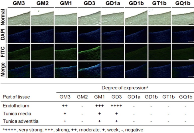
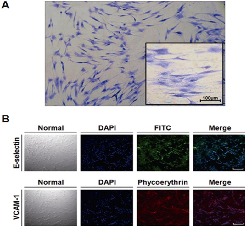
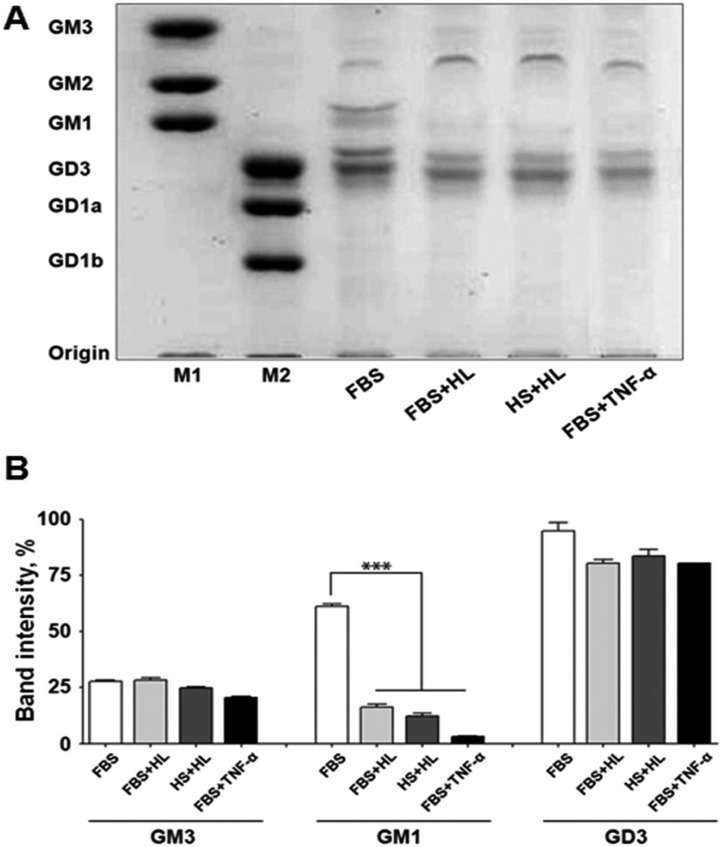
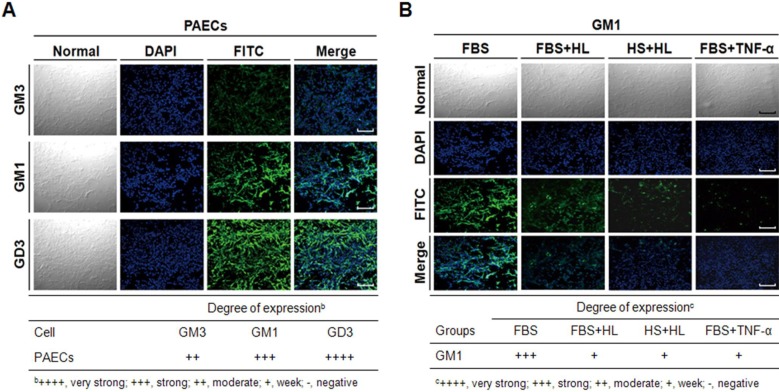
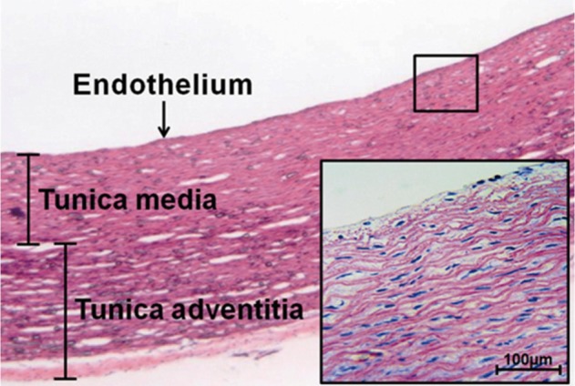
Gangliosides are ubiquitous components of the membranes of mammalian cells that are thought to play important roles in various cell functions such as cell-cell interaction, cell adhesion, cell differentiation, growth control, and signaling. However, the role that gangliosides play in the immune rejection response after xenotransplantation is not yet clearly understood. In this study, the regulatory effects of human leukocytes on ganglioside expression in primary cultured micro-pig aortic endothelial cells (PAECs) were investigated. To determine the impact of human leukocytes on the expression of gangliosides in PAECs, we performed high-performance thin layer chromatography (HPTLC) in PAECs incubated with FBS, FBS containing human leukocytes, human serum containing human leukocytes, and FBS containing TNF-α. Both HPTLC and immunohistochemistry analyses revealed that PAECs incubated with FBS predominantly express the gangliosides GM3, GM1, and GD3. However, the expression of GM1 significantly decreased in PAECs incubated for 5 h with TNF-α (10 ng/mL), 10% human serum containing human leukocytes, and 10% FBS containing human leukocytes. Taken together, these results suggest that human leukocytes induced changes in the expression profile of ganglioside GM1 similar to those seen upon treatment of PAECs with TNF-α. This finding may be relevant for designing future therapeutic strategies intended to prolong xenograft survival.

Keywords: Micro-pig aortic endothelial cells; ganglioside GM1; human leukocyte; human serum; tumor necrosis factor-α.


Figures

Figure 1
Figure 1
Immunofluorescence staining of micro-pig aorta sections. Sections were immunostained with GMR6 (anti-GM3 MAb), GMB28 (anti-GM2 MAb), GMB16 (anti-GM1 MAb), GMR19 (anti-GD3 MAb), GMR17 (anti-GD1a MAb), GGR12 (anti-GD1b MAb), GMR5 (anti-GT1b MAb), GMR13 (anti-GQ1b MAb), and fluorescein isothiocyanate (FITC)-labeled goat anti-mouse IgM antibody, and then stained with Hoechst 33342 for DNA. The bar represents 50 µm.
Figure 2
Figure 2
Characterization and GIEMSA staining of micro-pig aortic endothelial cells. Isolated micro-pig aortic endothelial cells (PAECs) were identified as endothelial cells (A) based on their morphology after Giemsa staining, and (B) by the presence of E-selectin/CD106 and VCAM-1/CD62E, well-known endothelial cell markers. The bar represents 50 µm.
Figure 3
Figure 3
High-performance thin layer chromatography and quantitative analysis of ganglioside expression in micro-pig aortic endothelial cells. (A) Gangliosides isolated from micro-pig aortic endothelial cells (PAECs) were separated by high-performance thin layer chromatography (HPTLC) silica gel plates using chloroform:methanol:0.25% CaCl2 (50:40:10, v/v/v), and gangliosides were visualized using resorcinol solution. Lane 1 (M1) and 2 (M2), ganglioside standard markers; lane 3, 10% FBS; lane 4, 10% FBS containing human leukocytes; lane 5, 10% human serum containing human leukocytes; lane 6, 10% FBS containing TNF-α (10 ng/mL). The results are representative of at least 3 independent experiments. (B) Quantitative analysis of ganglioside expression was performed using with the Beta 4.0.3 densitometry program (Scion Image, Frederick, MD, USA). Data are expressed as mean±SD; n=3; ***P<0.001.
Figure 4
Figure 4
Immunofluorescence staining of micro-pig aortic endothelial cells. (A) Normal PAECs were immunostained with GMR6 (anti-GM3 MAb), GMB16 (anti-GM1 MAb), GMR19 (anti-GD3 MAb), and FITC-labeled goat anti-mouse IgM antibody and then stained with Hoechst 33342 to detect DNA. (B) Normal PAECs and PAECs-incubated with FBS containing human leukocytes, with leukocyte-containing 10% human serum, or with FBS containing TNF-α (10 ng/mL) were immunostained with GMB16 (anti-GM1 MAb), and FITC-labeled goat anti-mouse IgM antibody, and then stained with Hoechst 33342 to detect DNA. The bar represents 50 µm.
Supplement 1
Supplement 1
Paraffin section and hematoxylin and eosin staining. A vertical section (4 µm thickness) of micro-pig aorta was stained with hematoxylin and eosin to reveal the presence of the endothelium, tunica media, and tunica adventitia. The bar represents 100 µm.

Similar articles

Cited by

References

    1. Cozzi E, White DJ. Xenotransplantation. Curr Opin Nephrol Hypertens. 1996;5(6):514–518. - PubMed
    1. Hancock WW. The past, present, and future of renal xenotransplantation. Kidney Int. 1997;51(3):932–944. - PubMed
    1. Parker W, Saadi S, Lin SS, Holzknecht ZE, Bustos M, Platt JL. Transplantation of discordant xenografts: a challenge revisited. Immunol Today. 1996;17(8):373–378. - PubMed
    1. Platt JL. The prospects for xenotransplantation of the kidney. Curr Opin Nephrol Hypertens. 1997;6(3):284–291. - PubMed
    1. Bach FH, Robson SC, Winkler H, Ferran C, Stuhlmeier KM, Wrighton CJ, Hancock WW. Barriers to xenotransplantation. Nat Med. 1995;1(9):869–873. - PubMed