

SATB1 expression is associated with biologic behavior in colorectal carcinoma in vitro and in vivo
- PMID: 23326301
- PMCID: PMC3543436
- DOI: 10.1371/journal.pone.0047902
SATB1 expression is associated with biologic behavior in colorectal carcinoma in vitro and in vivo
Abstract
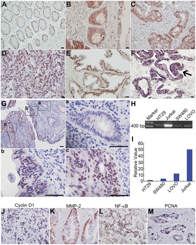
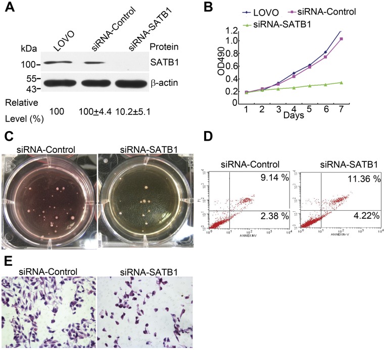
There is increasing evidence that Special AT-rich sequence-binding protein 1 (SATB1) is aberrantly expressed in several cancers and is correlated with clinicopathologic parameters in these tumors. In this study, we showed over-expression of SATB1 in 80 cases of colorectal cancer and in 3 colorectal cancer cell lines and found expression levels were strongly associated with tumor differentiation and stage. Expression levels of SATB1 protein were higher in poorly-differentiated as compared with well-differentiated cell lines, and both quantity and distribution patterns of SATB1 were associated with tumor differentiation and pTNM stage. Strikingly, we further investigated the effect of down regulation of SATB1 expression on malignant phenotypic features in colorectal cancer cells in vitro, and showed that SABT1 down-regulation negatively affected growth potential, anchorage-independent colony formation and cancer cell invasion, and resulted in increased apoptosis. SATB1 expression was positively associated with the expression of various biological and genetic markers, including Cyclin D1, MMP-2, NF-κB, and PCNA, and was associated with loss of APC and BRAF(V600E). These findings suggest that SATB1 is involved in the carcinogenesis, development and progression of colorectal cancer.
Conflict of interest statement
Figures
References
-
- Galande S, Purbey PK, Notani D, Kumar PP (2007) The third dimension of gene regulation: organization of dynamic chromatin loopscape by SATB1. Curr Opin Genet Dev 17: 408–414. - PubMed
-
- Kouzarides T (1999) Histone acetylases and deacetylases in cell proliferation. Curr Opin Genet Dev 9: 40–48. - PubMed
-
- Pavan Kumar P, Purbey PK, Sinha CK, Notani D, Limaye A, et al. (2006) Phosphorylation of SATB1, a global gene regulator, acts as a molecular switch regulating its transcriptional activity in vivo. Mol Cell 22: 231–243. - PubMed
-
- Wen J, Huang S, Rogers H, Dickinson LA, Kohwi-Shigematsu T, et al. (2005) SATB1 family protein expressed during early erythroid differentiation modifies globin gene expression. Blood 105: 3330–3339. - PubMed
Publication types
MeSH terms
Substances
LinkOut - more resources
Full Text Sources
Other Literature Sources
Medical
Research Materials
Miscellaneous
