

Expression of chicken DEC205 reflects the unique structure and function of the avian immune system
- PMID: 23326318
- PMCID: PMC3541370
- DOI: 10.1371/journal.pone.0051799
Expression of chicken DEC205 reflects the unique structure and function of the avian immune system
Abstract
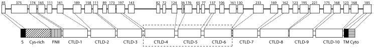
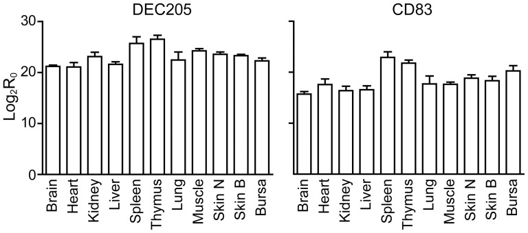
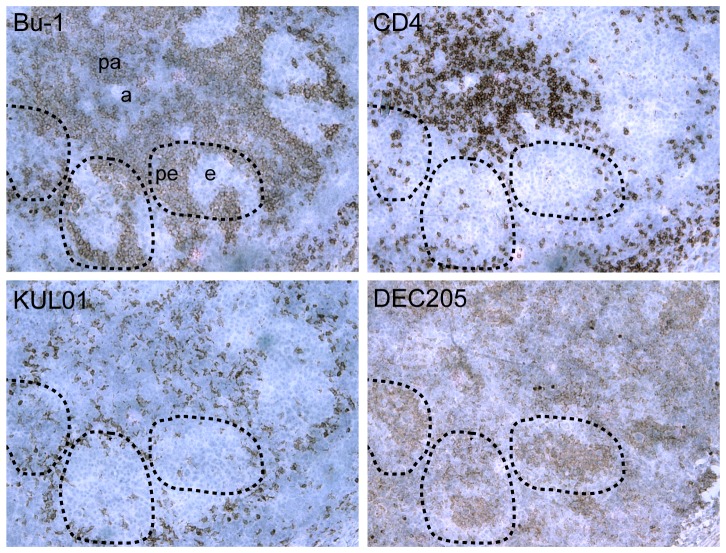
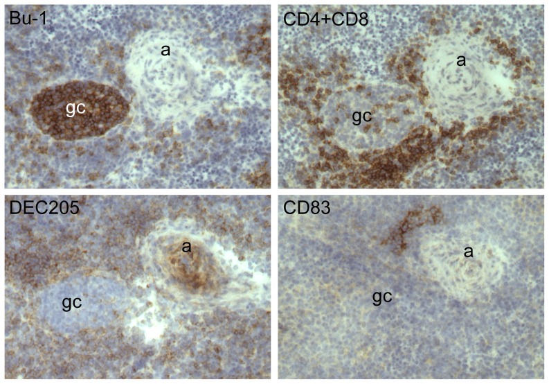
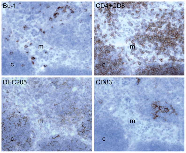
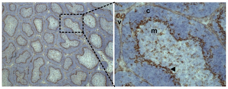
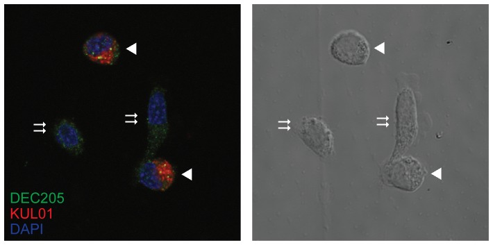
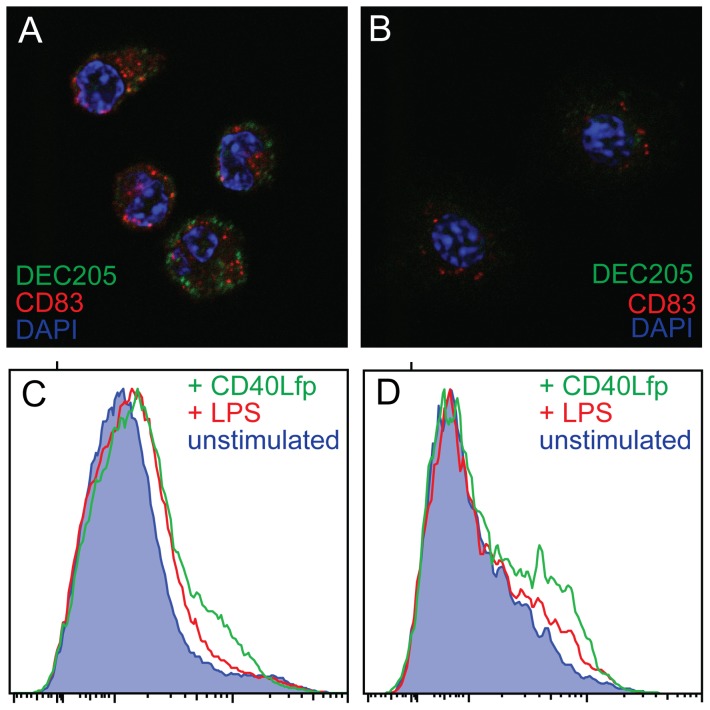
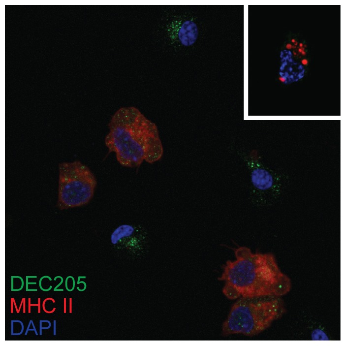
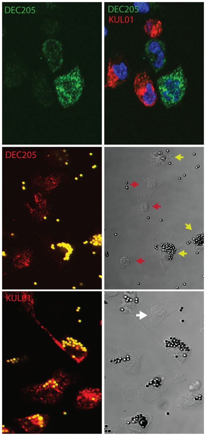
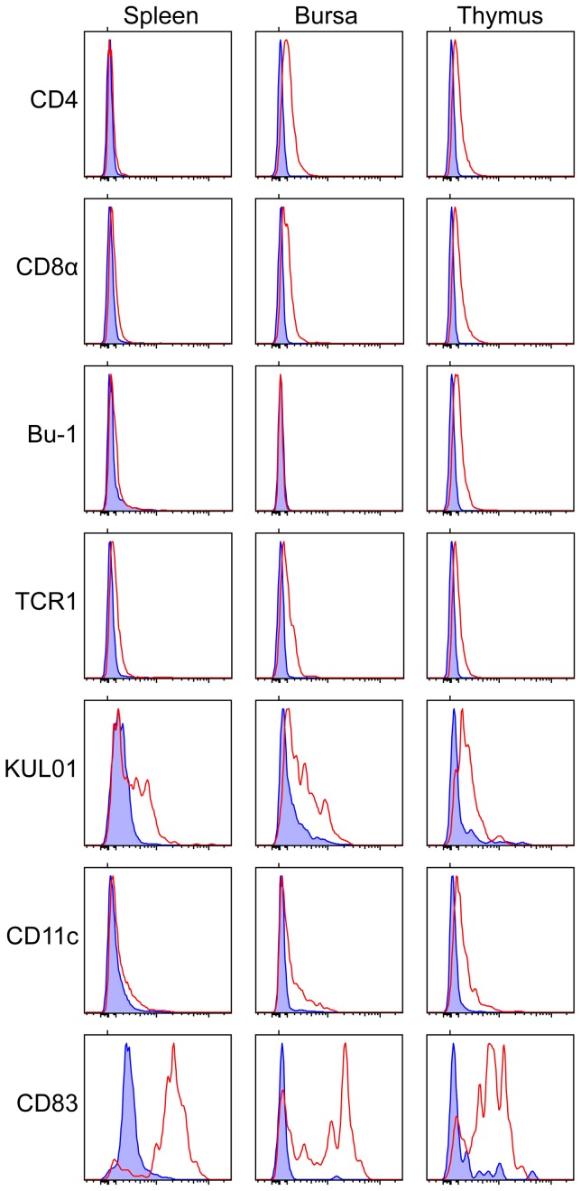
The generation of appropriate adaptive immune responses relies critically on dendritic cells, about which relatively little is known in chickens, a vital livestock species, in comparison with man and mouse. We cloned and sequenced chicken DEC205 cDNA and used this knowledge to produce quantitative PCR assays and monoclonal antibodies to study expression of DEC205 as well as CD83. The gene structure of DEC205 was identical to those of other species. Transcripts of both genes were found at higher levels in lymphoid tissues and the expression of DEC205 in normal birds had a characteristic distribution in the primary lymphoid organs. In spleen, DEC205 was seen on cells ideally located to trap antigen. In thymus it was found on cells thought to participate in the education of T cells, and in the bursa on cells that may be involved in presentation of antigen to B cells and regulation of B cell migration. The expression of DEC205 on cells other than antigen presenting cells (APC) is also described. Isolated splenocytes strongly expressing DEC205 but not the KUL01 antigen have morphology similar to mammalian dendritic cells and the distinct expression of DEC205 within the avian-specific Bursa of Fabricius alludes to a unique function in this organ of B cell diversification.
Conflict of interest statement
Figures
Similar articles
-
Unique features and distribution of the chicken CD83+ cell.J Immunol. 2007 Oct 15;179(8):5117-25. doi: 10.4049/jimmunol.179.8.5117. J Immunol. 2007. PMID: 17911597
-
Glycans from avian influenza virus are recognized by chicken dendritic cells and are targets for the humoral immune response in chicken.Mol Immunol. 2013 Dec;56(4):452-62. doi: 10.1016/j.molimm.2013.06.007. Epub 2013 Aug 1. Mol Immunol. 2013. PMID: 23911401
-
Full-length cDNAs from chicken bursal lymphocytes to facilitate gene function analysis.Genome Biol. 2005;6(1):R6. doi: 10.1186/gb-2004-6-1-r6. Epub 2004 Dec 23. Genome Biol. 2005. PMID: 15642098 Free PMC article.
-
Somatic diversification of the chicken immunoglobulin light-chain gene.Adv Immunol. 1990;48:41-67. doi: 10.1016/s0065-2776(08)60751-8. Adv Immunol. 1990. PMID: 2112303 Review.
-
Antibodies, immunoglobulin genes and the bursa of Fabricius in chicken B cell development.Dev Comp Immunol. 2006;30(1-2):101-18. doi: 10.1016/j.dci.2005.06.018. Dev Comp Immunol. 2006. PMID: 16139886 Review.
Cited by
-
Evolution of an expanded mannose receptor gene family.PLoS One. 2014 Nov 12;9(11):e110330. doi: 10.1371/journal.pone.0110330. eCollection 2014. PLoS One. 2014. PMID: 25390371 Free PMC article.
-
Enhancing Protective Efficacy of Poultry Vaccines through Targeted Delivery of Antigens to Antigen-Presenting Cells.Vaccines (Basel). 2018 Nov 15;6(4):75. doi: 10.3390/vaccines6040075. Vaccines (Basel). 2018. PMID: 30445683 Free PMC article. Review.
-
Re-sequencing data for refining candidate genes and polymorphisms in QTL regions affecting adiposity in chicken.PLoS One. 2014 Oct 21;9(10):e111299. doi: 10.1371/journal.pone.0111299. eCollection 2014. PLoS One. 2014. PMID: 25333370 Free PMC article.
-
The Functional Mechanism of BP9 in Promoting B Cell Differentiation and Inducing Antigen Presentation.Vaccines (Basel). 2024 Jun 1;12(6):607. doi: 10.3390/vaccines12060607. Vaccines (Basel). 2024. PMID: 38932336 Free PMC article.
-
The Well-Developed Mucosal Immune Systems of Birds and Mammals Allow for Similar Approaches of Mucosal Vaccination in Both Types of Animals.Front Nutr. 2018 Jul 12;5:60. doi: 10.3389/fnut.2018.00060. eCollection 2018. Front Nutr. 2018. PMID: 30050906 Free PMC article. Review.
References
-
- Steinman RM (1991) The dendritic cell system and its role in immunogenicity. Annu Rev Immunol 9: 271–296. - PubMed
-
- Kaspers B, Kothlow S, Butter C (2008) Avian Antigen Presenting Cells. In: Davison F, Kaspers B, Schat KA, editors. Avian Immunology. Ammsterdam: Academic Press. pp. 183–202.
-
- Wu Z, Kaiser P (2011) Antigen presenting cells in a non-mammalian model system, the chicken. Immunobiology 216: 1177–1183. - PubMed
-
- Wu Z, Hu T, Kaiser P (2011) Chicken CCR6 and CCR7 are markers for immature and mature dendritic cells respectively. Dev Comp Immunol 35: 563–567. - PubMed
Publication types
MeSH terms
Substances
Grants and funding
LinkOut - more resources
Full Text Sources
Other Literature Sources
Medical
