. 2013;8(1):e52287.
doi: 10.1371/journal.pone.0052287. Epub 2013 Jan 11.

Apolipoprotein(a) inhibits in vitro tube formation in endothelial cells: identification of roles for Kringle V and the plasminogen activation system

Affiliations

Apolipoprotein(a) inhibits in vitro tube formation in endothelial cells: identification of roles for Kringle V and the plasminogen activation system

Lei Liu et al. PLoS One. 2013.

Abstract

Elevated plasma concentrations of lipoprotein(a) are associated with increased risk for atherothrombotic diseases. Apolipoprotein(a), the unique glycoprotein component of lipoprotein(a), is characterized by the presence of multiple kringle domains, and shares a high degree of sequence homology with the serine protease zymogen plasminogen. It has been shown that angiostatin, a proteolytic fragment of plasminogen containing kringles 1-4, can effectively inhibit angiogenesis. Moreover, proteolytic fragments of plasminogen containing kringle 5 are even more potent inhibitors of angiogenesis than angiostatin. Despite its strong similarity with plasminogen, the role of apolipoprotein(a) in angiogenesis remains controversial, with both pro- and anti-angiogenic effects reported. In the current study, we evaluated the ability of apolipoprotein(a) to inhibit VEGF- and angiopoietin-induced tube formation in human umbilical cord endothelial cells. A 17 kringle-containing form of recombinant apo(a) (17K), corresponding to a well-characterized, physiologically-relevant form of the molecule, effectively inhibited tube formation induced by either VEGF or angiopoietin-1. Using additional recombinant apolipoprotein(a) (r-apo(a)) variants, we demonstrated that this effect was dependent on the presence of an intact lysine-binding site in kringle V domain of apo(a), but not on the presence of the functional lysine-binding site in apo(a) kringle IV type 10; sequences within in the amino-terminal half of the molecule were also not required for the inhibitory effects of apo(a). We also showed that the apo(a)-mediated inhibition tube formation could be reversed, in part by the addition of plasmin or urokinase plasminogen activator, or by removal of plasminogen from the system. Further, we demonstrated that apo(a) treated with glycosidases to remove sialic acid was significantly less effective in inhibiting tube formation. This is the first report of a functional role for the glycosylation of apo(a) although the mechanisms underlying this observation remain to be determined in the context of angiogenesis.


Conflict of interest statement

Competing Interests: The authors have declared that no competing interests exist.

Figures

Figure 1
Figure 1. Recombinant apo(a) variants used in the study.
The topology of the r-apo(a) variants used is shown in this schematic diagram. The top line represents the organization of the 17K r-apo(a) variant, which corresponds to a physiological apo(a) isoform and includes all 10 types of kringle IV sequences present in apo(a) isoforms, as well as the kringle V (V) and protease-like (P) domains. The black dot within a kringle denotes the presence of an amino acid substitution that inactivates the lysine binding site (LBS) in that kringle. The bar above KIV9 denotes the unpaired cysteine residue in this kringle that mediates covalent attachment to apoB-100 in LDL.
Figure 2
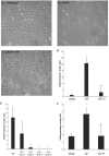
Figure 2. 17K r-apo(a) blocks capillary-like tube formation induced by VEGF and angiopoietin-1 in three-dimensional fibrin matrices.
The tube formation assay was conducted essentially as described by Babaei and colleagues . HUVECs were cultured on fibrin matrices in the absence (A) or presence (B) of VEGF (20 ng/mL), or with VEGF and 100 nM 17K r-apo(a) (C). After 24 hours, light microscopy images of six randomly preselected fields for each treatment were taken using a digital camera (10×) and analyzed by a computer-assisted morphometric analysis system. For each image, the differentiation index (DI) was calculated as the ratio of total tube (capillary-like structures >30 mm) length divided by the total area of residual EC monolayer for the same field. D–F: Quantitative analysis of capillary-like network formation calculated as DI. Scale bars represent the mean ± standard deviation of the DI value obtained for each culture well from six independent images; data shown are representative of three independent experiments. Asterisks: p<0.01 versus untreated; pilcrow sign (¶): p<0.01 versus VEGF or Ang-1 (100 ng/mL), as appropriate.
Figure 3
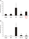
Figure 3. The effects of plasmin and MMP-9 on the 17K apo(a)-mediated inhibition of HUVEC tube formation.
HUVECs were cultured on fibrin matrices with the indicated treatments (VEGF: 20 ng/mL; 17K r-apo(a): 100 nM; plasmin (A): 50 nM; MMP-9 (B): 8 nM). Tube formation was measured as described in the legend to Figure 2. Scale bars represent the mean ± standard deviation of the DI value obtained for each culture well from six independent images; data shown are representative of three independent experiments. Asterisks: p<0.01 versus untreated; daggers: p<0.01 versus VEGF.
Figure 4
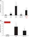
Figure 4. The effects of addition of uPA and depletion of plasminogen on the 17K apo(a)-mediated inhibition of HUVEC tube formation.
HUVECs were cultured on fibrin matrices with the indicated treatments (VEGF: 20 ng/mL; 17K r-apo(a): 100 nM; uPA (A): 0.5 nM). Experiments in B were conducted using plasminogen-depleted serum. Tube formation was measured as described in the legend to Figure 2. Scale bars represent the mean ± standard deviation of the DI value obtained for each culture well from six independent images; data shown are representative of three independent experiments. Asterisks: p<0.01 versus untreated; daggers: p<0.01 versus VEGF.
Figure 5
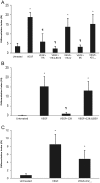
Figure 5. Effects of different r-apo(a) variants on HUVEC tube formation.
HUVECs were cultured on fibrin matrices with the indicated treatments (VEGF: 20 ng/mL; 17K r-apo(a) and all other variants: 100 nM). Tube formation was measured as described in the legend to Figure 2. Scale bars represent the mean ± standard deviation of the DI value obtained for each culture well from six independent images; data shown are representative of three independent experiments. Asterisks: p<0.01 versus untreated; daggers: p<0.01 versus VEGF.
Figure 6
Figure 6. Effect of deglycosylation on the ability of r-apo(a) to inhibit HUVEC tube formation.
17K r-apo(a) was incubated with CPS overnight at 37°C. The deglycosylated material was subjected to SDS-PAGE under non-reducing (NR) or reducing conditions, followed by silver-staining (inset to graph). HUVECs were cultured on fibrin matrices with the indicated treatments (VEGF: 20 ng/mL; 17K r-apo(a), either untreated or treated with CPS: 100 nM; CPS: 0.14 U/mL, equivalent to the amount of CPS added along with the CPS-treated apo(a)). Tube formation was measured as described in the legend to Figure 2. Scale bars represent the mean ± standard deviation of the DI value obtained for each culture well from six independent images; data shown are representative of three independent experiments. Asterisks: p<0.01 versus untreated; daggers: p<0.01 versus VEGF.
Figure 7
Figure 7. Complex effects of apo(a) on endothelial cell function.
(1) Through a mechanism involving, in a manner yet to be defined, integrin αVβ3, apo(a) induces Src and MAP kinase activation that results in increased migration and proliferation . (2) Through binding to a cell surface receptor that is currently undefined, apo(a) results in activation of Rho, which through its effector Rho kinase (RhoK) ultimately results in actin/myosin stress fiber formation and increased endothelial cell contraction and vascular permeability . In addition, through the dissociation of β-catenin form adherens junction and the promotion of β-catenin nuclear translocation through activation of Akt, apo(a) elicits an increase in the expression of cyclooxygenase-2 which results in increased synthesis of prostaglandin E2 (PGE2) (unpublished observations). (3) Apo(a) inhibits pericellular plasminogen activation that is likely dependent on uPA and the uPA receptor (uPAR), an effect that results in a decrease in tube formation (current study). (4) Through unknown mechanisms, apo(a) results in increases in the expression of several adhesion molecule genes including ICAM-1 and E-selectin .

Similar articles

Cited by

References

    1. Tsimikas S, Hall JL (2012) Lipoprotein(a) as a potential causal genetic risk factor of cardiovascular disease. J Am Coll Cardiol 60: 716–721. - PubMed
    1. Koschinsky ML, Côté GP, Gabel B, van der Hoek YY (1993) Identification of the cysteine residue in apolipoprotein(a) that mediates extracellular coupling with apolipoprotein B-100. J Biol Chem 268: 19819–19825. - PubMed
    1. McLean JW, Tomlinson JE, Kuang WJ, Eaton DL, Chen EY, et al. (1987) cDNA sequence of human apolipoprotein(a) is homologous to plasminogen. Nature 330: 132–137. - PubMed
    1. Koschinsky ML, Marcovina SM (2009) Lipoprotein(a). In Clinical Lipidology: A Companion to Braunwald's Heart Disease. (ed. C.M. Ballantyne), Saunders (Elsevier), Philadelphia. pp. 130–143.
    1. van der Hoek YY, Wittekoek ME, Beisiegel U, Kastelein JJ, Koschinsky ML (1993) The apolipoprotein(a) kringle IV repeats which differ from the major repeat kringle are present in variably-sized isoforms. Hum Mol Genet 2: 361–366. - PubMed

Publication types

MeSH terms

LinkOut - more resources